. 2020 Jan 21;117(3):1779-1787.
doi: 10.1073/pnas.1915943117. Epub 2020 Jan 6.

Specialized stellate cells offer a privileged route for rapid water flux in Drosophila renal tubule

Affiliations

Specialized stellate cells offer a privileged route for rapid water flux in Drosophila renal tubule

Pablo Cabrero et al. Proc Natl Acad Sci U S A. .

Abstract

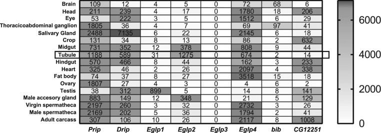
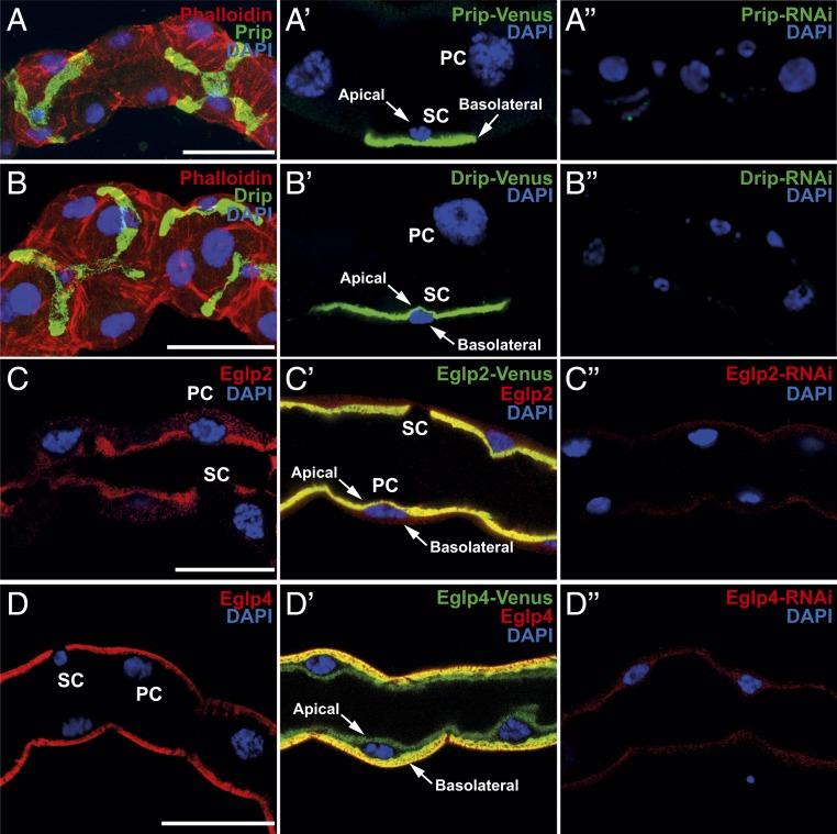
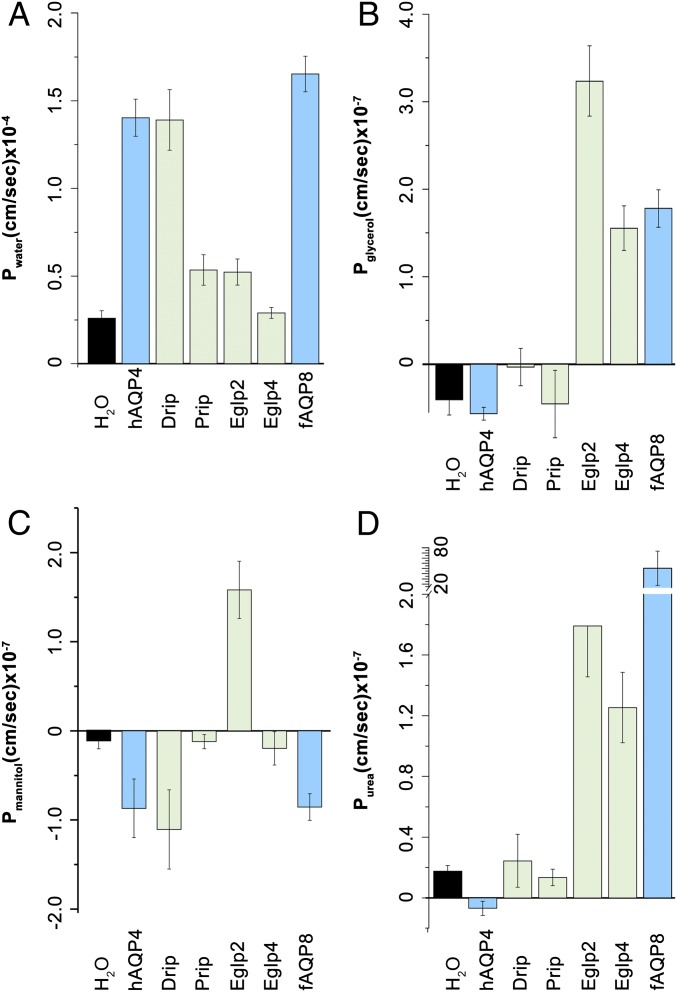
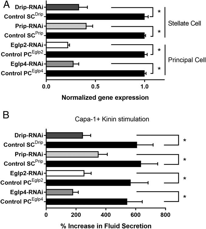
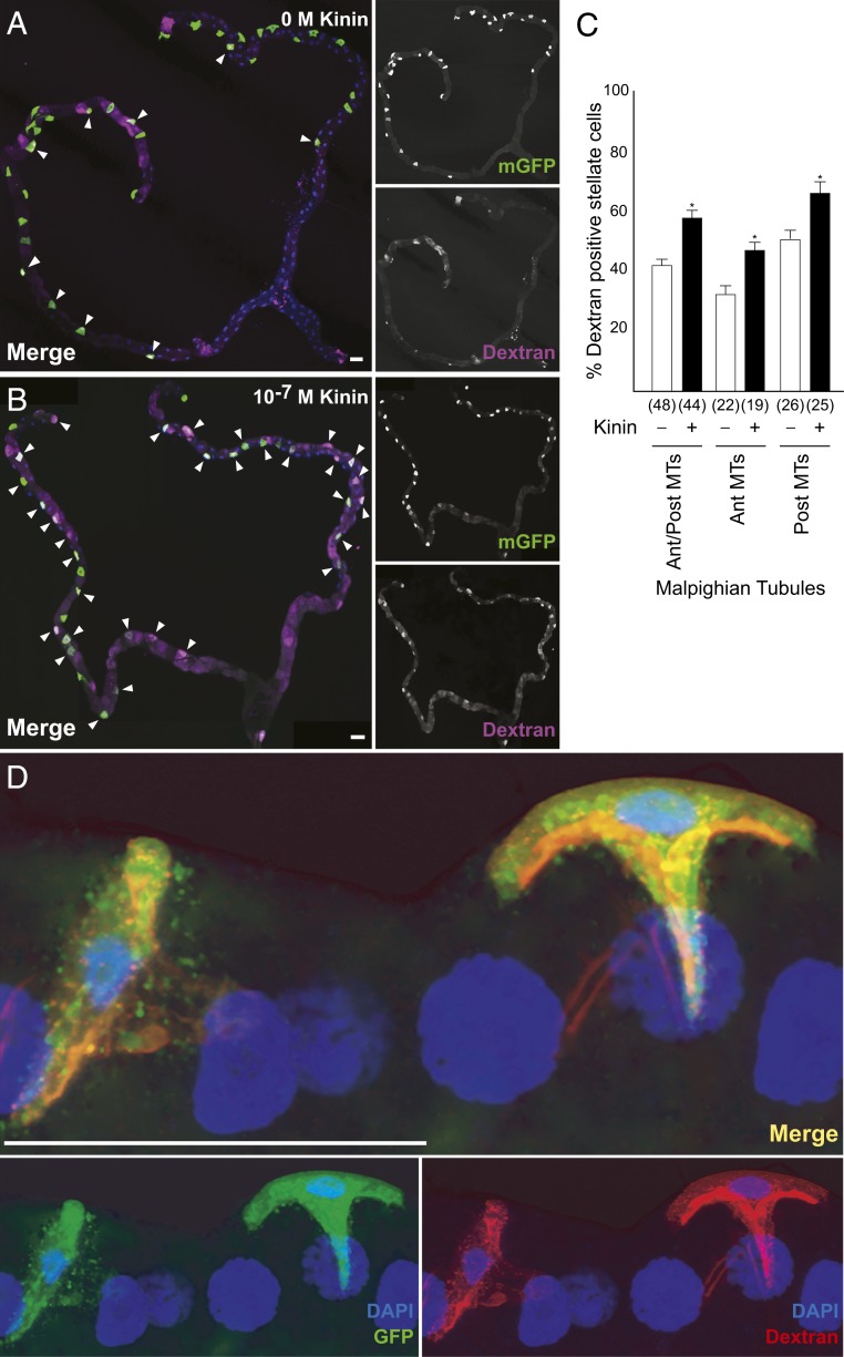
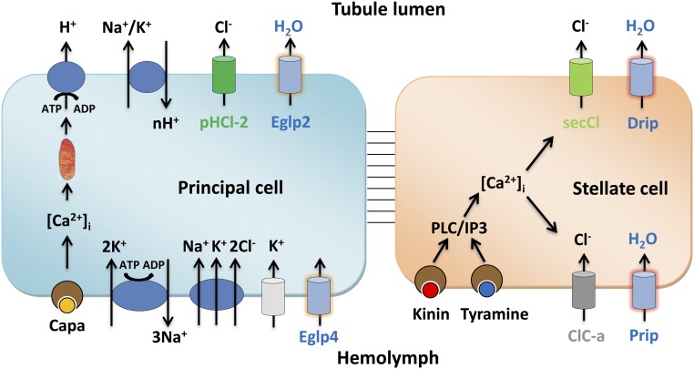
Insects are highly successful, in part through an excellent ability to osmoregulate. The renal (Malpighian) tubules can secrete fluid faster on a per-cell basis than any other epithelium, but the route for these remarkable water fluxes has not been established. In Drosophila melanogaster, we show that 4 genes of the major intrinsic protein family are expressed at a very high level in the fly renal tissue: the aquaporins (AQPs) Drip and Prip and the aquaglyceroporins Eglp2 and Eglp4 As predicted from their structure, and by their transport function by expressing these proteins in Xenopus oocytes, Drip, Prip, and Eglp2 show significant and specific water permeability, whereas Eglp2 and Eglp4 show very high permeability to glycerol and urea. Knockdowns of any of these genes result in impaired hormone-induced fluid secretion. The Drosophila tubule has 2 main secretory cell types: active cation-transporting principal cells, wherein the aquaglyceroporins localize to opposite plasma membranes, and small stellate cells, the site of the chloride shunt conductance, with these AQPs localizing to opposite plasma membranes. This suggests a model in which osmotically obliged water flows through the stellate cells. Consistent with this model, fluorescently labeled dextran, an in vivo marker of membrane water permeability, is trapped in the basal infoldings of the stellate cells after kinin diuretic peptide stimulation, confirming that these cells provide the major route for transepithelial water flux. The spatial segregation of these components of epithelial water transport may help to explain the unique success of the higher insects in regulating their internal environments.

Keywords: Drosophila melanogaster; Malpighian tubule; Xenopus oocyte; aquaporin; stellate cell.


Conflict of interest statement

The authors declare no competing interest.

Figures

Fig. 1.
Fig. 1.
MIP family expression in Drosophila melanogaster. Data mining of FlyAtlas.org identified 4 MIP genes (Prip, Drip, Eglp2, and Eglp4) with highly abundant expression in adult Malpighian tubules. FlyAtlas expression levels are derived from normalized Affymetrix microarray data, and are shaded according to the scale on the right.
Fig. 2.
Fig. 2.
Subcellular localization of MIPs in the Drosophila tubule. (A and B) Prip and Drip localize to opposite plasma membranes of the stellate cell (SC). (A) Prip is expressed in basolateral membrane (green) and is in contact with the outside of the tubule, or hemolymph and (B) Drip in apical membrane (green) and faces the lumen of the tubule. DAPI (blue) staining for nuclei and phalloidin (red) staining for actin are shown. Blue (DAPI) nuclei are shown to allow distinction between basal and apical distribution of MIPs, emphasized with arrows. (A′ and B′) Overexpression of Prip and Drip labeled with Venus (eYFP) recapitulate the pattern of expression observed by immunocytochemistry. (C and D) Eglp2 and Eglp4 localize to opposite, plasma membranes of the principal cell (PC). (C) Eglp2 is expressed in apical membrane (red) and (D) Eglp4 is expressed in basolateral membrane (red), and DAPI (blue). (C′ and D′) Colocalization (yellow) (C′) between Eglp2-Venus and Eglp2 to the apical membrane and (D′) between Eglp4-Venus and Eglp4 to the basolateral membrane. (A″D″) Down-regulation of MIPs in specific cell types using RNAi reduces protein levels. (Scale bar, 40 µm.)
Fig. 3.
Fig. 3.
Transport specificity of Drosophila tubule-enriched MIPs. Water-injected control oocytes or oocytes expressing Drosophila MIPs (Drip, Prip, Eglp2, and Eglp4), human AQP4 (hAQP4, a control classical AQP), and mefugu AQP8 (fAQP8, a control aquaglyceroporin) were tested for permeability of (A) water (Pf, PH2O), (B) urea (Purea), (C) glycerol (Pglycerol), and (D) mannitol (Pmannitol).
Fig. 4.
Fig. 4.
Validation of MIPs knockdowns and impact of cell-specific down-regulation of MIPs on fluid secretion. (A) Effects of knockdowns on tubule messenger RNA (mRNA) levels for MIPs, validated by qPCR. Cell-specific down-regulation of Eglp2 and Eglp4 in principal cells and Drip and Prip in stellate cells using their respective UAS-dsRNA lines (protein knockdown achieved by these lines is shown in Fig. 2). Data are expressed as mean fold change compared to parental controls ± SEM (n = 3). *P < 0.05 (Student’s t test). (B) Impact of cell-specific knockdowns of MIPs on stimulated fluid secretion by tubules in response to Capa-1 and Kinin at 10−7 M. Data are expressed as percentage increase from basal fluid secretion compared to parental controls ±SEM (n = 6 to 10). *P < 0.05 (Student’s t test).
Fig. 5.
Fig. 5.
Dextran labeling demonstrates water flux specific to the stellate cells. Accumulation of dextran (A) of unstimulated and (B) following application of Kinin (10−7 M) in tubule expressing mGFP in stellate cells. Because of the length of the tubule, B is a photomontage of 3 fields, all captured with the same microscope settings. (C) Quantification of dextran labeling. Data are expressed as percentage of dextran-positive stellate cells in response to 10−7 M Kinin compared to unstimulated tubule ± SEM (n = 44 to 48). *P < 0.05 (Student’s t test). (D) Maximum Z projection of tubules after application of 40 kDa of dextran conjugated to TRITC dye (red) to tubules in which stellate cells are expressing GFP (green) confirmed the accumulation of dextran to the stellate cell; DAPI, blue. (Scale bars, 50 μm.)
Fig. 6.
Fig. 6.
Dextran labeling as a tool to probe insect biodiversity. The dextran labeling protocol employed for D. melanogaster was applied to other insect species, selected from the major insect orders of exopterygotes and endopterygotes. The distribution of known stellate cells and Kinin labeling (diagnostic of the route of chloride shunt conductance) is adapted from ref. ; green denotes positive (gene is present, or kinin receptor localization confined to stellate cells); red denotes absence, and gray denotes not known or ambiguous. In all species, the main region [in Manduca, the distal ileac plexus (67, 68)] of the tubule is shown.
Fig. 7.
Fig. 7.
Model for tubule function. The mitochondria-rich principal cell is specialized for metabolically intensive cation and solute transport. The apical V-ATPase sets up a proton electrochemical gradient which drives net K+ secretion via NHA or NHE exchangers. Basolateral K+ entry is afforded by inward rectifier K+ channels, Na+,K+ ATPase and an Na+,K+,2Cl cotransport. The resulting lumen-positive potential drives a chloride shunt conductance, mainly via basolateral ClC-a and apical secCl channels in the stellate cell. The net transport of KCl drives osmotically obliged water, which is primarily via basolateral Prip and apical Drip in the stellate cells. In this way, the metabolically active principal cell is sheltered from the required high flux rates of water.

References

    1. Maddrell S. H. P., The fastest fluid-secreting cell known: The upper Malpighian tubule cell of Rhodnius. BioEssays 13, 357–362 (1991).
    1. Dow J. A. T., et al. , The Malpighian tubules of Drosophila melanogaster: A novel phenotype for studies of fluid secretion and its control. J. Exp. Biol. 197, 421–428 (1994). - PubMed
    1. O’Donnell M. J., Aldis G. K., Maddrell S. H. P., Measurements of osmotic permeability in the Malpighian tubules of an insect, Rhodnius prolixus Stal. Proc. R. Soc. Lond. B Biol. Sci. 216, 267–277 (1982).
    1. Sözen M. A., Armstrong J. D., Yang M., Kaiser K., Dow J. A. T., Functional domains are specified to single-cell resolution in a Drosophila epithelium. Proc. Natl. Acad. Sci. U.S.A. 94, 5207–5212 (1997). - PMC - PubMed
    1. Beyenbach K. W., Skaer H., Dow J. A. T., The developmental, molecular, and transport biology of Malpighian tubules. Annu. Rev. Entomol. 55, 351–374 (2010). - PubMed

Publication types

MeSH terms