. 2020 Jan;15(1):41-46.
doi: 10.1038/s41565-019-0600-1. Epub 2020 Jan 6.

Vitamin lipid nanoparticles enable adoptive macrophage transfer for the treatment of multidrug-resistant bacterial sepsis

Affiliations

Vitamin lipid nanoparticles enable adoptive macrophage transfer for the treatment of multidrug-resistant bacterial sepsis

Xucheng Hou et al. Nat Nanotechnol. 2020 Jan.

Erratum in

Abstract

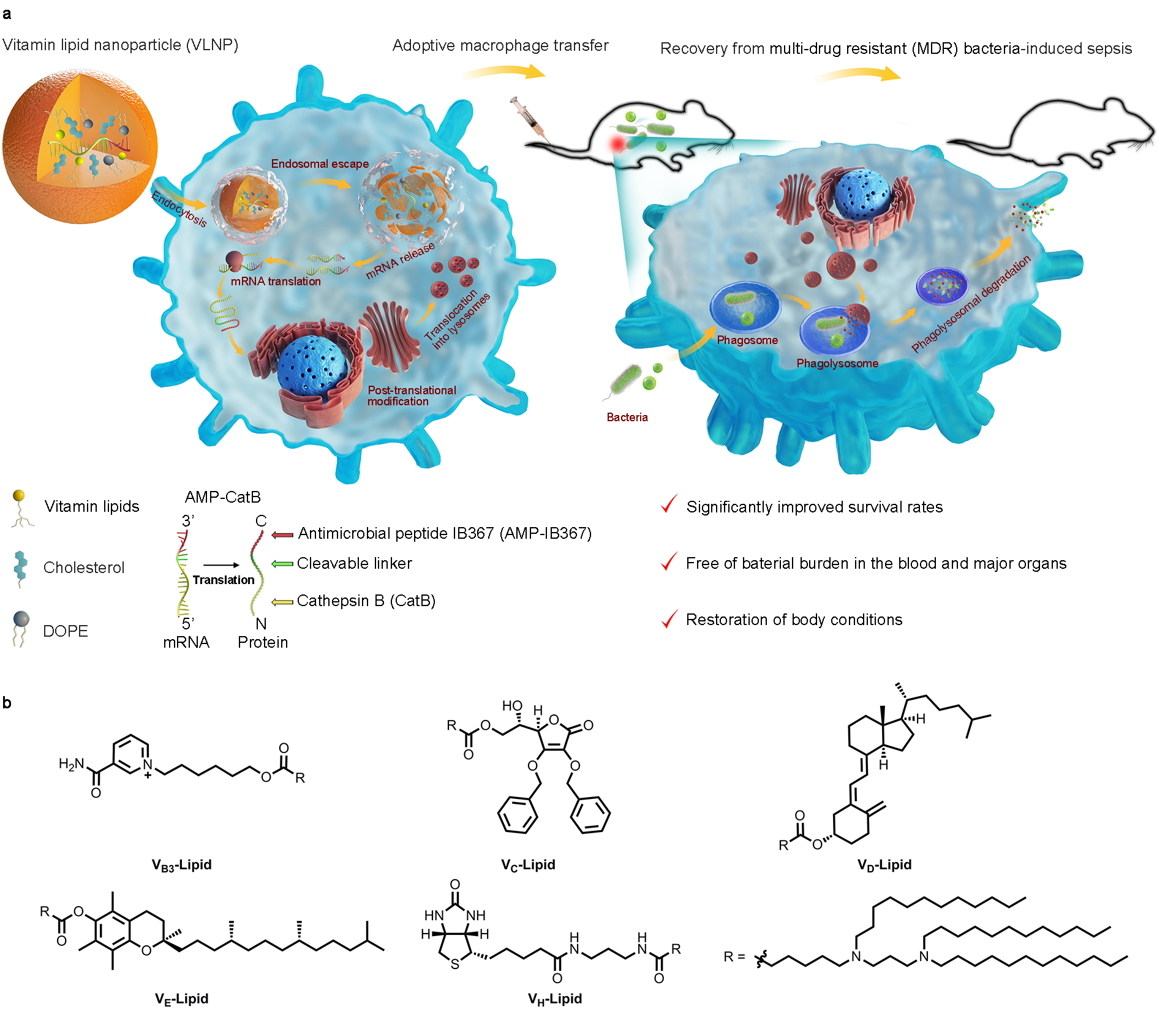
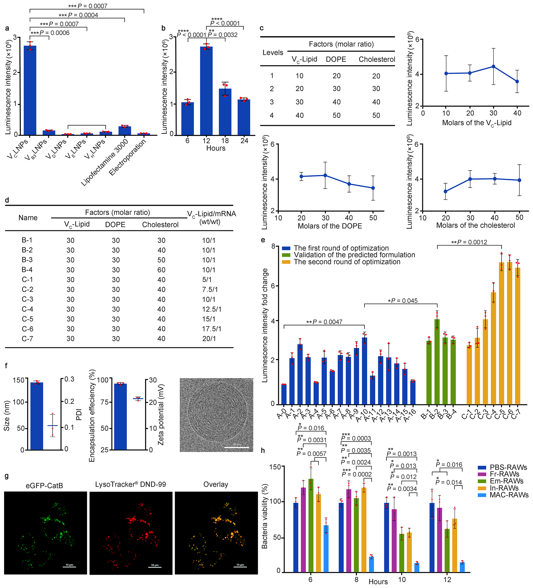
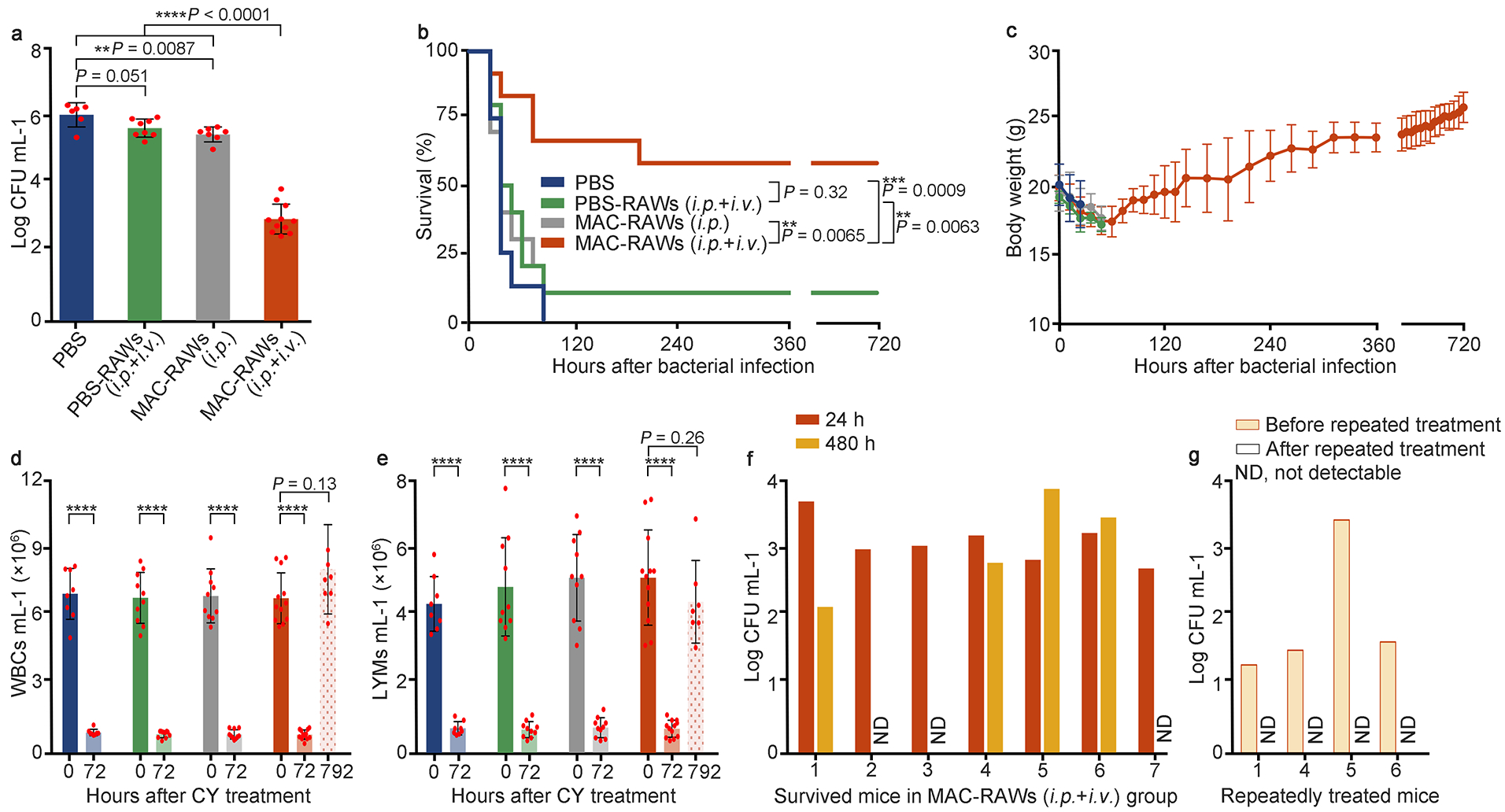
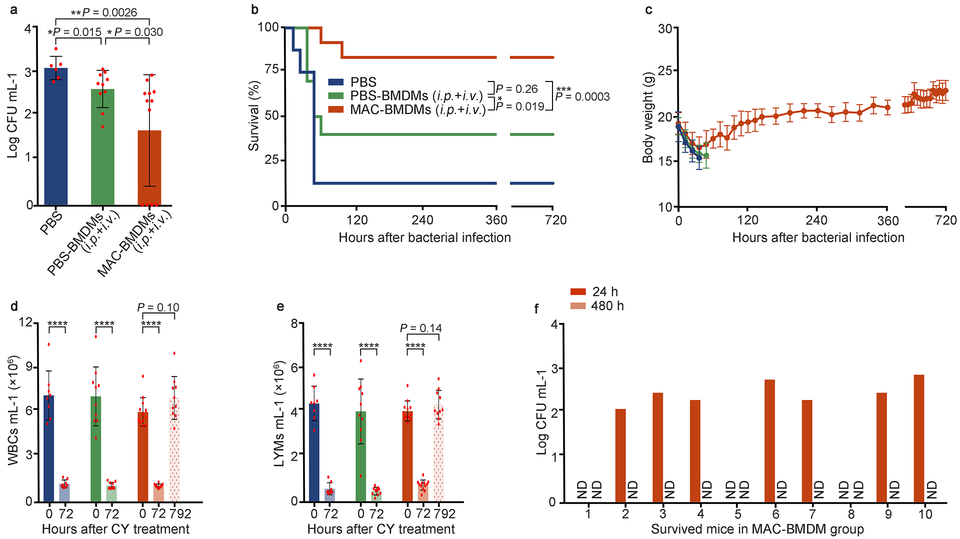
Sepsis, a condition caused by severe infections, affects more than 30 million people worldwide every year and remains the leading cause of death in hospitals1,2. Moreover, antimicrobial resistance has become an additional challenge in the treatment of sepsis3, and thus, alternative therapeutic approaches are urgently needed2,3. Here, we show that adoptive transfer of macrophages containing antimicrobial peptides linked to cathepsin B in the lysosomes (MACs) can be applied for the treatment of multidrug-resistant bacteria-induced sepsis in mice with immunosuppression. The MACs are constructed by transfection of vitamin C lipid nanoparticles that deliver antimicrobial peptide and cathepsin B (AMP-CatB) mRNA. The vitamin C lipid nanoparticles allow the specific accumulation of AMP-CatB in macrophage lysosomes, which is the key location for bactericidal activities. Our results demonstrate that adoptive MAC transfer leads to the elimination of multidrug-resistant bacteria, including Staphylococcus aureus and Escherichia coli, leading to the complete recovery of immunocompromised septic mice. Our work provides an alternative strategy for overcoming multidrug-resistant bacteria-induced sepsis and opens up possibilities for the development of nanoparticle-enabled cell therapy for infectious diseases.


Conflict of interest statement

Competing interests

The authors have no competing interests to declare.

Figures

Fig. 1 |
Fig. 1 |. Schematic illustration of adoptive macrophage transfer and chemical structures of the vitamin-derived lipids.
a, Construction of MACs for sepsis therapy. MACs stands for macrophages containing antimicrobial peptides linked to cathepsin B in the lysosomes (MACs). The AMP-CatB mRNA is encapsulated in the vitamin C lipid nanoparticle (VCLNP) and delivered to the macrophage where the mRNA is translated in the cytoplasm and translocated into the lysosomes. Within the lysosomes, the cleavable linker is cleaved by the lysosomal CatB, releasing the AMP-IB367. After phagosomes carrying MDR bacteria fuse with the lysosomes, the ingested MDR bacteria are eradicated by the pre-stored AMP-IB367. b, Chemical structures of vitamin-derived lipids including VB3-Lipid, VC-Lipid, VD-Lipid, VE-Lipid, and VH-Lipid.
Fig. 2 |
Fig. 2 |. Screening, optimization, and characterization of vitamin lipid nanoparticles (VLNPs).
a, mRNA delivery efficiency of VLNPs in RAW264.7 cells. b, Expression kinetics of mRNA delivered by VCLNPs in RAW264.7 cells. c, The first round of optimization: four levels and impact trend of each VCLNP’s component. d, Formulation table for the validation of the predicted formulation, and the second round of optimization: the mass ratio of VC-Lipid:mRNA. e, Fold changes of luminescence intensity in the two rounds of optimization. f, Characterization of the optimal VCLNP formulation, including size, polydispersity index (PDI), encapsulation efficiency, zeta potential, and Cryo-TEM image (Scale bar = 50 nm). g, Confocal imagines of the RAW264.7 cells incubated with VCLNPs encapsulating eGFP-CatB mRNA (Scale bar = 10 μm). Data in g are representative images from n = 3 independent experiments. h, Intracellular survival of MDR Staphylococcus aureus (MDRSA) in the RAW264.7 cells. Data in a, b, c, e, and h are from n = 3 biologically independent samples. All data are presented as mean ± s.d. Statistical significance was analyzed by the two-tailed Student’s t-test. *P < 0.05, **P < 0.01, ***P < 0.001, ****P < 0.0001.
Fig. 3 |
Fig. 3 |. Therapeutic effects of MAC-RAWs in MDRSA-induced sepsis mice with immunosuppression.
a, Bacterial burden in blood at 24 h after cell transfer. n = 6, 8, 7, and 11 live mice for PBS, PBS-RAWs (i.p. + i.v.), MAC-RAWs (i.p.), and MAC-RAWs (i.p. + i.v.) groups, respectively. b, Percentage survival of mice with sepsis induced by i.p. bacterial inoculation. c-e, The body weights (BWs), white blood cells (WBCs), and lymphocytes (LYMs) of mice. c, BWs; d, WBCs; e, LYMs. Data in b, c, d, and e (except time point 792 h), n = 8, 10, 10, and 12 for PBS, PBS-RAWs (i.p. + i.v.), MAC-RAWs (i.p.), and MAC-RAWs (i.p. + i.v.) groups, respectively. f, g, Bacterial burden in blood of each survived mouse treated by MAC-RAWs (i.p. + i.v.). Data in a, c, d, and e are the mean ± s.d. Statistical significance was analyzed by the two-tailed Student’s t-test. Data in b were analyzed by the log-rank (Mantel–Cox) test. *P < 0.05, **P < 0.01, ***P < 0.001; ns, not significant; ND, not detectable.
Fig. 4 |
Fig. 4 |. Therapeutic effects of MAC-BMDMs in mixed MDR bacteria (Staphylococcus aureus and Escherichia coli)-induced sepsis mice with immunosuppression.
a, Bacterial burden in blood at 24 h after cell transfer. n = 6, 10, and 12 live mice for PBS, PBS-BMDMs (i.p. + i.v.), and MAC-BMDMs (i.p. + i.v.) groups, respectively. b, Percentage survival of mice with sepsis induced by i.p. bacterial inoculation. c-e, The BWs, WBCs, and LYMs of mice. c, BWs; d, WBCs; e, LYMs. Data in b, c, d, and e (except time point 792 h), n = 8, 10, and 12 for PBS, PBS-BMDMs (i.p. + i.v.), and MAC-RAWs (i.p. + i.v.) groups, respectively. f, Bacterial burden in blood of each survived mouse treated by MAC-BMDMs. Data in a, c, d, and e are the mean ± s.d. Statistical significance was analyzed by the two-tailed Student’s t-test. Data in b were analyzed by the log-rank (Mantel–Cox) test. *P < 0.05, **P < 0.01, ***P < 0.001; ns, not significant; ND, not detectable.

Similar articles

Cited by

References

    1. Reinhart K, et al. Recognizing sepsis as a global health priority—a WHO resolution. New England Journal of Medicine 377, 414–417 (2017). - PubMed
    1. van der Poll T Immunotherapy of sepsis. The Lancet infectious diseases 1, 165–174 (2001). - PubMed
    1. Huttunen R & Aittoniemi J New concepts in the pathogenesis, diagnosis and treatment of bacteremia and sepsis. Journal of Infection 63, 407–419 (2011). - PubMed
    1. Hotchkiss RS & Karl IE The pathophysiology and treatment of sepsis. New England Journal of Medicine 348, 138–150 (2003). - PubMed
    1. Otto GP, et al. The late phase of sepsis is characterized by an increased microbiological burden and death rate. Critical care 15, R183 (2011). - PMC - PubMed

Publication types

MeSH terms