. 2020 Jan;26(1):52-58.
doi: 10.1038/s41591-019-0715-9. Epub 2020 Jan 6.

Near real-time intraoperative brain tumor diagnosis using stimulated Raman histology and deep neural networks

Affiliations

Near real-time intraoperative brain tumor diagnosis using stimulated Raman histology and deep neural networks

Todd C Hollon et al. Nat Med. 2020 Jan.

Abstract

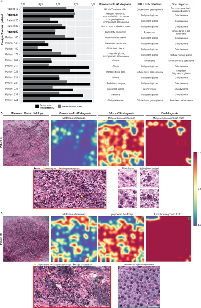
Intraoperative diagnosis is essential for providing safe and effective care during cancer surgery1. The existing workflow for intraoperative diagnosis based on hematoxylin and eosin staining of processed tissue is time, resource and labor intensive2,3. Moreover, interpretation of intraoperative histologic images is dependent on a contracting, unevenly distributed, pathology workforce4. In the present study, we report a parallel workflow that combines stimulated Raman histology (SRH)5-7, a label-free optical imaging method and deep convolutional neural networks (CNNs) to predict diagnosis at the bedside in near real-time in an automated fashion. Specifically, our CNNs, trained on over 2.5 million SRH images, predict brain tumor diagnosis in the operating room in under 150 s, an order of magnitude faster than conventional techniques (for example, 20-30 min)2. In a multicenter, prospective clinical trial (n = 278), we demonstrated that CNN-based diagnosis of SRH images was noninferior to pathologist-based interpretation of conventional histologic images (overall accuracy, 94.6% versus 93.9%). Our CNNs learned a hierarchy of recognizable histologic feature representations to classify the major histopathologic classes of brain tumors. In addition, we implemented a semantic segmentation method to identify tumor-infiltrated diagnostic regions within SRH images. These results demonstrate how intraoperative cancer diagnosis can be streamlined, creating a complementary pathway for tissue diagnosis that is independent of a traditional pathology laboratory.


Conflict of interest statement

Competing interests: D.A.O. is an advisor and shareholder of Invenio Imaging, Inc., a company developing SRH microscopy systems. C.W.F., Z.U.F., and J.T. are employees and shareholders of Invenio Imaging, Inc.

Figures

Extended Data Figure 1:
Extended Data Figure 1:. SRH image dataset and CNN training
The class distribution of (a) training and (b) validation set images are shown as number of patches and patients. Class imbalance results from different incidence rates among human central nervous system tumors. The training set contains over 50 patients for each of the five most common tumor types (malignant gliomas, meningioma, metastasis, pituitary adenoma, and diffuse lower grade gliomas). In order to maximize the number of training images, no cases from medulloblastoma or pilocytic astrocytoma were included in the validation set and oversampling was used to augment the underrepresented class during CNN training. c, Training and validation categorical cross entropy loss and patch-level accuracy is plotted for the training session that yielded the model used for our prospective clinical trial. Training accuracy converges to near-perfect with a peak validation accuracy of 86.4% following epoch 8. Training procedure was repeated 10 times with similar accuracy and cross entropy convergence. Additional training did not result in better validation accuracy and early stopping criteria were reached.
Extended Data Figure 2:
Extended Data Figure 2:. A taxonomy of intraoperative SRH diagnostic classes to inform intraoperative decision making
a, Representative example SRH images from each of the 13 diagnostic class are shown. Both diffuse astrocytoma and oligodendroglioma are shown as examples of diffuse lower grade gliomas. Classic histologic features (i.e., piloid process in pilocytic astrocytomas, whorls in meningioma, and microvascular proliferation in glioblastoma) can be appreciated, in addition to features unique to SRH images (e.g., axons in gliomas and normal brain tissue). Scale bar, 50 μm. b, A taxonomy of diagnostic classes was selected specifically to inform intraoperative decision making, rather than to match WHO classification. Essential intraoperative distinctions, such as tumoral versus nontumoral tissue or surgical versus nonsurgical tumors, allow for safer and more effective surgical treatment. Inference node probabilities inform intraoperative distinctions by providing coarse classification with potentially higher accuracy due to summation of daughter node probabilities. The probability of any inference node is the sum of all of its daughter node probabilities.
Extended Data Figure 3:
Extended Data Figure 3:. Inference algorithm for patient-level brain tumor diagnosis
A patch-based classifier that uses high-magnification, high-resolution images for diagnosis requires a method to aggregate patch-level predictions into a single intraoperative diagnosis. Our inference algorithm performs a feedforward pass on each patch from a patient, filters the nondiagnostic patches (line 12), and stores the output softmax vectors in an RN x array. Each column of the array, corresponding to each class, is summed and renormalized (line 22) to produce a probability distribution. We then used a thresholding procedure such that if greater than 90% of the probability density is nontumor/normal, that probability distribution is returned. Otherwise, the normal/nontumor class (grey matter, white matter, gliosis) probabilities are set to zero (line 31), the distribution renormalized, and returned. This algorithm leverages the observation that normal brain and nondiagnostic tissue imaged using SRH have similar features across patients resulting in high patch-level classification accuracy. Using the expected value of the renormalized patient-level probability distribution for the intraoperative diagnosis eliminates the need to train an additional classifier based on patch predictions.
Extended Data Figure 4.
Extended Data Figure 4.. Prospective clinical trial design and recruitment
a, Minimum sample size was calculated under the assumption that pathologists’ multiclass diagnostic accuracy ranges from 93% to 97% based on our previous experiments and that a clinically significant lower accuracy bound was less than 91%. We, therefore, selected an expected accuracy of 96% and equivalence/non-inferiority limit, or delta, of 5%, yielding a non-inferiority threshold accuracy of 91% or greater. Minimum sample size was 264 (black point) patients using an alpha of 0.05 and a power of 0.9 (beta = 0.1). b, Flowchart of specimen processing in both the control and experimental arms is shown. c, A total of 302 patients met inclusion criteria and were enrolled for intraoperative SRH imaging. Eleven patients were excluded at the time of surgery due to specimens that were below the necessary quality for SRH imaging. A total of 291 patients were imaged intraoperatively and 13 patients were subsequently excluded due to a Mahalanobis distance-based confidence score (See Extended Data Figure 5), resulting in a total of 278 patients included. d, Meningioma, pituitary adenomas, and malignant gliomas were the most common diagnoses in our prospective cohort. University of Michigan, University of Miami, and Columbia University recruited 55.0%, 26.6%, 18.4% of the total patients, respectively.
Extended Data Figure 5.
Extended Data Figure 5.. Mahalanobis distance-based confidence score
a, Pairwise comparison and b, principal component analysis of class conditional Mahalanobis distance-based confidence score for each layer output included in the ensemble. The confidence score from the mid- and high-level hidden features are correlated, which demonstrate that out-of-distribution samples result in greater Mahalanobis distances throughout the network. As previously described and observed in our results, out-of-distribution (i.e. rare tumors) are better detected in the representation space of deep neural networks, rather than the “label-overfitted” output space of the softmax layer. c, Specimen-level predictions (black hashes, n = 478) and kernel density estimate from the trained LDA classifier for all specimens imaged during the trial period projected onto the linear discriminant axis. Trial and rare tumor cases were linearly separable resulting in all 13 rare tumor cases imaged during the trial period correctly identified. d, SRH mosaics of rare tumors imaged during the trial period are shown. Germinomas show classic large round neoplastic cells with abundant cytoplasm and fibrovascular septae with mature lymphocytic infiltrate. Choroid plexus papilloma shows fibrovascular cores lined with columnar cuboidal epithelium. Papillary craniopharyngioma have fibrovascular cores with well-differentiated monotonous squamous epithelium. Clival chordoma has unique bubbly cytoplasm (i.e., physaliferous cells). Scale bar, 50 μm.
Extended Data Figure 6.
Extended Data Figure 6.. Error analysis of pathologist-based classification of brain tumors
a, The true class probability and intersection over union values for each of the prospective clinical trial patients incorrectly classified by the pathologists. All 17 were correctly classified using SRH plus CNN. All incorrect cases underwent secondary review by two board-certified neuropathologists (S.C.P., P.C.) to ensure the specimens were 1) of sufficient quality to make a diagnosis and 2) contained tumor tissue. b, SRH mosaic from patient 21 (glioblastoma, WHO IV) is shown. Pathologist classification was metastatic carcinoma; however, CNN metastasis heatmap does not show high probability. Malignant glioma probability heatmap shows high probability over the majority of the SRH mosaic, with a 73.4% probability of patient-level malignant glioma diagnosis. High-magnification views show regions of hypercellularity due to tumor infiltration of brain parenchyma with damaged axons, activated lipid-laden microglia, mitotic figures, and multinucleated cells. c, SRH mosaic from patient 52 diagnosed with diffuse large B-cell lymphoma predicted to be metastatic carcinoma by pathologist. While CNN identified patchy areas of metastatic features within the specimen, the majority of the image was correctly classified as lymphoma. High-magnification views show atypical lymphoid cells with macrophage infiltration. Regions with large neoplastic cells share cytologic features with metastatic brain tumors, as shown in Figure 3. Scale bar, 50 μm.
Extended Data Figure 7:
Extended Data Figure 7:. Activation maximization to elucidate SRH feature extraction using Inception-ResNet-v2
a, Schematic diagram of Inception-ResNet-v2 shown with repeated residual blocks compressed. Residual connections and increased depth resulted in better overall performance compared to previous Inception architectures. b, To elucidate the learned feature representations produced by training the CNN using SRH images, we used activation maximization. Images that maximally activate the specified filters from the 159th convolutional layer are shown as a time series of iterations of gradient ascent. A stable and qualitatively interpretable image results after 500 iterations, both for the CNN trained on SRH images and for ImageNet images. The same set of filters from the CNN trained on ImageNet are shown in order to provide direct comparison of the trained feature extractor for SRH versus natural image classification. c, Activation maximization images are shown for filters from the 5th, 10th, and 159th convolutional layers for CNN trained using SRH images only, SRH images after pretraining on ImageNet images, and ImageNet images only. The resulting activation maximization images for the ImageNet dataset are qualitatively similar to those found in previous publications using similar methods. CNN trained using only SRH images produced similar classification accuracy compared to pretraining and activation maximization images that are more interpretable compared to those generated using a network pretrained on ImageNet weights.
Extended Data Figure 8.
Extended Data Figure 8.. t-SNE plot of internal CNN feature representations for clinical trial patients
We used the 1536-dimensional feature vector from the final hidden layer of the Inception-ResNet-v2 network to determine how individual patches and patients are represented by the CNN using t-distributed stochastic neighbor embedding (t-SNE), an unsupervised clustering method to visualize high-dimensional data. a, One hundred representative patches from each trial patient (n = 278) were sampled for t-SNE and are shown in the above plot as small, semi-transparent points. Each trial patient is plotted as a large point located at their respective mean patch position. Recognizable clusters form that correspond to individual diagnostic classes, indicating that tumor types have similar internal CNN representations. b, Grey and white matter form separable clusters from tumoral tissue, but also from each other. lipid-laden myelin in white matter has significantly different SRH features compared to grey matter with axons and glial cells in a neuropil background. c, Diagnostic classes that share cytologic and histoarchitectural features form neighboring clusters, such as malignant glioma, pilocytic astrocytoma, and diffuse lower grade glioma (i.e., glial tumors). Lymphoma and medulloblastoma are adjacent and share similar features of hypercellularity, high nuclear:cytoplasmic ratios, and little to no glial background in dense tumor.
Extended Data Figure 9.
Extended Data Figure 9.. Methods and results of SRH segmentation
a, A 1000×1000-pixel SRH image is shown with the corresponding grid of probability heatmap pixels that results from using a 300×300-pixel sliding window with 100-pixel step size in both horizontal and vertical directions. Scale bar, 50 μm. b, An advantage of this method is that the majority of the heatmap pixels are contained within multiple image patches and the probability distribution assigned to each heatmap pixel results from a renormalized sum of overlapping patch predictions. This has the effect of pooling the local prediction probabilities and generates a smoother prediction heatmap. c, For our example, each pixel of the inner 6×6 grid has 9 overlapping patches from which the probability distribution is determined. d, An SRH image of a meningioma, WHO grade I, from our prospective trial is shown as an example. Scale bar, 50 μm. e, The meningioma probability heatmap is shown after bicubic interpolation to scale image to the original size. Nondiagnostic prediction and ground truth is for the same SRH mosaic and is shown. f, The SRH semantic segmentation results of the full prospective cohort (n = 278) are plotted. The upper plot shows the mean IOU and standard deviation (i.e., averaged over SRH mosaics from each patient) for ground truth class (i.e., output classes). Note that the more homogenous or monotonous histologic classes (e.g., pituitary adenoma, white matter, diffuse lower grade gliomas) had higher IOU values compared to heterogeneous classes (e.g., malignant glioma, pilocytic astrocytoma). The lower plot shows the mean inference class IOU and standard deviation (i.e., either tumor or normal inference class) for each trial patient. Mean normal inference class IOU for the full prospective cohort was 91.1 ± 10.8 and mean tumor inference class IOU was 86.4 ± 19.0. g, As expected, mean ground truth class IOU values for the prospective patient cohort (n = 278) were correlated with patient-level true class probability (Pearson correlation coefficient, 0.811).
Extended Data Figure 10.
Extended Data Figure 10.. Localization of metastatic brain tumor infiltration in SRH images
a, Full SRH mosaic of a specimen collected at the brain-tumor margin of a patient with a metastatic brain tumor (non-small cell lung adenocarcinoma). b, Metastatic rests with glandular formation are dispersed among gliotic brain with normal neuropil. c, Three-channel RGB CNN-prediction transparency is overlaid on the SRH image for pathologist review intraoperatively with associated (d) patient-level diagnostic class probabilities. e, Class probability heatmap for metastatic brain tumor (IOU 0.51), nontumor (IOU 0.86), and nondiagnostic (IOU 0.93) regions within the SRH image are shown with ground truth segmentation. Scale bar, 50 μm.
Figure 1.
Figure 1.. Intraoperative diagnostic pipeline using SRH and deep learning
The intraoperative workflows for both conventional hematoxylin and eosin-staining (H&E) histology and stimulated Raman histology (SRH) plus convolutional neural networks (CNN) are shown in parallel. (1) Freshly excised specimens are loaded directly into an SRH imager for image acquisition. Operation of the SRH imager is performed by a single user, who loads tissue into a carrier and interacts with a simple touch-screen interface to initate imaging. Images are sequentially acquired at two Raman shifts, 2845 cm−1 and 2930 cm−1, as strips. After strip stitching, the two image channels are registered and virtual H&E provides SRH mosaics for intraoperative review by surgeons and pathologists. Time to acquire a 1×1-mm SRH image is approximately 2 minutes. (2) Image processing starts by using a dense sliding window algorithm with valid padding over the 2845 cm−1 and 2930 cm−1 images concurrently. Registered 2845 cm−1 and 2930 cm−1 image patches are subtracted pixelwise to generate a third image channel (2930 cm−1-2845 cm−1) that highlights nuclear contrast and cellular density. Each image channel is postprocessed to enhance image contrast and concatenated to produce a single three-channel RGB image for CNN input. (3) To provide an intraoperative prediction of brain tumor diagnosis, each patch undergoes a feedforward pass through the trained CNN and takes approximately 15 secs using a single GPU for 1×1-mm SRH image. Our inference algorithm (Extended Data Figure 3) for patient-level diagnosis acts by retaining the high probability tumor regions within the image based on patch-level predictions, and filtering the nondiagnostic and normal areas. Patch-level predictions from tumor regions are then summed and renormalized to generate a patient-level probability distribution over the diagnostic classes. Our pipeline is able to provide a diagnosis in less than 2.5 minutes using a 1×1-mm image, which corresponds to more than a 10x speedup in time-to-diagnosis compared to conventional intraoperative histology. Scale bar, 50 μm.
Figure 2.
Figure 2.. Prospective clinical trial of SRH plus CNN versus conventional H&E histology
a, The prediction probabilities for the ground truth classes are plotted in descending order by medical center with indication of correct (green) or incorrect (red) classification. b, Multiclass confusion matrices for both the control arm and experimental arm. Mistakes in the control arm, traditional H&E histology with pathologist, were mostly misclassification of malignant gliomas (10/17). The glial tumors had the highest error rate in the SRH plus CNN arm (9/14). Less common tumors, including ependymoma, medulloblastoma, and pilocytic astrocytomas were also misclassified, likely due to insufficient number of cases for model training, resulting in lower mean class accuracy compared to the control arm. These errors are likely to improve with additional SRH training data. Model performance on cases misclassified using conventional H&E histology can be found in Extended Data Figure 6. The glioma inference class was used for the clinical trial in the setting where the control arm pathologist did not specify glioma grade at the time of surgery, thereby allowing for one-to-one comparison between study arms. *No gliosis/treatment effect cases were enrolled during the clinical trial. This row is included because gliosis was a predicted label and to maintain the convention of square confusion matrices.
Figure 3.
Figure 3.. Activation maximization reveals a hierarchy of learned SRH feature representations
a, Images that maximize the activation of select filters from layers 5, 10, and 159 are shown. (Activation maximization images for each layer’s filter bank can be found in Extended Data Figure 7.) A hierarchy of increasingly complex and recognizable histologic feature representations can be observed. b, The activation maximization images for the 148th, 12th, and 101st filter in the 159th layer are shown as column headings. These filters were selected because they are maximally active for the grey matter, malignant glioma, and metastatic brain tumor class, respectively, with example images from each class shown as row labels. A spatial map of the rectified linear unit (ReLU) values for the class example images and corresponding mean ReLU value (± standard deviation) is shown in each cell of the grid. Each cell also contains the distribution of mean activation values for 1000 images randomly sampled from each diagnostic class. High magnification crops from the example images which maximally activate each neuron are shown. Activation maximization images show interpretable image features for each diagnostic class, such as axons (neuron 148), hypercellularity with lipid droplets and high nuclear:cytoplastic ratios (neuron 12), and large cells with prominent nucleoli and cytoplasmic vesicles (neuron 101). Example image scale bar, 50 μm; maximum ReLU activation area image scale bar, 20 μm.
Figure 4.
Figure 4.. Semantic segmentation of SRH images identifies tumor-infiltrated and diagnostic regions
a, Full SRH mosaic of a specimen collected at the brain-tumor interface of a patient diagnosed with glioblastoma, WHO IV. b, Dense hypercellular glial tumor with nuclear atypia is seen diffusely on the left and peritumoral gliotic brain with reactive astrocytes on the right of the specimen. SRH imaging of fresh specimens without tissue processing preserves both the cytologic and histoarchitectural features allowing for visualization of the brain-tumor margin. c, Three-channel RGB CNN-prediction transparency is overlaid on the SRH image for surgeon and pathologist review intraoperatively with associated (d) patient-level diagnostic class probabilities. e, Inference class probability heatmap for tumor (IOU 0.869), nontumor (IOU 0.738), and nondiagnostic (IOU 0.400) regions within the SRH image are shown with ground truth segmentation. The brain-tumor interface is well delineated using CNN semantic segmentation and can be used in operating room to identify diagnostic regions, residual tumor burden and tumor margins. Scale bar, 50 μm.

Comment in

References

    1. Sullivan R et al. Global cancer surgery: delivering safe, affordable, and timely cancer surgery. Lancet Oncol. 16, 1193–1224 (2015). - PubMed
    1. Novis DA & Zarbo RJ Interinstitutional comparison of frozen section turnaround time. A College of American Pathologists Q-Probes study of 32868 frozen sections in 700 hospitals. Arch. Pathol. Lab. Med 121, 559–567 (1997). - PubMed
    1. Gal AA & Cagle PT The 100-year anniversary of the description of the frozen section procedure. JAMA 294, 3135–3137 (2005). - PubMed
    1. Robboy SJ et al. Pathologist workforce in the United States: I. Development of a predictive model to examine factors influencing supply. Arch. Pathol. Lab. Med 137, 1723–1732 (2013). - PubMed
    1. Freudiger CW et al. Label-free biomedical imaging with high sensitivity by stimulated Raman scattering microscopy. Science 322, 1857–1861 (2008). - PMC - PubMed

Publication types