

Trichoscopy of Tinea Capitis: A Systematic Review
- PMID: 31907867
- PMCID: PMC6994564
- DOI: 10.1007/s13555-019-00350-1
Trichoscopy of Tinea Capitis: A Systematic Review
Abstract
Introduction: An increased incidence of tinea capitis has been observed over the last few decades. Trichoscopy is a non-invasive, in-office method helpful in establishing the correct diagnosis in patients with hair loss and inflammatory hair disorders. The objective was to review and analyze current data on the trichoscopy of tinea capitis.
Methods: A systematic review of the literature was conducted using the PubMed, EBSCO and Scopus databases. The search terms included 'tinea capitis' combined with 'trichoscopy', 'dermatoscopy', 'dermoscopy', 'videodermatoscopy' or 'videodermoscopy'.
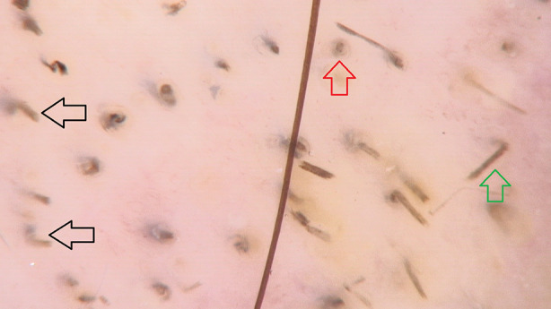
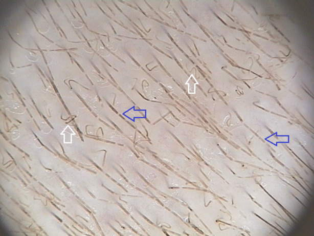
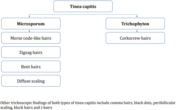
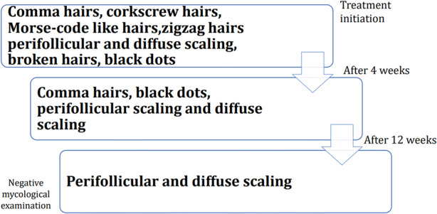
Results: Of 326 articles, 37 were considered eligible for the quantitative analysis. The most characteristic (with a high predictive value) trichoscopic findings of tinea capitis included comma hairs (51%), corkscrew hairs (32%), Morse code-like hairs (22%), zigzag hairs (21%), bent hairs (27%), block hairs (10%) and i-hairs (10%). Other common, but not characteristic, trichoscopic features were broken hairs (57%), black dots (34%), perifollicular scaling (59%) and diffuse scaling (89%). Morse code-like hairs, zigzag hairs, bent hairs and diffuse scaling were only observed in Microsporum tinea capitis (8/29, 28%; 6/29, 21%; 4/29, 14% and 4/29, 14%, respectively). In Trichophyton tinea capitis, corkscrew hairs were more commonly detected compared to Microsporum tinea capitis (21/38, 55% vs 3/29, 10%).
Conclusion: The presence of characteristic trichoscopic features of tinea capitis is sufficient to establish the initial diagnosis and introduce treatment before culture results are available. Trichoscopy may be useful in distinguishing between Microsporum and Trichophyton tinea capitis.
Keywords: Alopecia; Dermatoscopy; Dermoscopy; Fungal infection; Hair disorders; Hair loss; Scalp; Tinea; Tinea capitis; Trichoscopy.
Conflict of interest statement
Anna Waśkiel-Burnat, Adriana Rakowska, Mariusz Sikora, Piotr Ciechanowicz, Małgorzata Olszewska and Lidia Rudnicka have nothing to disclose. Adriana Rakowska is a member of the journals Editorial Board.
Figures
References
Publication types
LinkOut - more resources
Full Text Sources
