Review
. 2020 Apr;17(2):436-445.
doi: 10.1007/s13311-019-00829-x.

Aneurysmal Subarachnoid Hemorrhage: an Overview of Inflammation-Induced Cellular Changes

Affiliations
Review

Aneurysmal Subarachnoid Hemorrhage: an Overview of Inflammation-Induced Cellular Changes

A P Coulibaly et al. Neurotherapeutics. 2020 Apr.

Abstract

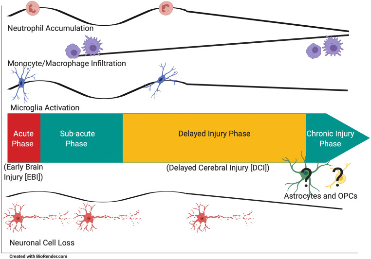
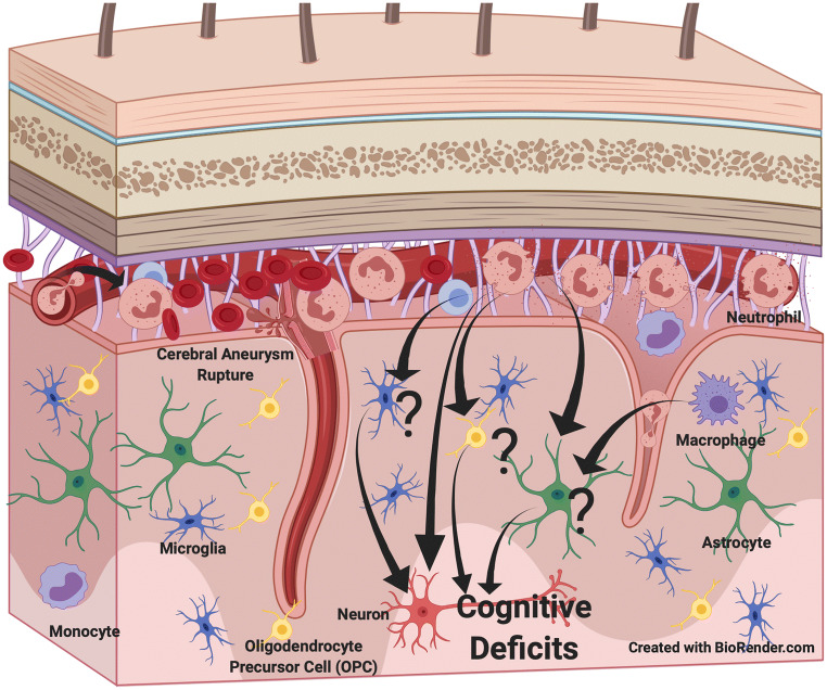
Aneurysmal subarachnoid hemorrhage (SAH) is a devastating disease that leads to poor neurological outcomes and is characterized by both vascular and neural pathologies. Recent evidence demonstrates that inflammation mediates many of the vascular and neural changes observed after SAH. Although most studies focus on inflammatory mediators such as cytokines, the ultimate effectors of inflammation in SAH are parenchymal brain and peripheral immune cells. As such, the present review will summarize our current understanding of the cellular changes of both CNS parenchymal and peripheral immune cells after SAH.

Keywords: Astrocytes; Delayed cerebral injury; Inflammation; Microglia; Neutrophils; Subarachnoid hemorrhage.


Figures

Fig. 1
Fig. 1
Temporal changes in cellular elements associated with SAH. Neutrophil accumulation, microglial activation, and neuronal cell loss appear to have two peaks (acute phase and delayed injury phase), whereas monocytes and macrophages appear to increase from the sub-acute phase to the recovery phase suggesting that they are important in recuperation and remodeling of the brain after injury
Fig. 2
Fig. 2
Possible cell interaction important in the development of delayed cerebral injury after SAH. Neutrophils and microglia appear to have a role in the pathophysiology that affects neuronal survival. The function of monocytes, macrophages, astrocytes, and oligodendrocyte precursor cells (OPCs) are less well studied

References

    1. Altay T, Smithason S, Volokh N, et al. A novel method for subarachnoid hemorrhage to induce vasospasm in mice. Journal of Neuroscience Methods. 2009;183:136–140. doi: 10.1016/j.jneumeth.2009.06.027. - DOI - PMC - PubMed
    1. Atangana E, Schneider UC, Blecharz K, et al. Intravascular Inflammation Triggers Intracerebral Activated Microglia and Contributes to Secondary Brain Injury After Experimental Subarachnoid Hemorrhage (eSAH) Translational Stroke Research. 2017;8:144–156. doi: 10.1007/s12975-016-0485-3. - DOI - PubMed
    1. Barz H, Schreiber A, Barz U. Demyelinating diseases as a result of cerebral edema? Medical Hypotheses. 2017;104:10–14. doi: 10.1016/j.mehy.2017.05.010. - DOI - PubMed
    1. Cahill J, Calvert JW, Solaroglu I, et al. Vasospasm and p53-induced apoptosis in an experimental model of subarachnoid hemorrhage. Stroke. 2006;37:1868–74. doi: 10.1161/01.STR.0000226995.27230.96. - DOI - PubMed
    1. Chaichana KL, Pradilla G, Huang J, et al. Role of inflammation (leukocyte-endothelial cell interactions) in vasospasm after subarachnoid hemorrhage. World Neurosurgery. 2010;73:22–41. doi: 10.1016/j.surneu.2009.05.027. - DOI - PubMed

Substances