. 2020 Feb;34(2):2730-2748.
doi: 10.1096/fj.201901830R. Epub 2019 Dec 19.

Control of Rab7a activity and localization through endosomal type Igamma PIP 5-kinase is required for endosome maturation and lysosome function

Affiliations

Control of Rab7a activity and localization through endosomal type Igamma PIP 5-kinase is required for endosome maturation and lysosome function

Ming Sun et al. FASEB J. 2020 Feb.

Abstract

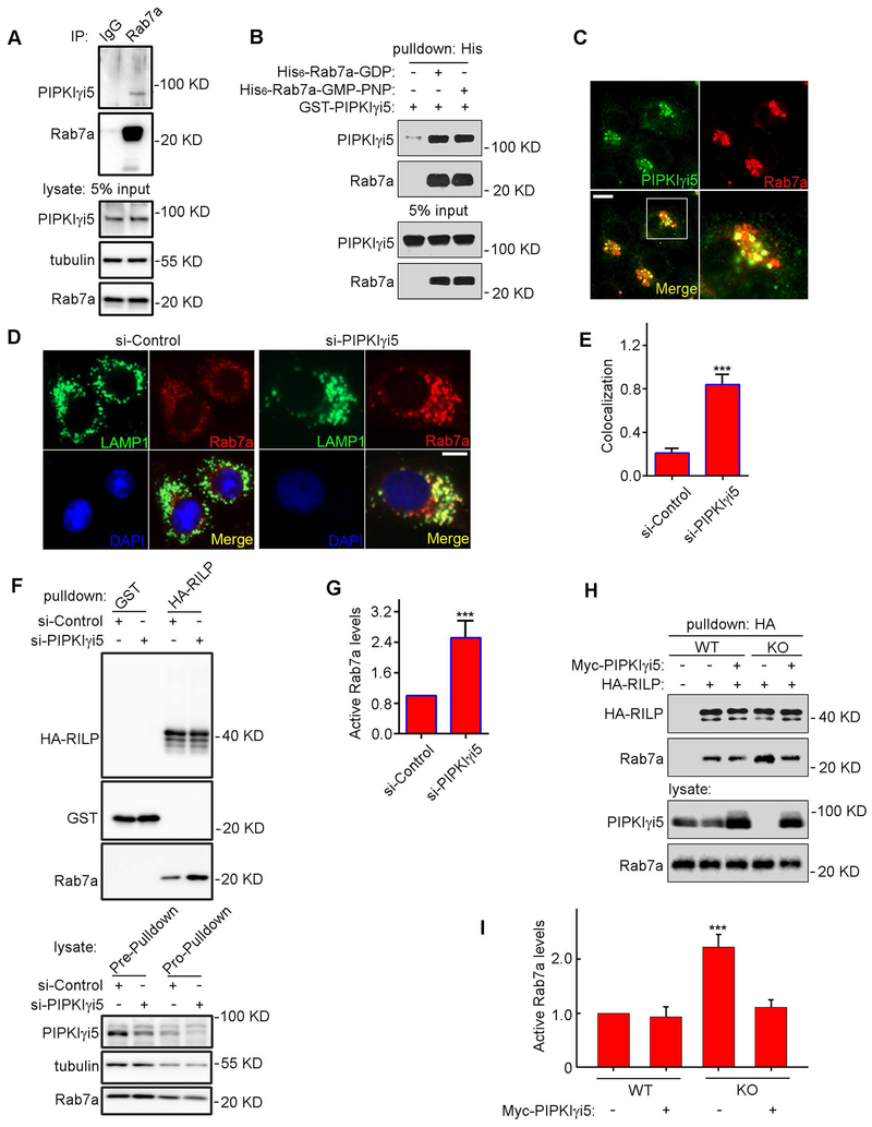
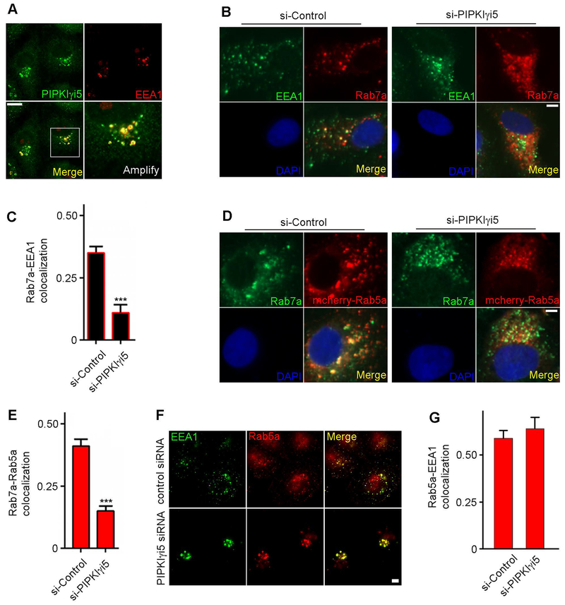
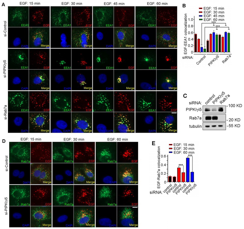
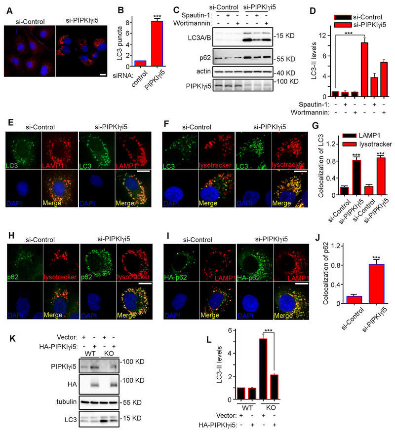
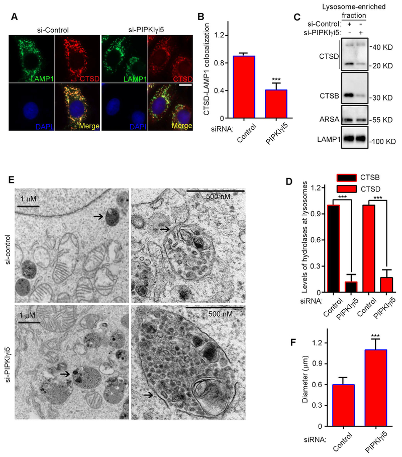
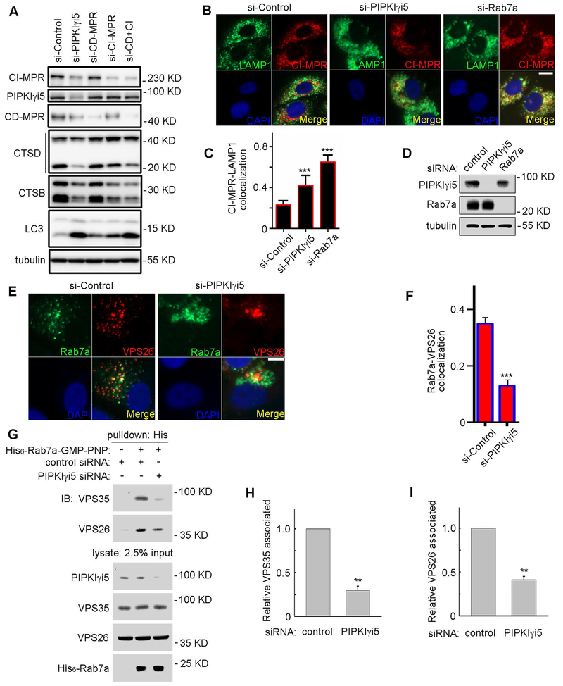
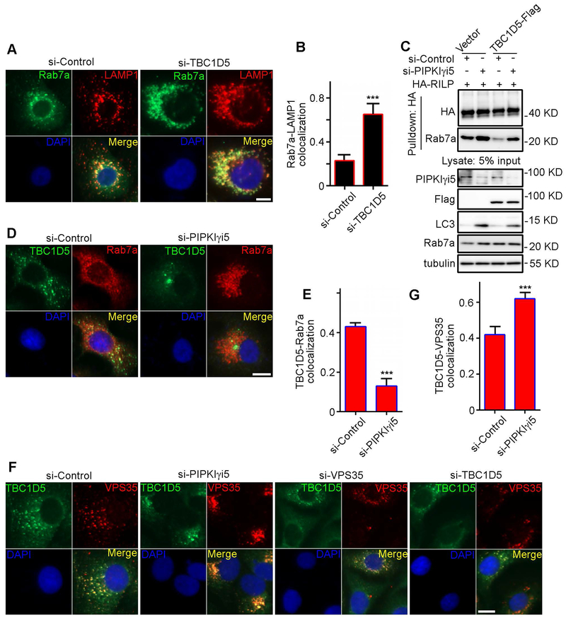
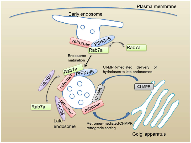
The small GTPase Ras-related protein Rab-7a (Rab7a) serves as a key organizer of the endosomal-lysosomal system. However, molecular mechanisms controlling Rab7a activation levels and subcellular translocation are still poorly defined. Here, we demonstrate that type Igamma phosphatidylinositol phosphate 5-kinase i5 (PIPKIγi5), an endosome-localized enzyme that produces phosphatidylinositol 4,5-bisphosphate, directly interacts with Rab7a and plays critical roles in the control of the endosomal-lysosomal system. The loss of PIPKIγi5 blocks Rab7a recruitment to early endosomes, which prevents the maturation of early to late endosomes. PIPKIγi5 loss disturbs retromer complex connection with Rab7a, which blocks the retrograde sorting of Cation-independent Mannose 6-Phosphate Receptor from late endosomes. This leads to the decreased sorting of hydrolases to lysosomes and reduces the autophagic degradation. By modulating the retromer-Rab7a connection, PIPKIγi5 is also required for the recruitment of the GTPase-activating protein TBC1 domain family member 5 to late endosomes, which controls the conversion of Rab7a from the active state to the inactive state. Thus, PIPKIγi5 is critical for the modulation of Rab7a activity, localization, and function in the endosomal-lysosomal system.

Keywords: PIPKIγi5; Rab7a; autophagy; endosome maturation; lysosome.


Figures

Figure 1.
Figure 1.. PIPKIγi5 interacts with Rab7a and controls Rab7a activation levels.
(A) UMSCC1 cells were subjected to immunoprecipitation with Rab7a antibody and then immunoblotted with antibodies as indicated. (B) Recombinant His6-Rab7a and GST-PIPKIγi5 were purified from E. coli and subjected to His6-pulldown assays. His6-Rab7a was preloaded with GDP or GMP-PNP. (C) IF staining of HA-PIPKIγi5 (green) and endogenous Rab7a (red). Bar, 20 μm. (D) IF staining of endogenous LAMP1 (green) and Rab7a (red) in control or PIPKIγi5 knockdown UMSCC1 cells. Bar, 20 μm. (E) Quantification of LAMP1-Rab7a colocalization (n=50 cells from three independent experiments). (F) HA-RALP was used to pull down Rab7a-GTP in control or PIPKIγi5 knockdown UMSCC1 cells. (G) Quantification of active Rab7a (Rab7a-GTP) levels in control or PIPKIγi5 knockdown UMSCC1 cells. (H) Myc-PIPKIγi5 expression was introduced in PIPKIγi5+/+ (WT) or PIPKIγi5−/− (KO) MEFs via lentivirus. An empty lentivirus vector was used as a control. Levels of PIPKIγi5, total Rab7a (in whole cell lysate), and active Rab7a (pulled down by HA-RILP) in PIPKIγi5+/+ (WT) and PIPKIγi5−/− (KO) MEFs were detected via Western blot. (I) Quantification of active Rab7a levels in WT or KO MEFs transfected with or without PIPKIγi5. The error bars indicate the mean ± SEM from three independent experiments. An unpaired Student’s two-tailed t-test was used. ***, P < 0.0001.
Figure 2.
Figure 2.. PIPKIγi5 is required for Rab7a recruitment to early endosomes.
(A) IF staining of HA-PIPKIγi5 (green) and EEA1 (red) in UMSCC1 cells. (B) Costaining of EEA1 and Rab7a in control or PIPKIγi5 knockdown UMSCC1 cells. (C) Quantification of Rab7a-EEA1 colocalization. (D) IF staining of Rab7a in mCherry-Rab5a-expressing control or PIPKIγi5 knockdown UMSCC1 cells. (E) Quantification of mCherry-Rab5a-Rab7a colocalization. (F) IF staining of EEA1 and Rab5a in control or PIPKIγi5 knockdown UMSCC1 cells. (G) Quantification of EEA1-Rab5a colocalization. The error bars indicate the mean ± SEM. (n=50 cells from three independent experiments). An unpaired Student’s two-tailed t-test was used. ***, P < 0.0001. Bar in IF images, 20 μm.
Figure 3.
Figure 3.. PIPKIγi5 and Rab7a are required for early to late endosome maturation.
(A) Costaining of EEA1 (green) and Alexa Fluor 555-EGF (red) in control, PIPKIγi5 knockdown, or Rab7a knockdown UMSCC1 cells. (B) Quantification of Alexa Fluor 555-EGF and EEA1 (n=50 cells from three independent experiments). One-way analysis of variance (ANOVA) with Tukey’s post hoc test was performed. ***, P < 0.0001. (C) PIPKIγi5 and Rab7a protein levels in control, PIPKIγi5 knockdown, or Rab7a knockdown UMSCC1 cells were detected by Western blot. (D) Costaining of Rab7a (green) and Alexa Fluor 555-EGF (red) in control or PIPKIγi5 knockdown UMSCC1 cells. (E) Quantification of Alexa Fluor 555-EGF and Rab7a. The error bars indicate the mean ± SEM (n=50 cells from three independent experiments). An unpaired Student’s two-tailed t-test was used. ***, P < 0.0001. Bar in IF images, 20 μm.
Figure 4.
Figure 4.. PIPKIγi5 controls autophagic flux.
(A) IF staining of LC3-II in control or PIPKIγi5 knockdown UMSCC1 cells. (B) Quantification of LC3-II puncta (n=50 cells from three independent experiments). (C) UMSCC1 cells were transfected with control or PIPKIγi5 siRNA and then treated with or without spautin-1 or wortmannin. The indicated protein levels were detected by Western blotting. (D) Quantification of LC3-II protein levels. (E) Costaining of LC3 and LAMP1 in control or PIPKIγi5 knockdown UMSCC1 cells. (F) Costaining of LC3 and LysoTracker. (G) Quantification of LC3 colocalization with LAMP1 or LysoTracker (n=50 cells from three independent experiments). (H) Costaining of endogenous p62 and LysoTracker. (I) Costaining of HA-p62 and LAMP1. (J) Quantification of p62 colocalization with LysoTracker (n=50 cells from three independent experiments). (K) HA-PIPKIγi5 expression was introduced in PIPKIγi5+/+ (WT) or PIPKIγi5−/− (KO) MEFs via lentivirus. An empty lentivirus vector was used as a control. Then, the PIPKIγi5 and LC3-II levels were detected by Western blot. (L) Quantification of LC3-II levels in MEFs. The error bars indicate the mean ± SEM from three independent experiments. An unpaired Student’s two-tailed t-test was used. ***, P < 0.0001. Bar in IF images, 20 μm.
Figure 5.
Figure 5.. PIPKIγi5 regulates hydrolase sorting to lysosomes.
(A) Costaining of CTSD (red) and LAMP1 (green) in control or PIPKIγi5 knockdown UMSCC1 cells. Bar, 20 μm. (B) Quantification of CTSD-LAMP1 colocalization (n=50 cells from three independent experiments). (C) The lysosome components were enriched using a lysosome isolation kit, and then mature CTSD and CTSB levels were detected by Western blotting. (D) Quantification of mature CTSD and CTSB levels in control or PIPKIγi5 knockdown UMSCC1 cells. (E) The morphology of lysosomes (indicated in EM images by arrows) was detected by electron microscopy (EM). (F) The lysosome diameters in control or PIPKIγi5 knockdown UMSCC1 cells was quantified (n=50 cells from three independent experiments). The error bars indicate the mean ± SEM from three independent experiments. An unpaired Student’s two-tailed t-test was used. ***, P < 0.0001.
Figure 6.
Figure 6.. PIPKIγi5 controls the colocalization of the retromer complex with Rab7a and regulates CI-MPR subcellular localization.
(A) The indicated siRNAs were transfected into UMSCC1 cells, and then the levels of the indicated proteins were detected by Western blotting. (B) IF staining of CI-MPR (red) and LAMP1 (green) in control, PIPKIγi5 knockdown, or Rab7a knockdown UMSCC1 cells. (C) Quantification of CI-MPR-LAMP1 colocalization (n=50 cells from three independent experiments). (D) PIPKIγi5 and Rab7a protein levels in control, PIPKIγi5 knockdown, or Rab7a knockdown UMSCC1 cells were detected by Western blot. (E) IF staining of Rab7a (green) and VPS26 (red) in control or PIPKIγi5 knockdown UMSCC1 cells. (F) Quantification of Rab7a-VPS26 colocalization (n=50 cells from three independent experiments). The error bars indicate the mean ± SEM. An unpaired Student’s two-tailed t-test was used. ***, P < 0.0001. Bar in IF images, 20 μm. (G) Same amount (200 ng) of His6-Rab7a loaded with GMP-PNP was used to pulldown retromer components in cell lysate from control or PIPKIγi5-knockdown UMSCC1 cells. The indicated proteins in cell lysate or pulled down by His6-Rab7a-GMP-PNP were detected by Western blot. (H) Quantification of VPS35 associated with His6-Rab7a-GMP-PNP in control or PIPKIγi5-knockdown UMSCC1 cells. (I) Quantification of VPS26 associated with His6-Rab7a-GMP-PNP in control or PIPKIγi5-knockdown UMSCC1 cells. The error bars indicate the mean ± SEM from three independent experiments. An unpaired Student’s two-tailed t-test was used. **, P < 0.001.
Figure 7.
Figure 7.. PIPKIγi5 is required for TBC1D5 recruitment to late endosomes.
(A) IF staining of Rab7a (green) and LAMP1 (red) in control or TBC1D5 knockdown UMSCC1 cells. (B) Quantification of Rab7a-LAMP1 colocalization (n=50 cells from three independent experiments). (C) Flag-TBC1D5 was transfected into control or PIPKIγi5 knockdown UMSCC1 cells, and then the levels of active Rab7a were measured. (D) IF staining of TBC1D5 (green) and Rab7a (red) in control or PIPKIγi5 knockdown UMSCC1 cells. (E) Quantification of TBC1D5-Rab7a colocalization (n=50 cells from three independent experiments). (F) IF staining of TBC1D5 (green) and VPS35 (red) in control, PIPKIγi5 knockdown, VPS35 knockdown, or TBC1D5 knockdown UMSCC1 cells. (G) Quantification of TBC1D5-VPS35 colocalization (n=50 cells from three independent experiments). The error bars indicate the mean ± SEM. An unpaired Student’s two-tailed t-test was used. ***, P < 0.0001. Bar in IF images, 20 μm.
Figure 8.
Figure 8.. Model of the modulation of endosome maturation and lysosome function by PIPKIγi5 and Rab7a.
PIPKIγi5 interacts with Rab7a and is required for Rab7a recruitment to early endosomes, which is a critical step in the initiation of the maturation of early endosomes into late endosomes. PIPKIγi5 is also required for retromer translocation to Rab7a-positive late endosomes. The retromer recruits TBC1D5 to convert Rab7a-GTP into Rab7a-GDP, which leads to the release of Rab7a from late endosomes. The retromer also mediates the retrograde sorting of CI-MPR from late endosomes to the TGN, which facilitates CI-MPR-dependent hydrolase delivery to late endosome/lysosomes.

Similar articles

Cited by

References

    1. Cullen PJ, and Steinberg F (2018) To degrade or not to degrade: mechanisms and significance of endocytic recycling. Nature reviews. Molecular cell biology 19, 679–696 - PubMed
    1. McNally KE, and Cullen PJ (2018) Endosomal Retrieval of Cargo: Retromer Is Not Alone. Trends in cell biology 28, 807–822 - PubMed
    1. Goldenring JR (2015) Recycling endosomes. Current opinion in cell biology 35, 117–122 - PMC - PubMed
    1. Podinovskaia M, and Spang A (2018) The Endosomal Network: Mediators and Regulators of Endosome Maturation. Progress in molecular and subcellular biology 57, 1–38 - PubMed
    1. Braulke T, and Bonifacino JS (2009) Sorting of lysosomal proteins. Biochimica et biophysica acta 1793, 605–614 - PubMed

Publication types

MeSH terms

LinkOut - more resources