GALE Promotes the Proliferation and Migration of Glioblastoma Cells and Is Regulated by miR-let-7i-5p
- PMID: 31908526
- PMCID: PMC6924591
- DOI: 10.2147/CMAR.S221585
GALE Promotes the Proliferation and Migration of Glioblastoma Cells and Is Regulated by miR-let-7i-5p
Abstract
Purpose: Glioma is the most common and lethal type of brain tumor. While GALE (UDP-galactose-4-epimerase) has been shown to be overexpressed in some kinds of cancers, its expression in gliomas has not been reported. MicroRNAs (miRNAs) function as tumor suppressors in many cancers, and online datasets can be used to predict whether GALE is regulated by miR-let-7i-5p. In this investigation, we explored the effect and regulatory mechanisms of GALE on glioblastoma growth via miR-let-7i-5p.
Methods: We used a Cox proportional hazards model and publicly available datasets to examine the relationship between GALE and the survival rates of glioma patients. Bioinformatics predicted the targeting of GALE by miR-let-7i-5p. The proliferation, migration, cell cycle and apoptosis of human glioblastoma cells were assessed by relevant assays. We also demonstrated the effect of GALE on glioblastoma multiforme [GBM] tumor growth using an in vivo orthotopic xenograft model.
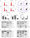
Results: GALE was upregulated in human gliomas, especially in high-grade gliomas (e.g., GBM). An obvious decline in GALE expression was observed in human glioblastoma cell lines (U87 and U251) following treatment with a small interfering RNA (siRNA) targeting GALE or miR-let-7i-5p mimics. Knockdown of GALE or overexpression of miR-let-7i-5p (with miR-let-7i-5p mimics) inhibited U87 and U251 cell growth. miR-let-7i-5p significantly restrained the migration ability of human glioblastoma cells in vascular mimic (VM), wound healing and transwell assays, and GALE promoted glioblastoma growth in vivo.
Conclusion: Our findings confirm that GALE plays an important role in promoting the development of human glioma and that GALE can be regulated by miR-let-7i-5p to inhibit human glioblastoma growth.
Implications for cancer survivors: Our data show that cancer survivors have low GALE expression, which indicates that GALE may be a diagnostic biomarker and a promising therapeutic target in glioblastoma.
Keywords: GALE; glioblastoma; miR-let-7i-5p; migration; proliferation.
© 2019 Sun et al.
Conflict of interest statement
The authors have no potential conflicts of interest to disclose.
Figures


Similar articles
-
Let-7i-5p enhances cell proliferation, migration and invasion of ccRCC by targeting HABP4.BMC Urol. 2021 Mar 28;21(1):49. doi: 10.1186/s12894-021-00820-9. BMC Urol. 2021. PMID: 33775245 Free PMC article.
-
Mir-758-5p Suppresses Glioblastoma Proliferation, Migration and Invasion by Targeting ZBTB20.Cell Physiol Biochem. 2018;48(5):2074-2083. doi: 10.1159/000492545. Epub 2018 Aug 10. Cell Physiol Biochem. 2018. PMID: 30099442
-
Verbascoside inhibits progression of glioblastoma cells by promoting Let-7g-5p and down-regulating HMGA2 via Wnt/beta-catenin signalling blockade.J Cell Mol Med. 2020 Mar;24(5):2901-2916. doi: 10.1111/jcmm.14884. Epub 2020 Jan 30. J Cell Mol Med. 2020. PMID: 32000296 Free PMC article.
-
Dual mechanism of Let-7i in tumor progression.Front Oncol. 2023 Sep 27;13:1253191. doi: 10.3389/fonc.2023.1253191. eCollection 2023. Front Oncol. 2023. PMID: 37829341 Free PMC article. Review.
-
A review shows that ATG10 has been identified as a potential prognostic marker and therapeutic target for cancer patients based on real-world studies.Front Oncol. 2025 Apr 17;15:1573378. doi: 10.3389/fonc.2025.1573378. eCollection 2025. Front Oncol. 2025. PMID: 40313244 Free PMC article. Review.
Cited by
-
HELLPAR/RRM2 axis related to HMMR as novel prognostic biomarker in gliomas.BMC Cancer. 2023 Feb 7;23(1):125. doi: 10.1186/s12885-023-10596-w. BMC Cancer. 2023. PMID: 36750807 Free PMC article.
-
Pan-cancer analysis of UDP-glucose 6-dehydrogenase in human tumors and its function in hepatocellular carcinoma.World J Gastrointest Oncol. 2025 Jul 15;17(7):105981. doi: 10.4251/wjgo.v17.i7.105981. World J Gastrointest Oncol. 2025. PMID: 40697240 Free PMC article.
-
lnc001776 Affects CPB2 Toxin-Induced Excessive Injury of Porcine Intestinal Epithelial Cells via Activating JNK/NF-kB Pathway through ssc-let-7i-5p/IL-6 Axis.Cells. 2023 Mar 29;12(7):1036. doi: 10.3390/cells12071036. Cells. 2023. PMID: 37048109 Free PMC article.
-
Epigenetic Modifications Associated With Wildland-Urban Interface (WUI) Firefighting.Environ Mol Mutagen. 2025 Jan-Feb;66(1-2):22-33. doi: 10.1002/em.70002. Epub 2025 Feb 19. Environ Mol Mutagen. 2025. PMID: 39968828 Free PMC article.
-
Therapeutic Applications of Poly-miRNAs and miRNA Sponges.Int J Mol Sci. 2025 May 9;26(10):4535. doi: 10.3390/ijms26104535. Int J Mol Sci. 2025. PMID: 40429680 Free PMC article. Review.
References
LinkOut - more resources
Full Text Sources

