. 2019 Dec 15;7(1):e000720.
doi: 10.1136/bmjdrc-2019-000720. eCollection 2019.

Senescence marker activin A is increased in human diabetic kidney disease: association with kidney function and potential implications for therapy

Affiliations

Senescence marker activin A is increased in human diabetic kidney disease: association with kidney function and potential implications for therapy

Xiaohui Bian et al. BMJ Open Diabetes Res Care. .

Abstract

Objective: Activin A, an inflammatory mediator implicated in cellular senescence-induced adipose tissue dysfunction and profibrotic kidney injury, may become a new target for the treatment of diabetic kidney disease (DKD) and chronic kidney diseases. We tested the hypothesis that human DKD-related injury leads to upregulation of activin A in blood and urine and in a human kidney cell model. We further hypothesized that circulating activin A parallels kidney injury markers in DKD.

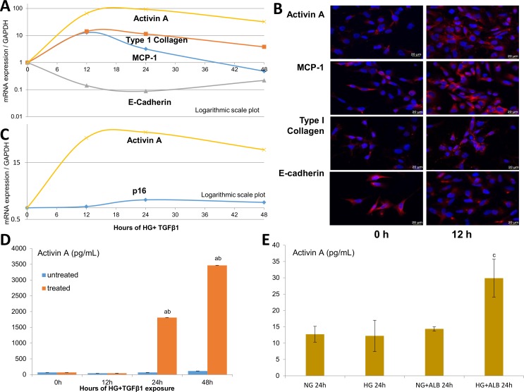
Research design and methods: In two adult diabetes cohorts and controls (Minnesota, USA; Galway, Ireland), the relationships between plasma (or urine) activin A, estimated glomerular filtration rate (eGFR) and DKD injury biomarkers were tested with logistic regression and correlation coefficients. Activin A, inflammatory, epithelial-mesenchymal-transition (EMT) and senescence markers were assayed in human kidney (HK-2) cells incubated in high glucose plus transforming growth factor-β1 or albumin.

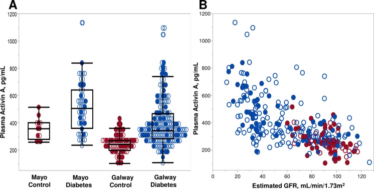
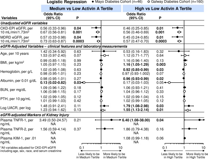
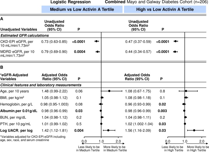
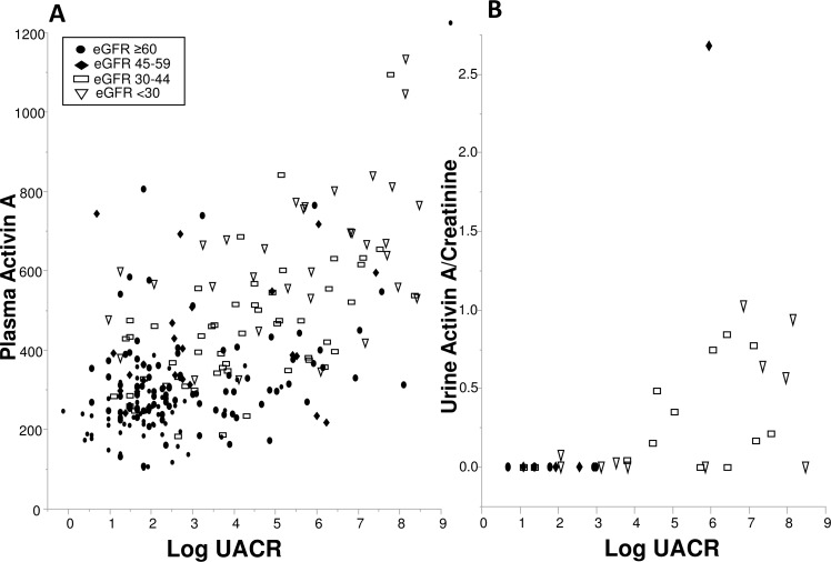
Results: Plasma activin A levels were elevated in diabetes (n=206) compared with controls (n=76; 418.1 vs 259.3 pg/mL; p<0.001) and correlated inversely with eGFR (rs=-0.61; p<0.001; diabetes). After eGFR adjustment, only albuminuria (OR 1.56, 95% CI 1.16 to 2.09) and tumor necrosis factor receptor-1 (OR 6.40, 95% CI 1.08 to 38.00) associated with the highest activin tertile. Albuminuria also related to urinary activin (rs=0.65; p<0.001). Following in vitro HK-2 injury, activin, inflammatory, EMT genes and supernatant activin levels were increased.

Conclusions: Circulating activin A is increased in human DKD and correlates with reduced kidney function and kidney injury markers. DKD-injured human renal tubule cells develop a profibrotic and inflammatory phenotype with activin A upregulation. These findings underscore the role of inflammation and provide a basis for further exploration of activin A as a diagnostic marker and therapeutic target in DKD.

Keywords: adipocytokine; clinical aspects of diabetes; clinical nephrology; renal fibrosis.


Conflict of interest statement

Competing interests: None declared.

Figures

Figure 1
Figure 1
(A) Distribution of plasma activin A concentrations by controls and diabetes (case) status in Mayo and Galway cohorts. Circulating activin concentrations were higher in the diabetes cases compared with controls (p=0.007 Mayo; p<0.0001 Galway). Sex distribution is shown for each group. (B) Individual plasma activin A concentrations by estimated glomerular filtration rate (eGFR) for diabetes cases and non-diabetes, non-chronic kidney disease (CKD) controls in Mayo and Galway cohorts. Spearman’s correlation (rs=−0.61; p<0.0001) for plasma activin A with eGFR. Sex distribution is shown for each group. Red: controls, individuals without diabetes mellitus or CKD; blue: cases, participants with diabetes mellitus with and without CKD; closed circles: female participants; open circles: male participants.
Figure 2
Figure 2
Unadjusted and eGFR-adjusted ORs (95% CIs) for tertiles of activin A per clinical characteristics and laboratory tests in Mayo and Galway cohorts with diabetes. Closed circles: OR for Mayo diabetes cohort; open circles: OR for Galway diabetes cohort. BMI, body mass index; BUN, blood urea nitrogen; CKD, chronic kidney disease; eGFR, estimated glomerular filtration rate; KIM-1, kidney injury molecule-1; MDRD, modification of diet renal disease; NA, not available; PTH, parathyroid hormone; TNFR, tumor necrosis factor receptor; UACR, urine albumin-to-creatinine ratio (Mayo diabetes cohort n=42; Galway n=160).
Figure 3
Figure 3
(A) Unadjusted and (B) eGFR-adjusted ORs (95% CIs) for tertiles of activin A per clinical characteristics and laboratory tests in combined cohorts with diabetes. BMI, body mass index; BUN, blood urea nitrogen; CKD, chronic kidney disease; eGFR, estimated glomerular filtration rate; KIM-1, kidney injury molecule-1; MDRD, modification of diet renal disease; NA, not available; PTH, parathyroid hormone; UACR, urine albumin-to-creatinine ratio (Mayo diabetes cohort n=42; Galway n=160).
Figure 4
Figure 4
(A) Individual plasma activin A and (B) urine activin A/creatinine concentrations by log urine albumin-to-creatinine ratio (UACR) for Mayo (and Galway; plasma only) diabetes cohort. Estimated glomerular filtration rate (eGFR) group distribution is shown for each. Spearman’s correlations: log UACR with plasma activin A (rs=0.48; p<0.0001) and log UACR with urine activin A/creatinine (rs=0.65; p<0.0001) concentrations. Closed circle: eGFR ≥60 mL/min/1.73 m2; closed diamond: eGFR 45–59 mL/min/1.73 m2; open rectangle: eGFR 30–45 mL/min/1.73 m2; open triangle: eGFR ≤30 mL/min/1.73 m2.
Figure 5
Figure 5
In vitro models of DKD-related injury. HK-2 studies following incubation with high glucose (HG, 25 mmol/L), transforming growth factor-β1 (TGF-β1; 5 ng/mL) and albumin (ALB, 5 mg/mL). Time-dependent effect of HG+TGF-β1 on the mRNA/gene expression of activin A, monocyte chemoattractant protein-1 (MCP-1; an inflammatory marker), type I collagen (mesenchymal marker) and E-cadherin (epithelial marker) in injured HK-2 cells (A) Immunofluorescent staining of HK-2 cells injured by HG+TGF-β1 at 12 hours (h) compared with baseline (B) Time-dependent effect for activin A and p16 expression in injured HK-2 cells (C) Protein levels of activin A in the supernatant of HK-2 treated with HG+TGF-β1 (D) and combinations of HG with or without ALB (E) at 24 hours. Values are mean±SEM. aP<0.05 compared with untreated HK-2 groups; bp<0.05 compared with the previous group; cp<0.05 compared with all prior groups. Scale bars: 20 µm.

References

    1. Ogurtsova K, da Rocha Fernandes JD, Huang Y, et al. . IDF diabetes atlas: global estimates for the prevalence of diabetes for 2015 and 2040. Diabetes Res Clin Pract 2017;128:40–50. 10.1016/j.diabres.2017.03.024 - DOI - PubMed
    1. Verzola D, Gandolfo MT, Gaetani G, et al. . Accelerated senescence in the kidneys of patients with type 2 diabetic nephropathy. Am J Physiol Renal Physiol 2008;295:F1563–73. 10.1152/ajprenal.90302.2008 - DOI - PubMed
    1. Minamino T, Orimo M, Shimizu I, et al. . A crucial role for adipose tissue p53 in the regulation of insulin resistance. Nat Med 2009;15:1082–7. 10.1038/nm.2014 - DOI - PubMed
    1. Liu J, Yang J-R, Chen X-M, et al. . Impact of ER stress-regulated ATF4/p16 signaling on the premature senescence of renal tubular epithelial cells in diabetic nephropathy. Am J Physiol Cell Physiol 2015;308:C621–30. 10.1152/ajpcell.00096.2014 - DOI - PubMed
    1. Melk A, Schmidt BMW, Takeuchi O, et al. . Expression of p16INK4a and other cell cycle regulator and senescence associated genes in aging human kidney. Kidney Int 2004;65:510–20. 10.1111/j.1523-1755.2004.00438.x - DOI - PubMed

Publication types

MeSH terms