Case Reports
. 2019 Dec 31;2019(12):513-515.
doi: 10.1093/omcr/omz126. eCollection 2019 Dec.

Linear dermatomyofibroma over the nape of neck: a report of an unusual case and literature review

Affiliations
Case Reports

Linear dermatomyofibroma over the nape of neck: a report of an unusual case and literature review

Lama R AlZamil et al. Oxf Med Case Reports. .

Abstract

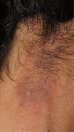
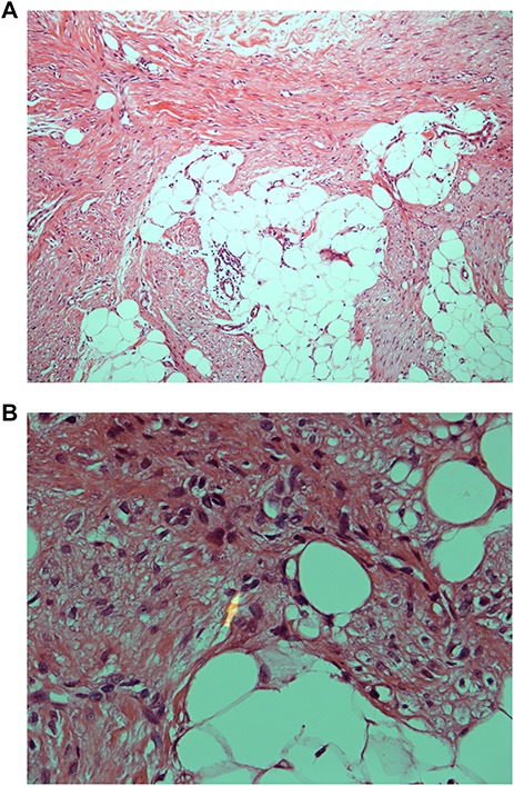
Dermatomyofibroma is a rare cutaneous mesenchymal tumor of benign fibroblastic and myofibroblastic derivations. It predominantly affects young women, and it usually presents as a reddish-brown plaque or nodule, which is commonly located over the upper trunk. We report the case of a 41-year-old female patient who presented with progressive linear dermatomyofibroma over the nape of her neck. This case report expands the knowledge about the clinical and histopathological features of this rare, benign and cutaneous tumor.

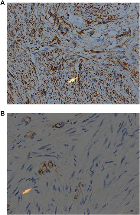
Keywords: cutaneous; dermatomyofibroma; immunohistochemistry; mesenchymal tumor; myofibroblasts.


Figures

Figure 1
Figure 1
Clinical presentation of dermatomyofibroma showing an ill-defined and linear reddish-brown indurated dermal plaque measuring approximately 7 × 3 cm over the nape of the neck.
Figure 2
Figure 2
(A) Low-power microscopic view of dermatomyofibroma showing the infiltrating fascicles of bland-looking spindle cells. Hematoxylin-eosin stain ×100. (B) High-power microscopic view of dermatomyofibroma showing the spindle- and oval-shaped nuclei of the tumor cells, which are mixed with small blood vessels and lipocytes. Note the absence of necrosis, pleomorphism and mitoses. Hematoxylin-eosin stain ×400
Figure 3
Figure 3
(A) IHC stain for h-caldesmon is strongly positive. This finding is in favor of dermatomyofibroma. The tumor was, however, negative for CD34, factor 13A and S100 protein. IHC stain ×200. (B) IHC stain for C-34 is negative. Note the presence of positive internal control in some blood vessels. IHC stain ×400.

Similar articles

  • Development of dermatomyofibroma in a male infant.
    Sim JH, Shin J, Vandersteen DP, Kim YC. Sim JH, et al. Ann Dermatol. 2011 Sep;23 Suppl 1(Suppl 1):S72-4. doi: 10.5021/ad.2011.23.S1.S72. Epub 2011 Sep 30. Ann Dermatol. 2011. PMID: 22028578 Free PMC article.
  • Dermatomyofibroma mimicking granuloma annulare.
    Cano Martínez N, Fernández-Antón Martínez C, Barchino Ortiz L, Lecona Echevarría M, Campos Domínguez M. Cano Martínez N, et al. Dermatol Online J. 2011 Jun 15;17(6):3. Dermatol Online J. 2011. PMID: 21696683
  • A rare case report for dermatomyofibroma in nasion.
    Fu S, Zhang J, Zhan Y, Zhang H, Herman EI, Lu Q, Zhou Y, Zhang G. Fu S, et al. Zhong Nan Da Xue Xue Bao Yi Xue Ban. 2018 Sep 28;43(9):1037-1040. doi: 10.11817/j.issn.1672-7347.2018.09.017. Zhong Nan Da Xue Xue Bao Yi Xue Ban. 2018. PMID: 30333298
  • Fibroblastic connective tissue nevus.
    Velez MJ, Billings SD, Weaver JA. Velez MJ, et al. J Cutan Pathol. 2016 Jan;43(1):75-9. doi: 10.1111/cup.12605. Epub 2015 Oct 21. J Cutan Pathol. 2016. PMID: 26268513 Review.
  • Dermatomyofibroma: case report and review.
    Rose C, Bröcker EB. Rose C, et al. Pediatr Dermatol. 1999 Nov-Dec;16(6):456-9. doi: 10.1046/j.1525-1470.1999.00117.x. Pediatr Dermatol. 1999. PMID: 10632944 Review.

References

    1. Hügel H. Die plaqueförmige dermale fibromatose. Hautarzt 1991;42:223–6. - PubMed
    1. Kamino H, Reddy VB, Gero M, Greco MA. Dermatomyofibroma: a benign cutaneous, plaque-like proliferation of fibroblasts and myofibroblasts in young adults. J Cutan Pathol 1992;19:85–93. doi: 10.1111/j.1600-0560.1992.tb01348.x. - DOI - PubMed
    1. Mentzel T, Kutzner H. Dermatomyofibroma: clinicopathologic and immunohistochemical analysis of 56 cases and reappraisal of a rare and distinct cutaneous neoplasm. Am J Dermatopathol 2009;31:44–9. doi: 10.1097/DAD.0b013e3181914e6f. - DOI - PubMed
    1. Viglizzo G, Occella C, Calonje E, Nozza P, Rongioletti F. A unique case of multiple dermatomyofibromas. Clin Exp Dermatol 2008;33:622–4. doi: 10.1111/j.1365-2230.2008.02788.x. - DOI - PubMed
    1. de LLAF, Lisboa GM, Filho EA da S, Rabelo G. Multiple dermatomyofibromas. An Bras Dermatol 2018;93:268–70. doi: 10.1590/abd1806-4841.20186748. - DOI - PMC - PubMed

Publication types