. 2019 Nov;16(4):743-755.
doi: 10.20892/j.issn.2095-3941.2019.0115.

Inactivation of FOXO1 induces T follicular cell polarization and involves angioimmunoblastic T cell lymphoma

Affiliations

Inactivation of FOXO1 induces T follicular cell polarization and involves angioimmunoblastic T cell lymphoma

Meifang Xu et al. Cancer Biol Med. 2019 Nov.

Erratum in

Abstract

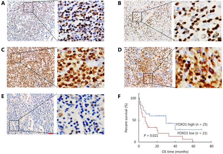
Objective: Angioimmunoblastic T cell lymphoma (AITL) is an aggressive form of non-Hodgkin lymphoma derived from mature T cells. However, the underlying pathogenesis of AITL remains unresolved. We aimed to explore the role of FOXO1-mediated signaling in the tumorigenesis and progression of AITL.

Methods: FOXO1 expression was assessed using immunohistochemistry on a total of 46 AITL tissue samples. Retroviruses encoding FOXO1 shRNA were used to knockdown FOXO1 expression in CD4+ T cells. Flow cytometric assays analyzed the proliferation and survival of FOXO1 knockdown CD4+ T cells. Furthermore, we performed adoptive T-cell transfer experiments to identify whether inactivation of FOXO1 induced neoplastic follicular-helper T (Tfh) cell polarization and function.

Results: Patients with low FOXO1 protein levels were prone to have an advanced tumor stage (P = 0.049), higher ECOG ps (P = 0.024), the presence of bone marrow invasion (P = 0.000), and higher IPI (P = 0.035). Additionally, the survival rates of patients in the FOXO1 high-expression group were significantly better than those in the FOXO1 low-expression group (χ2 = 5.346, P = 0.021). We also observed that inactivation of FOXO1 increased CD4+ T cell proliferation and altered the survival and cell-cycle progression of CD4+ T cells. Finally, we confirmed that inactivation of FOXO1 induces Tfh cell programing and function.

Conclusions: Inactivation of FOXO1 in AITL plays a key role in the tumorigenesis and progression of AITL. We propose that FOXO1 expression could be a useful prognostic marker in AITL patients to predict poor survival, and to design appropriate therapeutic strategies.

Keywords: Angioimmunoblastic T cell lymphoma; FOXO1; differentiation; inactivation.


Figures

1
1
FOXO1 expression exclusively in cytosol or negative in AITL samples correlates with poor prognosis. (A) FOXO1 was almost exclusively detected in the nuclei of lymphocytes in lymph nodes. (B) FOXO1 exhibited heterogeneous nuclear staining in some AITL patients. (C) FOXO1 displayed variegated staining in both the cytosol and nucleus in some AITL patients. (D) FOXO1 exerted dominant staining in the cytosol in some AITL patients. (E) FOXO1 was negative in some AITL patients. (F) Kaplan–Meier analysis of overall survival for AITL patients with high or low FOXO1 expression. EliVision Plus two-step immunohistochemical technique with 3-3’ diaminobenzidine (DAB) staining was used in (A-E)(400 ×; scale bar: 10 μm).
2
2
Inactivation of FOXO1 in CD4+ T cells increases cell proliferation. (A and B) Retroviral FOXO1 shRNA expression vectors and non-specific shRNA control vectors were transduced into CD4+ T cells. Knockdown of FOXO1 expression in CD4+ T cells was detected by RT-qPCR (A) and Western blot analysis (B). (C) In vitro CellTrace Violet (CTV) proliferation assay of CD4+ T cells transduced with retroviral FOXO1 shRNA or non-specific shRNA control vectors on day 3. The P value in (A) was calculated by ANOVA followed by Dunnett’s test using triplicate samples from two independent experiments. The P value in (C) was calculated by ANOVA followed by Dunnett’s test using triplicate samples from two independent experiments. Columns indicate means; bars are the standard error. **P ≤ 0.01.
3
3
Inactivation of FOXO1 alters the survival and cell-cycle progression of CD4+ T cells. (A and B) Retroviral FOXO1 shRNA expression vectors and non-specific shRNA control vectors were transduced into CD4+ T cells. Annexin V (A) and Fas (B) staining of transferring CD4+ T cells on day 3 were analyzed by flow cytometric analysis. (C) Immunoblots showing the detection of total caspase3 and cleaved caspase3 in transferred CD4+ T cells on day 3. β-actin was used as a loading control. (D) Cell cycle analysis with immunostaining of 7-AAD and BrdU in transferred CD4+ T cells on day 3. The plots represent cell cycle S phase. (E) Immunoblots showing the detection of p21 and cyclinD1 in transferred CD4+ T cells on day 3. The P values in (A, B, D) were calculated by ANOVA followed by Dunnett’s test using triplicate samples from three independent experiments. Columns indicate means; bars are the standard error. **P ≤ 0.01.
4
4
Inactivation of FOXO1 induces Tfh cell polarization and function. (A) Schematic representation of experimental design for adoptive T cell transfer experiments. (B) Representative photographs of spleens and lymph nodes from recipient TCRα-/- mice at 3 months after adoptive transfer of FOXO1 knockdown CD4+ T cells or control CD4+ T cells. (C and D) Tfh cells (C) and germinal center B cells (D) from TCRα-/- mice at 3 months after adoptive transfer of FOXO1 knockdown CD4+ T cells or control CD4+ T cells were analyzed by flow cytometry. (E) ELISA showing the detection of serum levels of cytokines from recipient TCRα-/- mice at 3 months after adoptive transfer of FOXO1 knockdown CD4+ T cells or control CD4+ T cells. The P values in (C and D) were calculated by ANOVA followed by Dunnett’s test using triplicate samples of 3 mice of each group in two independent experiments. The P value in (E) was calculated by ANOVA followed by Dunnett’s test for 6 mice of each group. Columns indicate means; bars are the standard error. **P ≤ 0.01.

Similar articles

Cited by

References

    1. De Leval L, Gisselbrecht C, Gaulard P Advances in the understanding and management of angioimmunoblastic T-cell lymphoma. Br J Haematol. 2010;148:673–89. doi: 10.1111/j.1365-2141.2009.08003.x. - DOI - PubMed
    1. Cortés JR, Palomero T The curious origins of angioimmunoblastic T-cell lymphoma. Curr Opin Hematol. 2016;23:434–43. doi: 10.1097/MOH.0000000000000261. - DOI - PMC - PubMed
    1. Lunning MA, Vose JM Angioimmunoblastic T-cell lymphoma: the many-faced lymphoma. Blood. 2017;129:1095–102. doi: 10.1182/blood-2016-09-692541. - DOI - PubMed
    1. Lemonnier F, Gaulard P, de Leval L New insights in the pathogenesis of T-cell lymphomas. Curr Opin Oncol. 2018;30:277–84. doi: 10.1097/CCO.0000000000000474. - DOI - PubMed
    1. Jaffe ES, Nicolae A, Pittaluga S Peripheral T-cell and NK-cell lymphomas in the WHO classification: pearls and pitfalls. Mod Pathol. 2013;26(Suppl 1):S71–87. - PMC - PubMed

Publication types

LinkOut - more resources