. 2019 Sep 5:20:56-66.
doi: 10.1016/j.jot.2019.08.001. eCollection 2020 Jan.

A novel calcium phosphate-based nanocomposite for the augmentation of cement-injectable cannulated pedicle screws fixation: A cadaver and biomechanical study

Affiliations

A novel calcium phosphate-based nanocomposite for the augmentation of cement-injectable cannulated pedicle screws fixation: A cadaver and biomechanical study

Haolin Sun et al. J Orthop Translat. .

Abstract

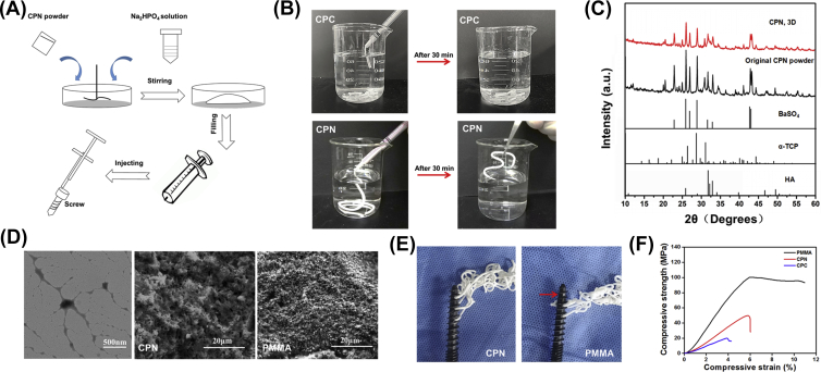
Background/objective: Both polymethylmethacrylate (PMMA) and traditional calcium phosphate-based cements have some deficiencies as augmentation materials for pedicle screw fixation. Here, a novel calcium phosphate-based nanocomposite (CPN) for the augmentation of pedicle screw fixation was developed based on previous study, and the handling properties, biomechanical performance, and biodegradation behaviour of CPN were evaluated and compared with clinical PMMA by means of a cadaver study and animal tests.

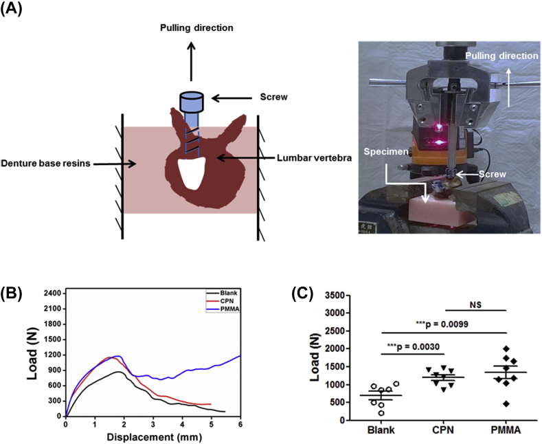
Methods: Bone mineral density of the lumbar vertebrae was tested. Pedicle screws were placed into the lumbar vertebrae under the guidance of three dimensionally printed templates; each of which was designed based on computed tomography (CT) reconstruction of each vertebrae ​and augmented with either PMMA or CPN. X-ray and CT scan were used to evaluate the accuracy of screw placement and dispersion as well as interdigitation of bone cement. The axial pull-out strength and maximum torque were tested using a mechanical testing machine. Degradation behaviour of CPN was evaluated by in vitro immersion tests for 8 weeks and in vivo rabbit femur defect model for up to 6 months, respectively.

Results: Standard mechanical tests revealed that PMMA was much stronger than CPN after setting (compressive strength 95 vs. 49 ​MPa, respectively, p ​< ​0.001). Results of the projection area and volume distribution of cement along the distal end of the screws revealed that CPN exhibited unique dispersing and interdigitation abilities compared with PMMA. Specifically, CPN dispersed uniformly and symmetrically along the screw, while PMMA was limited to the proximal part of the screw. Axial pull-out test results showed that the axial pull-out strengths of CPN- and PMMA-augmented pedicle screws were similar (1199 ​± ​225 ​N vs 1337 ​± ​483 ​N, respectively) and not significantly different (p ​= ​0.47), although CPN was an intrinsically weaker material than PMMA. Similarly, CPN showed average torque values of 0.72 ​± ​0.31 ​N·m slightly lower than those of PMMA (0.96 ​± ​0.23 ​N·m), but statistically there was no significant difference between CPN and PMMA (p ​= ​0.21). In a rabbit model of femoral bone defect, the implanted CPN maintained its clear boundary and there is no disintegration in the cement clump after 20 days and 24 weeks, and there was moderate bioabsorption of CPN and clearly new bone ingrowth at the absorbed sites after 24 weeks.

Conclusion: A new nanocomposite cement CPN, designed for replacing the nondegradable PMMA cement and overcoming the mechanical inferiority of calcium phosphate cement, was evaluated for its biomechanical and biodegradation behaviours in cement-injectable cannulated pedicle screws (CICPS) application. Although CPN is a mechanically weaker material than PMMA, CPN demonstrates similar biomechanical properties to PMMA in the application of augmentation for CICPS fixation in cadaveric vertebrae. This improvement in biomechanical property is attributed to a better dispersion and interdigitation mode of CPN. In addition, the animal study results suggest the in vivo absorption of CPN is slow enough and matches the bone ingrowth.

The translational potential of this article: This work reports a cadaveric and biomechanical study of novel CPN for the application in the augmentation of CICPS. The results suggest that CPN has equivalent or better biomechanical and interdigitation performance compared with PMMA. Together with the biodegradability and ossointegration capability, CPN reveals high translational potential as a new bone cements for load-bearing bone fixation and repair.

Keywords: Calcium phosphate–based cement; Injectable; Osteoporosis; PMMA; Pedicle screw.


Figures

Figure 1
Figure 1
Design of three dimensionally printed template and evaluation of the accuracy of CICPS placement. (A) Schematic revealing that design strategy of three dimensionally printed template is to ensure that the (1) screws are parallel on the sagittal plane, (2) the angle to the midline in coronal plane are same, and (3) the screws can be completely placed within the pedicle. (B) X-ray images showing that three points were selected for the transverse section of the pedicle in the CT scan to evaluated the accuracy of screw placement. The point A was a screw into the pedicle, point B was the midpoint of the pedicle, and point C was a screw into the vertebral body (C) The screw was located completely in the transverse section of the point A in pedicle, showing the accuracy of the screw placement guided by three dimensionally printed template. CICPS, cement-injectable cannulated pedicle screw
Figure 2
Figure 2
The CICPSs were inserted into the lumbar vertebra under the guidance of three dimensionally printed template. (A) Screw insertion instruments. ①Screw handle, ②T-handle, ③cannulated screw tap, ④cannulated drill bit, and ⑤Kirschner wire. (B) Insert the cannulated drill bit along the channel of the template. (C) Remove the inner core and insert the Kirschner wire in needle lumen. (D) Remove the cannulated drill bit. (E) Tap along the Kirschner wire. (F) Insert the screw. (G) The specimen with screw placement in all lumbar segments. CICPS, cement-injectable cannulated pedicle screw.
Figure 3
Figure 3
Radiological evaluation after augmentation with different cements. (A) A-P and lateral view of X-ray film of the specimen after inserting the screws. (B) A-P and lateral view of X-ray film of the specimen after screws augmentation. (C) A-P view and (D) axial view X-ray film of a single vertebral body after screws augmentation. (E) The dispersion model of CPN and PMMA was different. The CPN bone cement evenly surrounds all side holes of pedicle screw and the PMMA bone cement is before surrounding the proximal side holes. Point D is the location of the hole in the middle of the CICPSs. (F) The total volume (TV) of bone cement was divided into two parts. The yellow part was front volume (FV) and the blue part was rear volume (RV). CPN, calcium phosphate–based nanocomposite; PMMA, polymethylmethacrylate; CICPS, cement-injectable cannulated pedicle screw.
Figure 4
Figure 4
Preparation and properties of calcium phosphate-based nanocomposite. (A) Schematic revealing the preparation of injectable CPN cement; (B) CPN showed good injectability and anticollapsibility in water and self-setting behaviour in the water after 30 ​min. (C) X-ray diffraction patterns of CPN before and after setting, comparing with hydroxyapatite (HA), alfa-TCP, and BaSO4. (D) TEM image of nanoscale networks of gelatinized starch and SEM images of the microstructure of CPN and PMMA, respectively. (E) CPN could flow out from all the holes of the screw, while PMMA had no cement flowed out from the holes at the far end of the screw. (F) Representative compressive stress–strain curves of CPC, CPN, and PMMA. CPN, calcium phosphate–based nanocomposite; PMMA, polymethylmethacrylate; CPC, calcium phosphate cement; TCP, tricalcium phosphate; TEM, transmission electron microscopy; SEM, scanning electron microscopy.
Figure 5
Figure 5
Dispersing and interdigitation abilities of cements in the vertebrae. (A) Projected areas of cements in the A-P view and axial view, and there was no significant difference in the projected area of CPN and PMMA in all directions (for A-P view, p ​= ​0.089; axial view, p ​= ​0.098). (B) Dispersion volumes of CPN and PMMA (including total volume, front volume, and rear volume), and the front volume of CPN was higher than that of PMMA (p ​= ​0.008). CPN, calcium phosphate–based nanocomposite; PMMA, polymethylmethacrylate.
Figure 6
Figure 6
Results of axial pull-out tests on cement-augmented CICPS. (A) Schematic and photo showing the setup of axial pull-out tests and vertical alignment of screw in the vertebra. (B) Typical load–displacement curves of the axial pull-out tests of CICPS without cement (Blank) and with CPN and PMMA augmentation, respectively; (C) Statistical results of the axial pull-out strengths of augmented CICPS. The average value of CPN (1199 ​± ​225N) was slightly lower than that of PMMA (1337 ​± ​483N), but the difference is not significant (P ​= ​0.47), and both CPN and PMMA had significantly higher pull-out strengths than that of the screws without cement augmentation (693 ​± ​312 ​N) ​(P ​< ​0.01).Data are mean ​± ​standard deviation. CPN, calcium phosphate–based nanocomposite; PMMA, polymethylmethacrylate; CICPS, cement-injectable cannulated pedicle screw.
Figure 7
Figure 7
Results of torsion tests on cement-augmented CICPS. (A) Schematic and photo showing the setup of torsion tests and horizontal alignment of screw. (B) Representative torque–angle curves of the torsion tests of CICPS augmented without cement (blank) and with CPN and PMMA augmentation, respectively; (C) Statistical results of the maximum torque of augmented CICPS. PMMA showed a slightly higher average torque value than CPN, but the difference has no statistical significance (P ​= ​0.21). The screws without cement showed maximum torque values (0.29 ​± ​0.12 ​N ​m) much lower than those of CPN (0.72 ​± ​0.31 ​N ​m, p ​= ​0.0276) and PMMA (0.96 ​± ​0.23 ​N ​m, p ​= ​0.0003). Data are mean ​± ​standard deviation. CPN, calcium phosphate–based nanocomposite; PMMA, polymethylmethacrylate; CICPS, cement-injectable cannulated pedicle screw.
Figure 8
Figure 8
Histological analysis of the CPN after implanted in rabbit femur defect. (A) Schematic showing the surgical process and (B) the position of cements implanted into the femoral condyle of the rabbit. (C) X-ray image of the cement. (D)The boundary of the CPN samples kept its original cylindrical shape after 20 days, and (E) CPN almost did not degrade. (F) CPN significantly degraded in vivo after 24 weeks, and (G) bone ingrowth was observed. CPN, calcium phosphate–based nanocomposite.

References

    1. Renner S.M., Lim T.H., Kim W.J., Katolik L., An H.S., Andersson G.B. Augmentation of pedicle screw fixation strength using an injectable calcium phosphate cement as a function of injection timing and method. Spine. 2004;29(11):E212–E216. Phila Pa 1976. [eng] - PubMed
    1. Kim Y.J., Bridwell K.H., Lenke L.G., Rhim S., Cheh G. Pseudarthrosis in long adult spinal deformity instrumentation and fusion to the sacrum: prevalence and risk factor analysis of 144 cases. Spine. 2006;31(20):2329–2336. Phila Pa 1976. - PubMed
    1. Elder B.D., Lo S.F., Holmes C., Goodwin C.R., Kosztowski T.A., Lina I.A. The biomechanics of pedicle screw augmentation with cement. Spine J. 2015;15(6):1432–1445. - PubMed
    1. Saracen A., Kotwica Z. Complications of percutaneous vertebroplasty: an analysis of 1100 procedures performed in 616 patients. Medicine (Baltim) 2016;95(24):e3850. - PMC - PubMed
    1. Mclachlin S.D., Al S.K., Gurr K.R., Bailey S.I., Bailey C.S., Dunning C.E. Comparative assessment of sacral screw loosening augmented with PMMA versus a calcium triglyceride bone cement. Spine. 2010;36(11):699–704. - PubMed