. 2019 Aug 7:20:113-121.
doi: 10.1016/j.jot.2019.06.006. eCollection 2020 Jan.

Prediction on the plantar fascia strain offload upon Fascia taping and Low-Dye taping during running

Affiliations

Prediction on the plantar fascia strain offload upon Fascia taping and Low-Dye taping during running

Tony Lin-Wei Chen et al. J Orthop Translat. .

Abstract

Background: Taping is commonly prescribed to treat plantar fasciitis for runners by virtue of its alleged ability to offload the plantar fascia and facilitate positive injury prognosis. Our study aimed to investigate how different taping methods could change the loading on the plantar fascia during running using computational simulations.

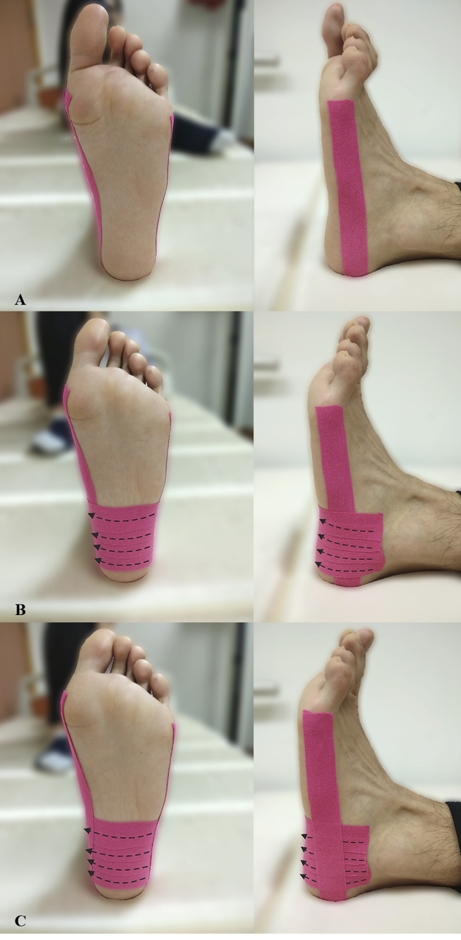
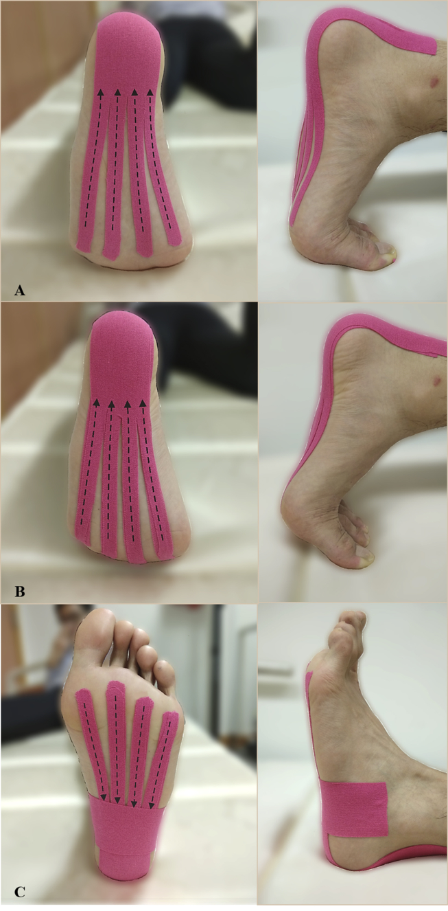
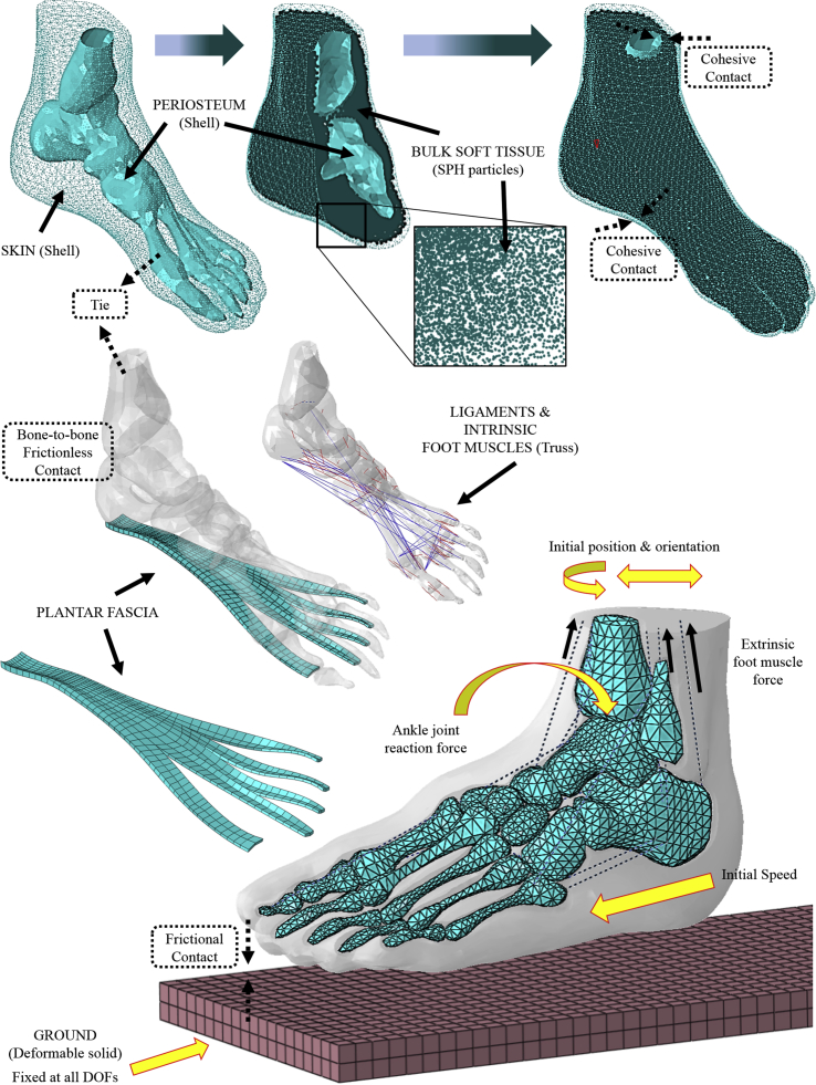
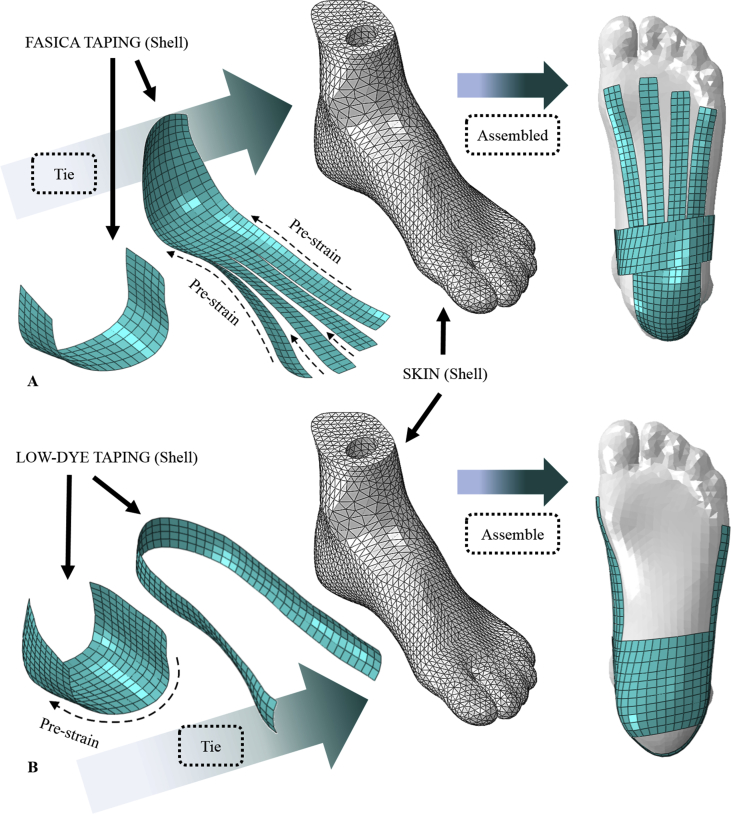
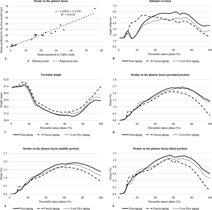
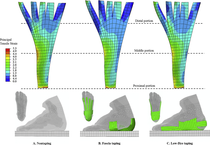
Methods: A finite element foot model was modified from a previous version to fit the study's purpose. The model featured twenty bones, bulk soft tissue, foot muscles, ligaments/tendons, and a solid part representing the plantar fascia. A runner performed several running trials under one untaped condition and two taped conditions-Low-Dye taping and Fascia taping, which were implemented by a physiotherapist using the Kinesio tapes. The captured motion data were processed to drive a scaled musculoskeletal model and calculate segmental kinematics, foot muscle force, and joint reaction force. These variables were then input as the boundary/loading conditions for finite element analyses of running. The principal tensile strain on the plantar fascia, subtalar eversion, and navicular height during the stance phase were averaged across five trials of each condition and compared using Friedman's test.

Results: Maximal subtalar eversion did not differ among conditions (p = 0.449). Fascia taping significantly reduced maximal strains on the fascia band (p = 0.034, Kendall's W = 0.64-0.76) and increased the navicular height (p = 0.013, Kendall's W = 0.84) compared with nontaping. There were no significant differences in all outcome variables between Low-Dye taping and nontaping (p = 0.173-0.618).

Conclusion: From a mechanical point of view, our study provided quantitative evidence to support the application of taping treatments for overstrained plantar fascia. The untensed fascia band by Fascia taping could be a potential indicator of pain relief for the runners. However, a prospective study targeting the patient population would be needed to address the point.

The translational potential of this article: The study quantified the loading status of the plantar fascia during running and provided mechanical evidence to support the usage of taping as a mean to reduce fascial strain, thus possibly controlling injury risks for the runners. The results of the study also highlighted the importance of selecting specific taping methods based on individuals' needs.

Keywords: Athletic tape; Biomechanics; Finite element analysis; Plantar fascia; Running.


Figures

Figure 1
Figure 1
(A) The first step of Low-Dye taping; (B) The second step of Low-Dye taping; (C) The third step of Low-Dye taping. In the first step, the first strap, which served as the anchor tape, was applied with gentle compression. It started at the medial first metatarsal head, stretched proximally along the medial border of the foot, went around the back of the heel, and attached to the lateral fifth metatarsal head. In the second step, a series of stirrups were applied when the rearfoot was slightly supinated. The first stirrup started distal to the lateral malleolus, pulled the calcaneus medially with a 50% stretch (50% tensile strain), and attached to just below the medial malleolus. The second, third, and fourth stirrup followed the same pattern with an overlap of approximately one-third of the tape width, moving in the distal direction. In the third step, the last strap was applied in the similar form of the first strap to secure the whole taping structure. Dash arrows denote the direction of stretch.
Figure 2
Figure 2
(A) The first step of Fascia taping; (B) The second step of Fascia taping; (C) The third step of Fascia taping. In the first step, after the metatarsophalangeal joints were dorsiflexed, the first strap was adhered firmly to the posterior heel at its proximal end. The other end of the strap was cut into four slices of equal width. Each slice was applied with a 50% stretch (50% tensile strain) and attached to the plantar forefoot. In the second step, another strap was applied following the same pattern and overlapped the first strap. In the third step, the last strap was applied with gentle compression across the bases of the four slices beneath the foot and wrapped around the rearfoot. Dashed arrows denote the direction of stretch.
Figure 3
Figure 3
Setup overview of the finite element foot model and boundary conditions. Solid black arrows denote the names of the parts. Solid maize arrows represent boundary/loading conditions. Dashed frames denote the interactions among parts. The bulk soft tissue was modelled as SPH particle elements and encapsulated in a shell unit that possessed an interior profundal fascia layer and exterior skin layer. The internal layer of the shell was tied to the skeletal structures. The plantar foot was connected by ligaments (truss unit), intrinsic foot muscles (truss unit), and the plantar fascia (three-dimensional solid). The ground plate was fully fixed, and the foot model was placed at an initial position/orientation. Three-dimensional ankle joint reaction force, extrinsic foot muscle force, and initial transitional velocity were applied to the model to drive the simulation. DOFs = degrees of freedom; SPH = smoothed-particle hydrodynamics.
Figure 4
Figure 4
(A) Modelling of Fascia taping in the simulations; (B) modelling of Low-Dye taping in the simulations. Dashed arrows denote the direction assigned with a prestrain of 50% to mimic stretch on the tapes. Solid black arrows denote the names of the parts. Shell units that represented the tape straps were tied to the skin surface.
Figure 5
Figure 5
(A) Results of model validation; (B) predicted subtalar eversion; (C) predicted navicular height; (D) predicted strains on the proximal plantar fascia; (E) predicted strains on the middle plantar fascia; (F) predicted strains on the distal plantar fascia. Each dot in the validation plot denotes the simulated and measured strain value of one measurement point as depicted in Clark's study. Parameters of the trend line (R2: 0.95, slope: 1.05, intercept: 0.17) indicated a good agreement between the model estimates and experimental outcomes; all outcome variables in the predictions are scaled to the percentile stance phase.
Figure 6
Figure 6
(A) Strain contour of plantar fascia for the non-taping condition; (B) Strain contour of plantar fascia for the Fascia taping condition; (C) Strain contour for the plantar fascia for the Low-Dye taping condition. The contour plots were extracted at the instant of maximal strain for a representative trial. The images are colour coded based on the distribution of principal tensile strains. Red means the highest strain, and blue means the lowest strain. Regions of the plantar fascia that are tied to the bony segments are removed from display, and the remainder is equally divided into three portions from proximal to distal. The figure shows that Fascia taping produced an apparently larger cool-tonal area, which indicates a lower strain level on average.

References

    1. Jamali B., Walker M., Hoke B., Echternach J. Windlass taping technique for symptomatic relief of plantar fasciitis. J Sport Rehabil. 2004;13:228–243.
    1. Riddle D.L., Pulisic M., Pidcoe P., Johnson R.E. Risk factors for Plantar fasciitis: a matched case-control study. J Bone Joint Surg Am. 2003;85-A 872–7. - PubMed
    1. Buchbinder R. Clinical practice. Plantar fasciitis. N Engl J Med. 2004;350:2159–2166. - PubMed
    1. Tong K.B., Furia J. Economic burden of plantar fasciitis treatment in the United States. Am J Orthoped. 2010;39:227–231. - PubMed
    1. Wearing S.C., Smeathers J.E., Urry S.R., Hennig E.M., Hills A.P. The pathomechanics of plantar fasciitis. Sport Med. 2006;36:585–611. - PubMed