. 2019 Jul-Dec;9(2):261-282.
doi: 10.4103/ams.ams_151_19.

Frontal Bone Fractures and Frontal Sinus Injuries: Treatment Paradigms

Affiliations

Frontal Bone Fractures and Frontal Sinus Injuries: Treatment Paradigms

Priya Jeyaraj. Ann Maxillofac Surg. 2019 Jul-Dec.

Abstract

Background: Timely, expeditious and appropriate management of Frontal bone fractures and associated Frontal Sinus (FS) injuries are both crucial as well as challenging. Treatment options vary considerably, depending upon the nature, extent and severity of these injuries as well as operator skill, expertise and experience. In cases of posterior table fractures of the Frontal Sinus, literature reports have in general, propounded direct visualization and exploration of the sinus via a bifrontal craniotomy, followed by sinus cranialization.

Aims and objectives: To review the standard protocols of management of Frontal bone fractures and Frontal Sinus injuries. To assess the efficacy of a more conservative approach in the management of outer and inner table fractures of the FS.

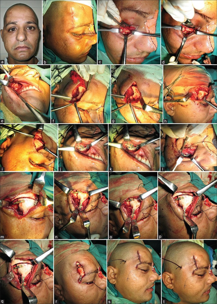
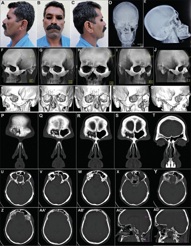
Materials and methods: Contemporary and evolving management protocols and changing treatment paradigms of different types and severities of frontal bone fractures and frontal sinus injuries, have been presented in this case series. A useful Treatment Algorithm has been proposed to efficiently and effectively manage these injuries.

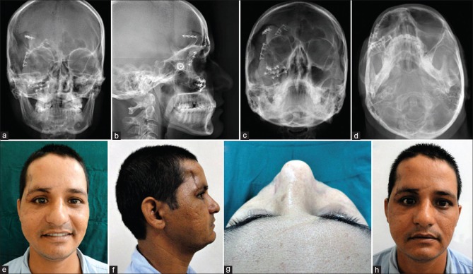
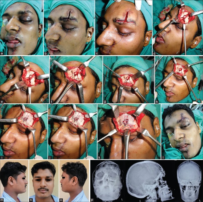
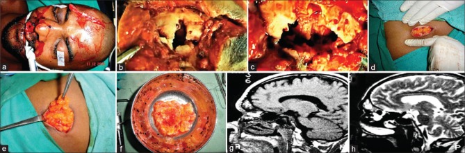
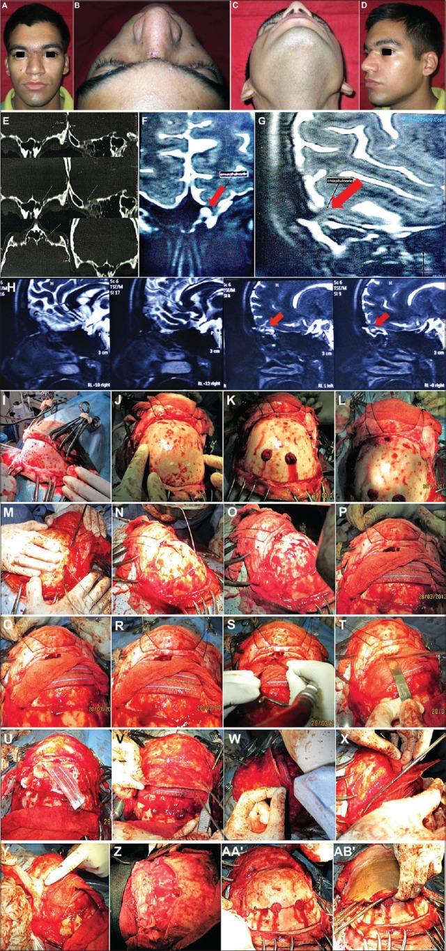
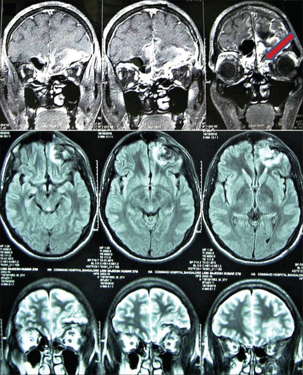
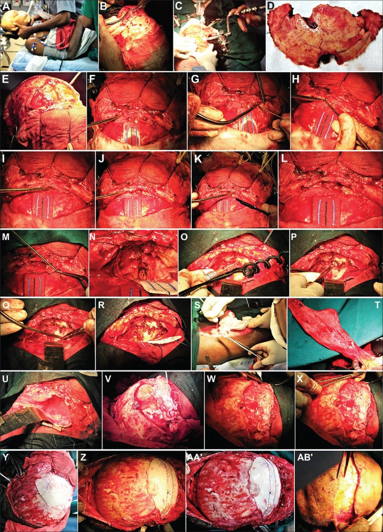
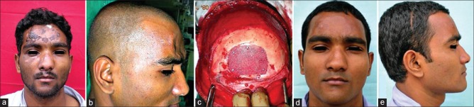
Results: In the present case series, effective and satisfactory results could be achieved in cases of significantly displaced inner and outer table fractures of the Frontal sinus by a more conservative protocol comprising of open reduction and internal fixation carried out via the existing scar of injury, without having to resort to the more radical intracranial approach and sinus cranialization. Nevertheless, presence of complicating factors such as cerebrospinal fluid rhinorrhea, evidence of meningitis or the development of encephalomeningocoeles necessitated the standard protocol of sinus exploration and its cranialization or obliteration.

Conclusion: Management protocols of Frontal Sinus injuries vary, based on aspects such as the timing of presentation and intervention, degree of injury sustained, concomitant associated Craniomaxillofacial injuries present, presence of complicating factors or Secondary/Residual deformities & Functional debility, and need to be decided upon on a case to case basis.

Keywords: Anterior and posterior table fractures of frontal sinus; frontal bone; frontal sinus; frontal sinus cranialization; frontal sinus obliteration; nasofrontal duct; nasofrontal outflow tract; onlay grafting.


Conflict of interest statement

There are no conflicts of interest.

Figures

Figure 1
Figure 1
(a and b) A 23-year-old male patient who sustained multiple abrasions, lacerations, and contusions over the face in a fall from a two-wheeler. (c-h) Noncontrast computed tomography scans (coronal and axial sections) revealed a minimally displaced fracture of the outer table of the frontal sinus with hemosinus. He was managed conservatively and developed no complications
Figure 2
Figure 2
(a-d) A 37-year-old male patient sustained panfacial trauma in a road traffic accident. He had fracture of the mandibular parasymphysis, a Le Forte 1 fracture of the right maxilla, and a linear, undisplaced fracture of outer table of the frontal sinus, which was revealed on a computed tomography scan. (e-g) Open reduction and internal fixation of maxilla and mandible carried out, frontal bone fracture was unaddressed. (h-j) Follow-up of 2 years showed no complications arising from the undisplaced frontal bone fracture and no residual forehead deformity or contour irregularity postoperatively
Figure 3
Figure 3
(a-c) A 29-year-old male patient with residual deformities resulting from a road traffic accident 3 months before. Hypoglobus and enophthalmos (right). (d) Arch bar fixation done. (e and f) Moderately displaced fracture of the frontal bone, superior orbital rim, and outer table of the frontal sinus, Le Forte II fracture of maxilla and right zygomaticomaxillary complex, large orbital floor defect (right). (g-j) Fractures exposed. (k-p) Herniated orbital contents restored and floor defect reconstructed using symphyseal bone graft. (q and r) Frontal bone and superior orbital rim fractures reduced and fixed. (s-v) Open reduction and internal fixation of fractures. (w-z) Satisfactory correction of residual deformities
Figure 4
Figure 4
(a-g) A 53-year-old male patient who sustained panfacial trauma in a road traffic accident. Moderately displaced fracture of the frontal bone with disjunctions at the frontozygomatic sutures, fracture left zygomaticomaxillary complex and mandibular body. (h-j) Open reduction and internal fixation of frontal bone via a bicoronal approach. Single screw placed through the anterior table bone to reduce and reposition the depressed fractured frontal bone fragments and stabilize them while rigid fixation was applied using titanium microplates and screws. (k and l) Good esthetic and functional results with nil postoperative complications
Figure 5
Figure 5
(a-d) A 26-year-old male patient with residual deformity resulting from a road traffic accident 1 month earlier. The patient was kept under neurological surveillance for a month and thereafter referred for maxillofacial surgical intervention. The patient presented with a distinct antimongoloid slant, drooping of entire right zygomaticomaxillary complex. (e-h) Noncontrast computed tomography scans revealed severely displaced fracture of the frontal bone and right zygomaticomaxillary complex, with disjunction of the entire segment from the cranium. (i-x) Fractures of frontal, temporal bones, and right greater wing of sphenoid evident on coronal, axial, and sagittal sections
Figure 6
Figure 6
(a-e) Open reduction and internal fixation under general anesthesia. (f-i) The existing scar above the right eyebrow was used to expose the fractured, displaced, and depressed frontal and temporal bone fragments, including the regions of frontozygomatic and zygomaticotemporal dysjunctions. (j-l) Shattered right zygomatic buttress, body of zygoma, and infraorbital rim reduced and fixed. (m-q) The fractured fragments of the frontal and temporal bones carefully reduced, reapproximated, and fixed with titanium plates and screws. (r-t) Closure was completed in layers after placement of a vacuum-assisted closed suction drain
Figure 7
Figure 7
(a-d) Radiographs postsurgery showing successful repositioning of the displaced fractured segments of the frontal and zygomatic bones with implants in situ. (e-h) Photographs showing a successful correction of the cosmetic deformity and antimongoloid slant caused by the severely displaced and sagging frontal bone and zygomatic complex
Figure 8
Figure 8
(A-D) A 29-year-old male patient who was struck on the face with a metal pole and presented with a lacerated wound across the forehead, left eyebrow, and across the face in region of ala of nose, lips, and chin on the left. (E-L) Radiographs revealed a vertical depressed fracture of the frontal bone on the left with comminution of the left supraorbital rim and fracture of the left maxilla. (M-AB’) Inferiorly displaced segment of the part of the frontal bone forming roof of the left orbit, apparent on coronal and axial sections of noncontrast computed tomography
Figure 9
Figure 9
(a-d) Existing scar used to expose fracture of the frontal bone and left supraorbital rim. (e-h) Displaced fragments reapproximated, repositioned, and fixed. Unsalvageable free fragments of crushed bone removed. (i-l) Defect of the frontal bone reconstructed using three-dimensional dynamic titanium mesh implant, followed by layer-wise closure. (m-o) Smooth postoperative recovery with a good esthetic as well as functional outcome. (p) Postoperative radiographs showing restoration of the displaced frontal bone and orbital roof and rim fractures with implants in situ
Figure 10
Figure 10
(a) Severely comminuted fracture of the outer table of the frontal sinus sustained by a 39-year-old male patient in a road traffic accident. (b) Existing laceration used to expose the fracture. (c) Free unsalvageable fragments of bone removed and Mucosal lining of the entire frontal sinus carefully extirpated. (d-f) Autologous fat harvested from the subcutaneous layer of the anterior abdominal wall and used to obturate, obliterate, and seal the frontal sinus, prior to replacement of the outer table augmented with titanium mesh. (g and h) Postoperative magnetic resonance imaging showing the healthy and viable fat tissue within the frontal sinus
Figure 11
Figure 11
(a-e) Postoperative appearance and plain radiographs of the patient showing a good restoration of the forehead contour. (f-o) Non-contrast computed tomography showing a good reconstruction of the severely comminuted outer table of the frontal sinus by the three-dimensional dynamic titanium mesh implant. (p-t) Coronal sections showing complete obliteration of the right frontal sinus with the titanium mesh implant in situ. (u-ad’) Axial and Sagittal sections showing good restoration of contour and integrity of the outer table of the FS
Figure 12
Figure 12
A 20-year-old male patient with isolated posterior table fracture (A-D) sustained in a horse-riding accident, initially managed conservatively. After 6 months developed recurrent episodes of fever, vomiting and headaches. (E-H) Computed tomography scans and magnetic resonance imaging cisternography revealed a 4 mm × 5 mm defect in frontal sinus posterior table and formation of an encephalomeningocoele. (I-Q) Bifrontal craniotomy exposing the frontal lobes and interior of the frontal sinus. (R-U) Frontal sinus cranialization carried out. (V-Z) Pericranial flap draped over denuded sinus floor, tucked beneath the frontal lobes, and sutured to dura. (AA’ and AB’) Bone flap replaced
Figure 13
Figure 13
(a-c) A 27-year-old male patient who sustained severe injuries to the upper third of his face in a road traffic accident and presented with cerebrospinal fluid rhinorrhea. (d-k) Noncontrast computed tomography craniomaxillofacial region revealed comminuted fracture of the frontal bone disrupting both the anterior and posterior tables of the sinus and comminution of the floor of the anterior cranial fossa (as depicted by the red arrows)
Figure 14
Figure 14
Magnetic resonance imaging brain showed dural tear, contusion, and herniation of the left frontal lobe through the defects in the posterior table of the frontal sinus and floor of the anterior cranial fossa
Figure 15
Figure 15
(A-H) Bifrontal craniotomy. Exposure of fractured floor of the anterior cranial fossa. (I-M) Debridement of free, unsalvageable bone fragments. Remaining bone of posterior table of frontal sinus bone removed, cranializing the frontal sinus. (N-R) Delicate fragments of cranial floor reapproximated, secured, and fixed. (S-V) Fascia lata graft layered over the sinus and cranial floor to separate and seal off the intracranial cavity above from the sinonasal tract below. (W and X) The free flap sutured with the dura. (Y-AB’) Bifrontal bone flap replaced
Figure 16
Figure 16
(a and b) A 32-year-old male patient, an old case of unaddressed depressed fracture of the bifrontal region, reported a year later for correction of a residual deformity of the forehead region. (c-e) Onlay grafting using Medpore implant was carried out to correct the contour irregularity with a satisfactory esthetic outcome

References

    1. Erdmann D, Follmar KE, Debruijn M, Bruno AD, Jung SH, Edelman D, et al. A retrospective analysis of facial fracture etiologies. Ann Plast Surg. 2008;60:398–403. - PubMed
    1. Strong EB, Pahlavan N, Saito D. Frontal sinus fractures: A 28-year retrospective review. Otolaryngol Head Neck Surg. 2006;135:774–9. - PubMed
    1. Friedman JA, Ebersold MJ, Quast LM. Persistent posttraumatic cerebrospinal fluid leakage. Neurosurg Focus. 2000;9:e1. - PubMed
    1. El Khatib K, Danino A, Malka G. The frontal sinus: A culprit or a victim? A review of 40 cases. J Craniomaxillofac Surg. 2004;32:314–7. - PubMed
    1. Salentijn EG, Peerdeman SM, Boffano P, van den Bergh B, Forouzanfar T. A ten-year analysis of the traumatic maxillofacial and brain injury patient in Amsterdam: Incidence and aetiology. J Craniomaxillofac Surg. 2014;42:705–10. - PubMed