Review
. 2020 Jan 3:6:3.
doi: 10.1038/s41531-019-0103-7. eCollection 2020.

Parkinson's disease: proteinopathy or lipidopathy?

Affiliations
Review

Parkinson's disease: proteinopathy or lipidopathy?

Saranna Fanning et al. NPJ Parkinsons Dis. .

Abstract

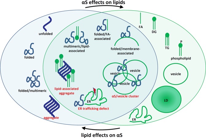
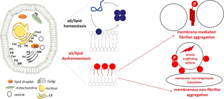
Lipids play a more significant role in Parkinson's disease and its related brain disorders than is currently recognized, supporting a "lipid cascade". The 14 kDa protein α-synuclein (αS) is strongly associated with Parkinson's disease (PD), dementia with Lewy bodies (DLB), other synucleinopathies such as multiple system atrophy, and even certain forms of Alzheimer's disease. Rigorously deciphering the biochemistry of αS in native systems is the key to developing treatments. αS is highly expressed in the brain, the second most lipid-rich organ, and has been proposed to be a lipid-binding protein that physiologically interacts with phospholipids and fatty acids (FAs). αS-rich cytoplasmic inclusions called Lewy bodies and Lewy neurites are the hallmark lesions of synucleinopathies. Excess αS-membrane interactions may trigger proteinaceous αS aggregation by stimulating its primary nucleation. However, αS may also exert its toxicity prior to or independent of its self-aggregation, e.g., via excessive membrane interactions, which may be promoted by certain lipids and FAs. A complex αS-lipid landscape exists, which comprises both physiological and pathological states of αS. As novel insights about the composition of Lewy lesions occur, new lipid-related PD drug candidates emerge, and genome-wide association studies (GWAS) increasingly validate new hits in lipid-associated pathways, it seems timely to review our current knowledge of lipids in PD and consider the roles for these pathways in synucleinopathies.Fig. 1αS ↔ lipid interplay: aspects of cellular αS homeostasis (blue oval), aspects of lipid homeostasis (green oval), and overlapping aspects.Pathological states are labeled in red. Simplified schematic of both select αS and select lipid species. Several existing publications suggest αS effects on lipids and vice versa, as indicated by arrows. DG diglyceride, ER endoplasmic reticulum, FA fatty acid, LD, lipid droplet, TG triglyceride.

Keywords: Cellular neuroscience; Parkinson's disease.


Conflict of interest statement

Competing interestsD.J.S. is a director and consultant to Prothena Biosciences. The other authors declare no conflict of interest.

Figures

Fig. 1
Fig. 1. αS ↔ lipid interplay: aspects of cellular αS homeostasis (blue oval), aspects of lipid homeostasis (green oval), and overlapping aspects.
Pathological states are labeled in red. Simplified schematic of both select αS and select lipid species. Several existing publications suggest αS effects on lipids and vice versa, as indicated by arrows. DG diglyceride, ER endoplasmic reticulum, FA fatty acid, LD, lipid droplet, TG triglyceride.
Fig. 2
Fig. 2. Electrostatic and hydrophobic interactions govern transient αS membrane binding.
a Amino acid sequence of human wt αS; residues that fully conform to the core repeat motif “KTKEGV” are highlighted in gray. b Amino acid sequence of human wt αS displayed by aligning the KTKEGV motifs. c Analogous to b, color-coded residues: black = uncharged, red = negatively charged, dark blue = positively charged, light blue = histidine, and purple = uncharged and polar. d Color-coded schematic of repeats 1–7 (omitting “ATVA” between repeats 4 and 5) in an 11/3 helical wheel, embedded in the outer leaflet of a lipid membrane. e Simplified schematics of membrane-induced αS helices: αS wt as well as 3 K (amplified E46K) and KLK (engineered highly hydrophobic). Top: wt. Middle: a proposed increased electrostatic interaction between excess positive charges of the lysines (highlighted in yellow) and phospholipid head groups is indicated by a blue line. Bottom: the KLK variant is stabilized by excess hydrophobicity in the hydrophobic half of the amphipathic helix, highlighted in yellow.
Fig. 3
Fig. 3. Contrasting αS fibrillar (“amyloid”) and (vesicle) membrane/lipid-rich aggregation forms.
They are not mutually exclusive. Membranous aggregates could, e.g., be precursors of fibrillar aggregates.
Fig. 4
Fig. 4. αS membrane-associated aggregation in LBs and models thereof.
a Vesicle-rich membranous αS aggregation in the αS::GFP expression model in yeast. Insert: immunogold staining for αS. Reprinted by permission from the National Academy of Sciences, USA. b Vesicle- and tubule-rich membranous αS aggregation in the αS 3K neuroblastoma cell model. Reprinted from Dettmer et al. (CC-BY license). c Vesicle/membrane/lipid-rich αS aggregation in human Lewy bodies. Reprinted by permission from the Springer Publishing Group.
Fig. 5
Fig. 5. Indications for an αS ↔ OA bidirectional pathogenic loop.
αS excess leads to increased OA levels; increases in OA disrupt αS homeostasis leading to αS + vesicle clusters, vesicle-trafficking defects, and possibly αS aggregates. Inhibition of the rate-limiting enzyme in OA production, (SCD), promises to mitigate these pathogenic events.
Fig. 6
Fig. 6. αS/lipid homeostasis and dyshomeostasis.
Left: cell with LDs, vesicles, ER, nucleus, mitochondria and annotated with some of the lipid species discussed in this review and relevant to PD. Parts are adapted from Van Meer et al. Cer ceramide, Chol cholesterol, PC phosphotidylcholine, PE phosphotidylethanolamine, PI phosphatidylinositol, PS phosphotidylserine, SM sphingomyelin, TG triglyceride. Middle: intact αS/lipid homeostasis (top: αS transiently binds to membranes and undergoes cycles of assembly and disassembly) and αS lipid dyshomeostasis (bottom: αS accumulates at membranes of altered composition). Right: αS in disequilibrium forms fibrillar (top) or membranous non-fibrillar aggregates, mediated by abnormal lipid interactions.

References

    1. Sastry PS. Lipids of nervous tissue: composition and metabolism. Prog. Lipid Res. 1985;24:69–176. doi: 10.1016/0163-7827(85)90011-6. - DOI - PubMed
    1. Stöckl M, Fischer P, Wanker E, Herrmann A. Alpha-synuclein selectively binds to anionic phospholipids embedded in liquid-disordered domains. J. Mol. Biol. 2008;375:1394–1404. doi: 10.1016/j.jmb.2007.11.051. - DOI - PubMed
    1. Bodner CR, Dobson CM, Bax A. Multiple tight phospholipid-binding modes of alpha-synuclein revealed by solution NMR spectroscopy. J. Mol. Biol. 2009;390:775–790. doi: 10.1016/j.jmb.2009.05.066. - DOI - PMC - PubMed
    1. Bodner CR, Maltsev AS, Dobson CM, Bax A. Differential phospholipid binding of alpha-synuclein variants implicated in Parkinson’s disease revealed by solution NMR spectroscopy. Biochemistry. 2010;49:862–871. doi: 10.1021/bi901723p. - DOI - PMC - PubMed
    1. Ruipérez V, Darios F, Davletov B. Alpha-synuclein, lipids and Parkinson’s disease. Prog. Lipid Res. 2010;49:420–428. doi: 10.1016/j.plipres.2010.05.004. - DOI - PubMed