. 2020 Jan 3:3:7.
doi: 10.1038/s42003-019-0737-3. eCollection 2020.

A dual fluorescent Plasmodium cynomolgi reporter line reveals in vitro malaria hypnozoite reactivation

Affiliations

A dual fluorescent Plasmodium cynomolgi reporter line reveals in vitro malaria hypnozoite reactivation

Annemarie M Voorberg-van der Wel et al. Commun Biol. .

Abstract

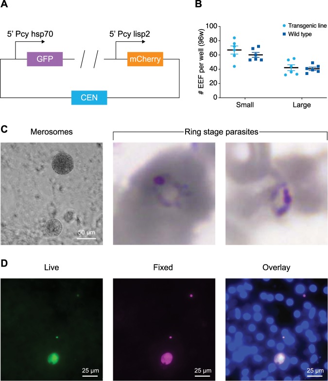
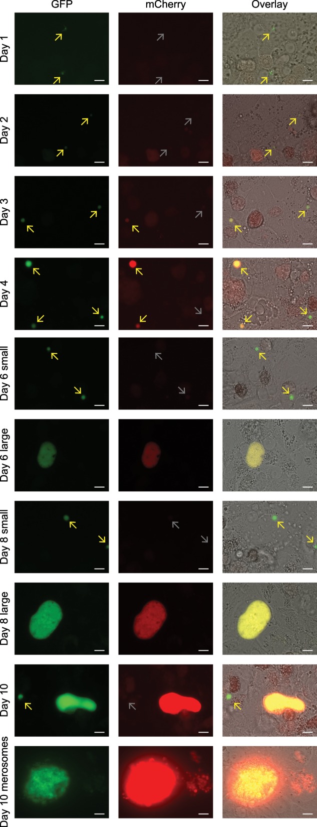
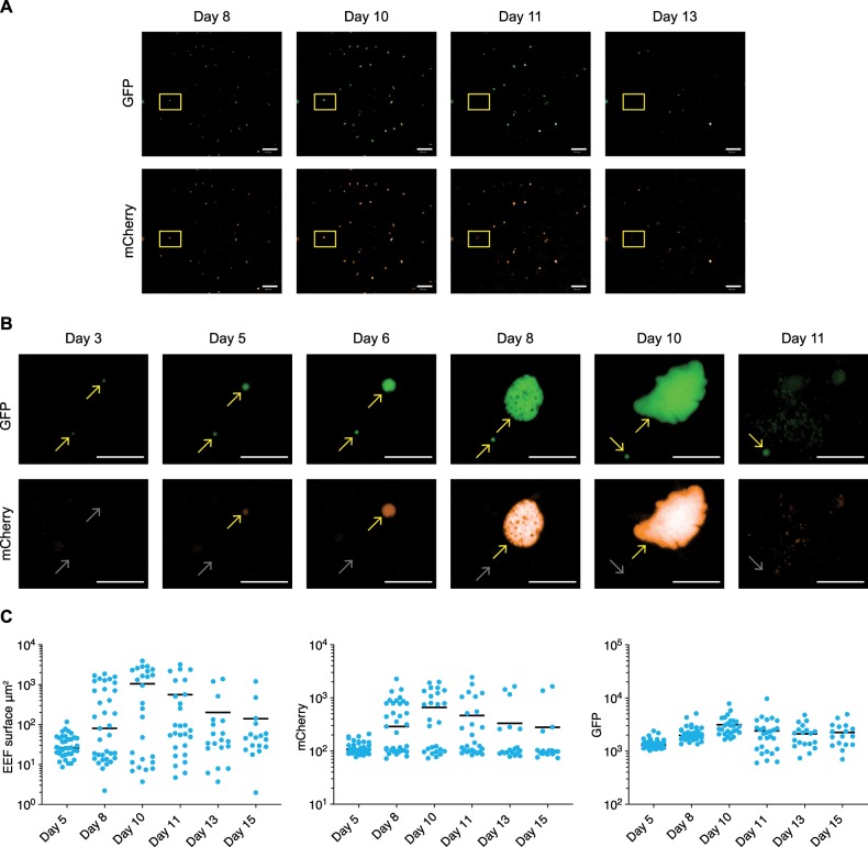
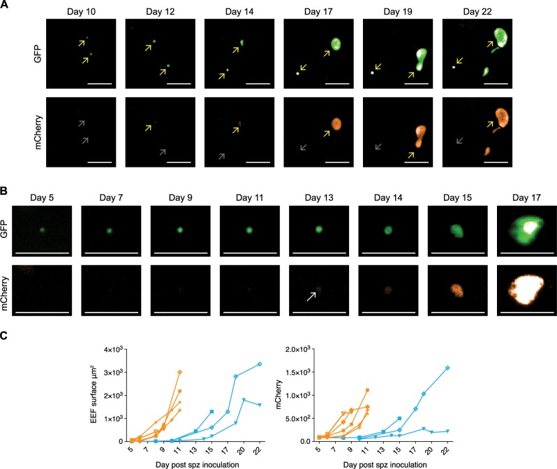
Plasmodium vivax malaria is characterized by repeated episodes of blood stage infection (relapses) resulting from activation of dormant stages in the liver, so-called hypnozoites. Transition of hypnozoites into developing schizonts has never been observed. A barrier for studying this has been the lack of a system in which to monitor growth of liver stages. Here, exploiting the unique strengths of the simian hypnozoite model P. cynomolgi, we have developed green-fluorescent (GFP) hypnozoites that turn on red-fluorescent (mCherry) upon activation. The transgenic parasites show full liver stage development, including merozoite release and red blood cell infection. We demonstrate that individual hypnozoites actually can activate and resume development after prolonged culture, providing the last missing evidence of the hypnozoite theory of relapse. The few events identified indicate that hypnozoite activation in vitro is infrequent. This system will further our understanding of the mechanisms of hypnozoite activation and may facilitate drug discovery approaches.

Keywords: Malaria; Parasite biology.


Conflict of interest statement

Competing interestsDevendra Kumar Gupta and Thierry Tidiane Diagana are employed by and/or are shareholders of Novartis Pharma AG. The other authors declare that no competing interests exist.

Figures

Fig. 1
Fig. 1. The P. cynomolgi dual reporter line fully recapitulates the complete life cycle.
a Schematic of the most important building blocks of the construct used for transfection; for details of the complete construct, see methods and the Supplementary Data file 1. The plasmid contains two fluorescent reporters, GFP driven by the hsp70 promoter and mCherry controlled by the lisp2 promoter. A centromere is included to maintain the construct throughout the life cycle. b Dot plot of the number of small and large exoerythrocytic forms (EEFs) per well in a 96-well plate with s.e.m. from n = 6 wells. Results derive from a side by side comparison of transgenic and wild-type parasites. c Left, P. cynomolgi merosomes (transgenic line) in culture at day 10 post sporozoite invasion. Scale bar, 50 µm. Right, Giemsa staining of red blood cells collected one day post overlay on a liver stage culture containing merosomes shows two ring stage blood forms. d GFP expression (live) in two small forms and one large form at day 6 post inoculation of a primary hepatocyte culture with transgenic P. cynomolgi sporozoites; the same image after fixation and IFA with anti-HSP70 antibodies, and an overlay with DAPI. Scale bars, 25 µm.
Fig. 2
Fig. 2. Lisp2-driven mCherry differentiates P. cynomolgi hypnozoites from developing forms.
GFP and mCherry expression at different time points of transgenic P. cynomolgi liver stage development. The panels at the righthand side show an overlay with brightfield images. Scale bars, 10 µm.
Fig. 3
Fig. 3. The dual fluorescent P. cynomolgi line reveals the real-time development of individual parasites.
a Images of parasites visualized live by fluorescence microscopy using an Operetta High Content imager. Overviews are shown from stitched images of the same well (96-well plate) at different time points, showing GFP-expressing parasites (upper panel) and mCherry-expressing parasites (lower panel). Parasites from the yellow inset are depicted in b. Scale bar, 500 µm. b Images of a small and a developing form expressing GFP (upper panel) and mCherry (lower panel) at different time points after sporozoite invasion. Yellow arrows mark expression, and grey arrows mark absence of fluorescence. At day 11, individual merozoites can be observed. Scale bar, 50 µm. c Operetta measurements of fluorescent EEF surface areas (left panel), mCherry intensity (middle panel), and GFP intensity (right panel) in individual parasites (blue dots) followed at different days p.i. (post infection) in ten fields of a single well. The bar represents the median values.
Fig. 4
Fig. 4. Monitoring individual parasites over time by fluorescence microscopy reveals hypnozoite reactivation.
a Hypnozoite reactivation resulting in a fully mature liver stage schizont releasing merosomes. Operetta images show GFP expression (upper panel) and mCherry expression (lower panel) of a hypnozoite (mCherry negative), and an activating hypnozoite at different time points after sporozoite invasion. Yellow arrows mark expression, and grey arrows mark absence of fluorescence. Scale bar, 50 µm. b Images showing GFP expression (upper panel) and mCherry expression (lower panel) in an activating hypnozoite at different time points after sporozoite invasion. A white arrow marks the onset of mCherry expression. Scale bar, 50 µm. c Operetta measurements of activated hypnozoites with respect to EEF fluorescent surfaces (left panel) and mCherry intensity (right panel) at different days post sporozoite (spz) inoculation (green lines). For reference, the measurements of five individual primary liver stage schizonts as already are also depicted (red lines).

References

    1. Robinson LJ, et al. Strategies for understanding and reducing the Plasmodium vivax and Plasmodium ovale hypnozoite reservoir in Papua New Guinean children: a randomised placebo-controlled trial and mathematical model. PLoS Med. 2015;12:e1001891. doi: 10.1371/journal.pmed.1001891. - DOI - PMC - PubMed
    1. Markus MB. New evidence for hypnozoite-ndependent Plasmodium vivax malarial recurrences. Trends Parasitol. 2018;34:1015–1016. doi: 10.1016/j.pt.2018.08.010. - DOI - PubMed
    1. Krotoski WA. Discovery of the hypnozoite and a new theory of malarial relapse. Trans. R. Soc. Trop. Med. Hyg. 1985;79:1–11. doi: 10.1016/0035-9203(85)90221-4. - DOI - PubMed
    1. Krotoski WA, et al. Relapses in primate malaria: discovery of two populations of exoerythrocytic stages. Br. Med. J. 1980;280:153–154. doi: 10.1136/bmj.280.6208.153-a. - DOI - PMC - PubMed
    1. Krotoski WA, et al. Observations on early and late post-sporozoite tissue stages in primate malaria. I. Discovery of a new latent form of Plasmodium cynomolgi (the hypnozoite), and failure to detect hepatic forms within the first 24 h after infection. Am. J. Trop. Med. Hyg. 1982;31:24–35. doi: 10.4269/ajtmh.1982.31.24. - DOI - PubMed

Publication types

MeSH terms

Substances