

Central giant cell granuloma of the mandibular condyle: A rare case and a literature review
- PMID: 31909260
- PMCID: PMC6939106
- DOI: 10.1016/j.heliyon.2019.e03085
Central giant cell granuloma of the mandibular condyle: A rare case and a literature review
Abstract
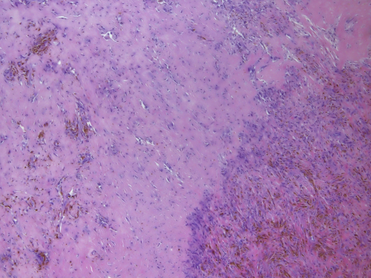
Introduction: Central giant cell granuloma is a benign intraosseous lesion; tumours in the condylar region are rarely reported.
Case presentation: We present the case of a 60-year-old woman with preauricular swelling, limitation of joint motion and pain on only the right side.
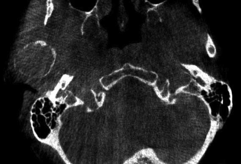
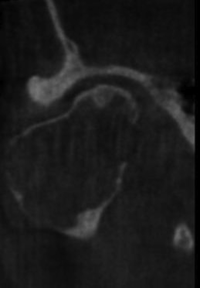
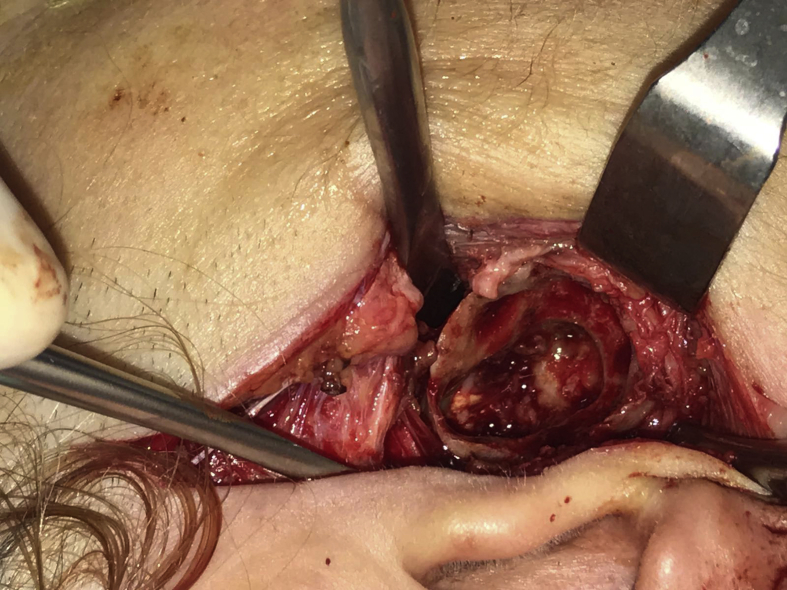
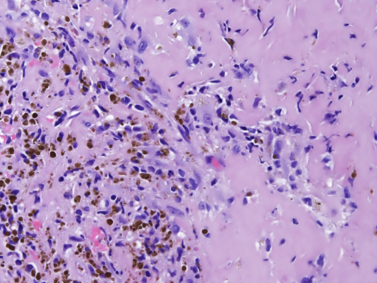
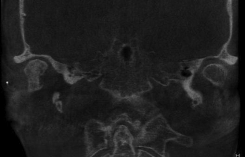
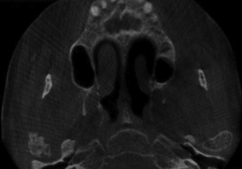
Discussion: The patient was evaluated based on her preoperative clinical manifestations, by orthopantomography and computed tomography (CT). CT revealed a lesion on the right condylar head. Surgery was scheduled based on this imaging finding, histological findings from an incisional biopsy specimen, and the patient's indications and symptoms.
Conclusion: Of all reported cases of central giantcell granuloma, only five (including this case) were located in the mandibular condyle.
Keywords: Bone; Central giant cell granuloma; Condyle; Dental surgery; Dentistry; Intraosseous; Mandible; Oral medicine; Surgery.
© 2019 The Author(s).
Figures
Similar articles
-
The first case of osteoma of the mandibular notch located both medially and laterally and treated with a transoral endoscopy assisted approach. A case report.Int J Surg Case Rep. 2018;49:70-73. doi: 10.1016/j.ijscr.2018.06.013. Epub 2018 Jun 26. Int J Surg Case Rep. 2018. PMID: 29966952 Free PMC article.
-
Central giant cell granuloma of the mandibular condyle: a case report and review of the literature.Dentomaxillofac Radiol. 2011 Jan;40(1):60-4. doi: 10.1259/dmfr/85668294. Dentomaxillofac Radiol. 2011. PMID: 21159917 Free PMC article. Review.
-
Peripheral giant cell granuloma of the mandibular condyle presenting as a preauricular mass.Eur Arch Otorhinolaryngol. 2005 Mar;262(3):178-81. doi: 10.1007/s00405-004-0758-4. Epub 2004 May 5. Eur Arch Otorhinolaryngol. 2005. PMID: 15133683
-
An aggressive central giant cell granuloma in a pediatric patient: case report and review of literature.J Otolaryngol Head Neck Surg. 2019 Jul 18;48(1):32. doi: 10.1186/s40463-019-0356-5. J Otolaryngol Head Neck Surg. 2019. PMID: 31319877 Free PMC article. Review.
-
A peripheral giant cell granuloma with extensive osseous metaplasia or a hybrid peripheral giant cell granuloma-peripheral ossifying fibroma: a case report.J Med Case Rep. 2015 Feb 4;9:14. doi: 10.1186/1752-1947-9-14. J Med Case Rep. 2015. PMID: 25649957 Free PMC article.
Cited by
-
Assessment of Tumor Angiogenesis by Expression of CD 105 in Ameloblastoma, Odontogenic Keratocyst and Central Giant Cell Lesion.Asian Pac J Cancer Prev. 2020 Nov 1;21(11):3373-3379. doi: 10.31557/APJCP.2020.21.11.3373. Asian Pac J Cancer Prev. 2020. PMID: 33247698 Free PMC article.
-
Evaluation of the relationship between the expression of AgNOR and Ki67 with the recurrence rate in central granulomatous giant cell lesions: A case-control.Clin Exp Dent Res. 2024 Apr;10(2):e870. doi: 10.1002/cre2.870. Clin Exp Dent Res. 2024. PMID: 38506305 Free PMC article.
-
Central Giant Cell Granuloma in the Mandibular Condyle in a Teenager. A Case Report with Literature Review.J Clin Med. 2022 Jul 21;11(14):4239. doi: 10.3390/jcm11144239. J Clin Med. 2022. PMID: 35888004 Free PMC article.
References
-
- Jaffe H. Giant-cell reparative granuloma, traumatic bone cyst, and fibrous (fibro-oseous) dysplasia of the jawbones. Oral Surg. Oral Med. Oral Pathol. 1953;6:159–175. - PubMed
-
- Kramer I.R., Pindborg J.J., Shear M. second ed. Springer-Verlag; Berlin, Germany: 1991. Histological typing of odontogenic tumors; p. 31.
-
- Austin L.T., Dahlin C.D., Royer Q.R. Central giant cell granuloma and related condition affecting the jawbone. Oral Surg. Oral Med. Oral Pathol. 1955;12:1259. - PubMed
-
- Chuong R., Kaban L., Kozakewich H., Perez-Atayde A. Central giant cell lesions of the jaws: a clinicopathologic study. J. Oral Maxillofac. Surg. 1986;44:708–713. - PubMed
-
- Ficarra G., Kaban L., Hansen L. Central giant cell lesions of the mandible and maxilla: a clinicopathologic and cytometric study. Oral Surg. Oral Med. Oral Pathol. 1987;64:44–49. - PubMed
Publication types
LinkOut - more resources
Full Text Sources
