. 2019 Dec 27;6(1):e03124.
doi: 10.1016/j.heliyon.2019.e03124. eCollection 2020 Jan.

Effects of PEGylated Fe-Fe3O4 core-shell nanoparticles on NIH3T3 and A549 cell lines

Affiliations

Effects of PEGylated Fe-Fe3O4 core-shell nanoparticles on NIH3T3 and A549 cell lines

B H Domac et al. Heliyon. .

Abstract

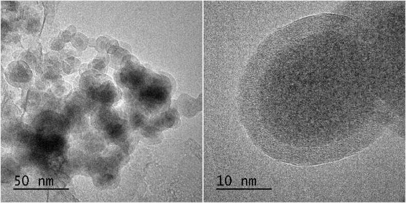
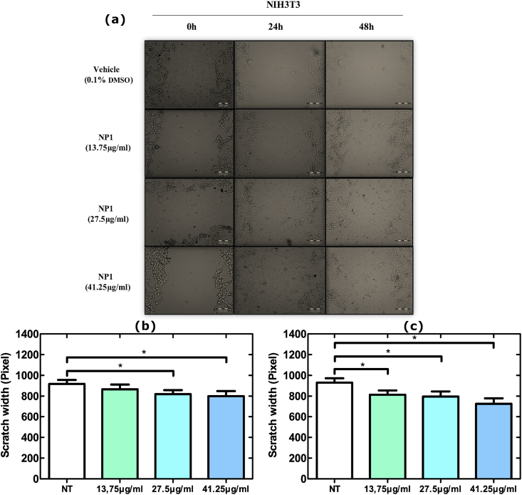
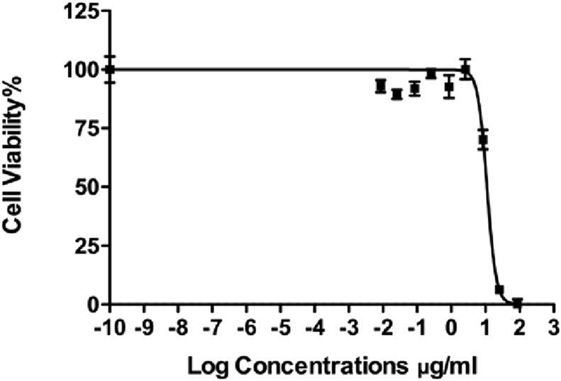
Magnetic nanoparticles are key components in many fields of science and industry. Especially in cancer diagnosis and therapy, they are involved in targeted drug delivery and hyperthermia applications due to their ability to be controlled remotely. In this study, a PEG-coated Fe/Fe3O4 core-shell nanoparticle with an average size of 20 nm and 13 nm and high room temperature coercivity (350 Oe) has been successfully synthesized. These nanoparticles were further tested for their effect on cellular toxicity (IC50) and proliferation by WST assay. In addition, their potential as anti-cancer agents were assessed using scratch assay in NIH3T3 mouse embryonic fibroblast and A549 non-small cell lung cancer cell lines. In previous reports, the IC50 values of the magnetite nanoparticles are reported at concentrations of 100 μg/ml and higher. In this study, IC50 value is observed to be at 1 μg/ml, which is significantly lower when compared to similar studies. In scratch assay, the Fe/Fe3O4 core-shell nanoparticle showed a higher inhibitory potential on cell motility in A549 lung cancer cells in comparison to the NIH3T3 cells mouse embryonic fibroblasts. This could be due to the accelerated release of free Fe ion from the Fe core, resulting in cell death. Consequently, data obtained from this study suggest that the synthesized nanoparticles can be a potential drug candidate with anti-cancer activity for chemotherapeutic treatment.

Keywords: Biophysics; Chemotherapy; Drug delivery; Fe–Fe3O4 nanoparticles; Magnetism; Nanoparticle synthesis; Nanotechnology; Targeted drug delivery.


Figures

Figure 1
Figure 1
Schematics of nanoparticle synthesis setup.
Figure 2
Figure 2
a. X-ray diffraction (Cu Ka1) and b. Vibrating Sample Magnetometer data of nanoparticles.
Figure 3
Figure 3
TEM bright-field image of NP1, showing the core-shell morphology.
Figure 4
Figure 4
a. and b. Cell viability versus concentration plots of the NP1 at 0, 100 ng/ml, 300 ng/ml, 1 μg/ml, 3 μg/ml, 10 μg/ml, 30 μg/ml and 100 μg/ml concentrations on NIH3T3 and A549 cells, respectively.
Figure 5
Figure 5
a. Microscopy images obtained in the wound healing experiment for three different NP1 concentrations (13.75 μg/ml, 27.5 μg/ml, and 41.25 μg/ml) in NIH3T3 cells. The measurement scale bar is 200μm. Scratch width pixel charts based on different concentrations of nanoparticles and treatment time (b. 24 h, c. 48 h) in NIH3T3 cells.
Figure 6
Figure 6
a. Microscopy images obtained in the scratch assay for three different NP1 concentrations (0.5 μg/ml, 1 μg/ml, and 2 μg/ml) in A549 cells. The measurement scale bar is 200μm. Scratch width pixel charts based on different concentrations and treatment time (b. 24 h, c. 48 h) of nanoparticles in A549 cells.
Figure 7
Figure 7
Cell viability versus concentration plots of the nanoparticles Paclitaxel at 0, 100 ng/ml, 300 ng/ml, 1 μg/ml, 3 μg/ml, 10 μg/ml, 30 μg/ml and100 μg/ml concentrations on A549 cells.
Figure 8
Figure 8
a. and b. Cell viability versus concentration plots of the nanoparticles NP2 at 0, 100 ng/ml, 300 ng/ml, 1 μg/ml, 3 μg/ml, 10 μg/ml, 30 μg/ml and100 μg/ml concentrations on NIH3T3 and A549 cells.

References

    1. Arruebo M., Vilaboa N., Sáez-Gutierrez B. Assessment of the evolution of cancer treatment therapies. Cancers. 2011;3(3):3279–3330. - PMC - PubMed
    1. Arias L.S., Pessan J.P., Vieira A.P.M., Lima T.M.T., Delbem A.C.B., Monteiro D.R. Iron oxide nanoparticles for biomedical applications: a perspective on synthesis, drugs, antimicrobial activity and toxicity. Antibiotics. 2018;7(2):46. - PMC - PubMed
    1. Yang H. Targeted nanosystems: advances in targeted dendrimers for cancer therapy. Nanomedicine. 2015;12(2):309–316. - PMC - PubMed
    1. Baetke S.C., Lammers T., Kiessling F. Applications of nanoparticles for diagnosis and therapy of cancer. Br. J. Radiol. 2015;88(1054):20150207. - PMC - PubMed
    1. Zhou Q., Zhang L., Wu H. Nanomaterials for cancer therapies. Nanotechnol. Rev. 2017;6(5):473–496.

LinkOut - more resources