. 2019 Jan-Dec:2:10.1177/2515256419895688.
doi: 10.1177/2515256419895688. Epub 2019 Dec 16.

Lipid droplet motility and organelle contacts

Affiliations

Lipid droplet motility and organelle contacts

Marcus D Kilwein et al. Contact (Thousand Oaks). 2019 Jan-Dec.

Abstract

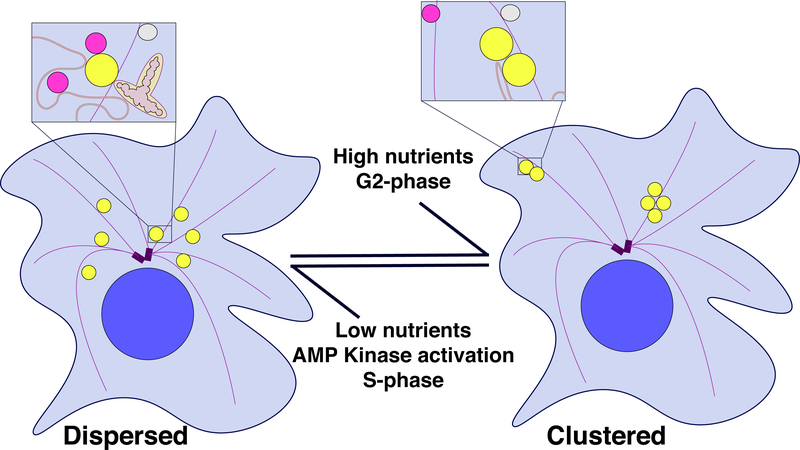
Lipid droplets (LDs) are fat storage organelles integral to energy homeostasis and a wide range of cellular processes. LDs physically and functionally interact with many partner organelles, including the ER, mitochondria, lysosomes, and peroxisomes. Recent findings suggest that the dynamics of LD inter-organelle contacts is in part controlled by LD intracellular motility. LDs can be transported directly by motor proteins along either actin filaments or microtubules, via Kinesin-1, Cytoplasmic Dynein, and type V Myosins. LDs can also be propelled indirectly, by hitchhiking on other organelles, cytoplasmic flows, and potentially actin polymerization. Although the anchors that attach motors to LDs remain elusive, other regulators of LD motility have been identified, ranging from modification of the tracks to motor co-factors to members of the perilipin family of LD proteins. Manipulating these regulatory pathways provides a tool to probe whether altered motility affects organelle contacts and has revealed that LD motility can promote interactions with numerous partners, with profound consequences for metabolism. LD motility can cause dramatic redistribution of LDs between a clustered and a dispersed state, resulting in altered organelle contacts and LD turnover. We propose that LD motility can thus promote switches in the metabolic state of a cell. Finally, LD motility is also important for LD allocation during cell division. In a number of animal embryos, uneven allocation results in a large difference in LD content in distinct daughter cells, suggesting cell-type specific LD needs.

Keywords: cell division; contact; cytoskeleton; lipid droplet; metabolism; molecular motor.


Figures

Figure 1:
Figure 1:
Mechanisms of LD motility. (A, B) LDs are transported as direct cargo by molecular motors along cytoskeletal filaments: transport along actin filaments is driven by plus-end directed Myosins (A); transport along microtubules can be unidirectional towards either the plus-or minus end (powered by Kinesin-1 or Cytoplasmic Dynein, respectively) or bidirectional, using both types of motors (B). (C) LDs hitchhike on other organelles that are transported by motors along microtubules. (D) Molecular motors promote bulk flow of the cytoplasm that drags many organelles, including LDs, along. (E) Polymerization of actin filaments may push LDs through the cytoplasm. (F) Myosin II may move LDs indirectly, by sliding actin filaments with attached LDs against each other. (G). LD moving by Brownian motion until it is captured by a tether on a target organelle.
Figure 2:
Figure 2:
Alternative LD distributions may control organelle contacts and cellular metabolism. The main cartoon depicts the two distributions LDs adopt in cells; insets highlight consequences for organelle contacts. The black arrows indicate the internal and external factors that can lead to switching between the two LD distributions. In the clustered distribution, LDs are stored away from interacting organelles and are thus preserved for later use. In the dispersed distribution, LDs have higher chance of contacting a partner organelle, allowing for LD utilization. For simplicity, the whole cell view displays a reduced number of cellular structures. The blown-up panels emphasize those structures implicated in affecting LD function through contacts: LDs (yellow), lysosomes (magenta), unidentified “other” vesicle (grey), mitochondria (oblong, two-tone grey), microtubule (purple filaments) emanating from centrosome (purple), nuclei (large blue circle), ER (red lines with a darkened interior lumen), and cytoplasm (light blue).

References

    1. Akhmanova A, and Hammer JA 3rd., (2010). Linking molecular motors to membrane cargo. Curr Opin Cell Biol 22, 479–487. doi: 10.1016/j.ceb.2010.04.008 - DOI - PMC - PubMed
    1. Akil A, Peng J, Omrane M, Gondeau C, Desterke C, Marin M, Tronchere H, Taveneau C, Sar S, Briolotti P, et al. (2016). Septin 9 induces lipid droplets growth by a phosphatidylinositol-5-phosphate and microtubule-dependent mechanism hijacked by HCV. Nat Commun 7, 12203. doi: 10.1038/ncomms12203 - DOI - PMC - PubMed
    1. Anand P, Cermelli S, Li Z, Kassan A, Bosch M, Sigua R, Huang L, Ouellette AJ, Pol A, Welte MA, and Gross SP. (2012). A novel role for lipid droplets in the organismal antibacterial response. Elife 1, e00003. doi: 10.7554/eLife.00003 - DOI - PMC - PubMed
    1. Ariu F, Strina A, Murrone O, Falchi L, Bebbere D, Ledda S, Zedda MT, Pau S, and Bogliolo L. (2016). Lipid droplet distribution of immature canine oocytes in relation to their size and the reproductive stage. Anim Sci J 87, 147–150. doi: 10.1111/asj.12432 - DOI - PubMed
    1. Arora GK, Tran SL, Rizzo N, Jain A, and Welte MA. (2016). Temporal control of bidirectional lipid-droplet motion in Drosophila depends on the ratio of kinesin-1 and its co-factor Halo. J Cell Sci 129, 1416–1428. doi: 10.1242/jcs.183426 - DOI - PMC - PubMed