Aging aggravates intervertebral disc degeneration by regulating transcription factors toward chondrogenesis
- PMID: 31909538
- PMCID: PMC7018543
- DOI: 10.1096/fj.201902109R
Aging aggravates intervertebral disc degeneration by regulating transcription factors toward chondrogenesis
Abstract
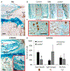
Osterix is a critical transcription factor of mesenchymal stem cell fate, where its loss or loss of Wnt signaling diverts differentiation to a chondrocytic lineage. Intervertebral disc (IVD) degeneration activates the differentiation of prehypertrophic chondrocyte-like cells and inactivates Wnt signaling, but its interactive role with osterix is unclear. First, compared to young-adult (5 mo), mechanical compression of old (18 mo) IVD induced greater IVD degeneration. Aging (5 vs 12 mo) and/or compression reduced the transcription of osterix and notochordal marker T by 40-75%. Compression elevated the transcription of hypertrophic chondrocyte marker MMP13 and pre-osterix transcription factor RUNX2, but less so in 12 mo IVD. Next, using an Ai9/td reporter and immunohistochemical staining, annulus fibrosus and nucleus pulposus cells of young-adult IVD expressed osterix, but aging and compression reduced its expression. Lastly, in vivo LRP5-deficiency in osterix-expressing cells inactivated Wnt signaling in the nucleus pulposus by 95%, degenerated the IVD to levels similar to aging and compression, reduced the biomechanical properties by 45-70%, and reduced the transcription of osterix, notochordal markers and chondrocytic markers by 60-80%. Overall, these data indicate that age-related inactivation of Wnt signaling in osterix-expressing cells may limit regeneration by depleting the progenitors and attenuating the expansion of chondrocyte-like cells.
Keywords: Wnt/β-catenin/LRPs; biomechanics; genetic animal models; osterix.
© 2019 Federation of American Societies for Experimental Biology.
Conflict of interest statement
Figures


Similar articles
-
Canonical Wnt signaling in the notochordal cell is upregulated in early intervertebral disk degeneration.J Orthop Res. 2012 Jun;30(6):950-7. doi: 10.1002/jor.22000. Epub 2011 Nov 14. J Orthop Res. 2012. PMID: 22083942
-
Loss of tenomodulin expression is a risk factor for age-related intervertebral disc degeneration.Aging Cell. 2020 Mar;19(3):e13091. doi: 10.1111/acel.13091. Epub 2020 Feb 21. Aging Cell. 2020. PMID: 32083813 Free PMC article.
-
A novel mouse model of intervertebral disc degeneration shows altered cell fate and matrix homeostasis.Matrix Biol. 2018 Sep;70:102-122. doi: 10.1016/j.matbio.2018.03.019. Epub 2018 Mar 29. Matrix Biol. 2018. PMID: 29605718 Free PMC article.
-
Cell Clusters Are Indicative of Stem Cell Activity in the Degenerate Intervertebral Disc: Can Their Properties Be Manipulated to Improve Intrinsic Repair of the Disc?Stem Cells Dev. 2018 Feb 1;27(3):147-165. doi: 10.1089/scd.2017.0213. Epub 2018 Jan 16. Stem Cells Dev. 2018. PMID: 29241405 Review.
-
Molecular mechanisms of cell death in intervertebral disc degeneration (Review).Int J Mol Med. 2016 Jun;37(6):1439-48. doi: 10.3892/ijmm.2016.2573. Epub 2016 Apr 21. Int J Mol Med. 2016. PMID: 27121482 Free PMC article. Review.
Cited by
-
Emerging role and therapeutic implications of p53 in intervertebral disc degeneration.Cell Death Discov. 2023 Dec 1;9(1):433. doi: 10.1038/s41420-023-01730-5. Cell Death Discov. 2023. PMID: 38040675 Free PMC article. Review.
-
Sox9 deletion causes severe intervertebral disc degeneration characterized by apoptosis, matrix remodeling, and compartment-specific transcriptomic changes.Matrix Biol. 2020 Dec;94:110-133. doi: 10.1016/j.matbio.2020.09.003. Epub 2020 Oct 4. Matrix Biol. 2020. PMID: 33027692 Free PMC article.
-
Macrophage Changes and High-Throughput Sequencing in Aging Mouse Intervertebral Disks.JOR Spine. 2025 Apr 8;8(2):e70061. doi: 10.1002/jsp2.70061. eCollection 2025 Jun. JOR Spine. 2025. PMID: 40201536 Free PMC article.
-
Unveiling the role of TCF19 in intervertebral disc degeneration with single-cell and bulk RNA sequencing.Sci Rep. 2025 May 8;15(1):16043. doi: 10.1038/s41598-025-01180-2. Sci Rep. 2025. PMID: 40341079 Free PMC article.
-
ERBB2-PTGS2 axis promotes intervertebral disc degeneration by regulating senescence of nucleus pulposus cells.BMC Musculoskelet Disord. 2023 Jun 20;24(1):504. doi: 10.1186/s12891-023-06625-1. BMC Musculoskelet Disord. 2023. PMID: 37340393 Free PMC article.
References
-
- Rutges JP, Duit RA, Kummer JA, Oner FC, van Rijen MH, Verbout AJ, Castelein RM, Dhert WJ, and Creemers LB (2010) Hypertrophic differentiation and calcification during intervertebral disc degeneration. Osteoarthritis Cartilage 18, 1487–1495 - PubMed
-
- Boos N, Nerlich AG, Wiest I, von der Mark K, and Aebi M (1997) Immunolocalization of type X collagen in human lumbar intervertebral discs during ageing and degeneration. Histochem Cell Biol 108, 471–480 - PubMed
-
- Antoniou J, Steffen T, Nelson F, Winterbottom N, Hollander AP, Poole RA, Aebi M, and Alini M (1996) The human lumbar intervertebral disc: evidence for changes in the biosynthesis and denaturation of the extracellular matrix with growth, maturation, ageing, and degeneration. The Journal of clinical investigation 98, 996–1003 - PMC - PubMed
Publication types
MeSH terms
Substances
Grants and funding
LinkOut - more resources
Full Text Sources
Medical
Molecular Biology Databases

