. 2020 Mar 18;31(3):656-662.
doi: 10.1021/acs.bioconjchem.9b00804. Epub 2020 Jan 22.

Bioluminescent Antibodies through Photoconjugation of Protein G-Luciferase Fusion Proteins

Affiliations

Bioluminescent Antibodies through Photoconjugation of Protein G-Luciferase Fusion Proteins

Simone F A Wouters et al. Bioconjug Chem. .

Abstract

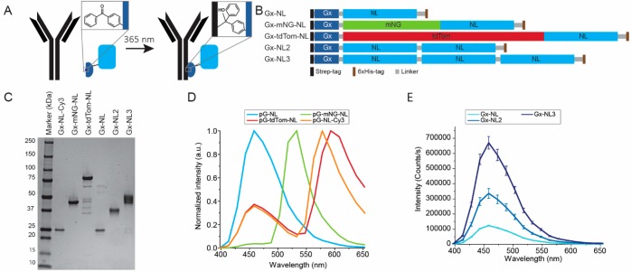
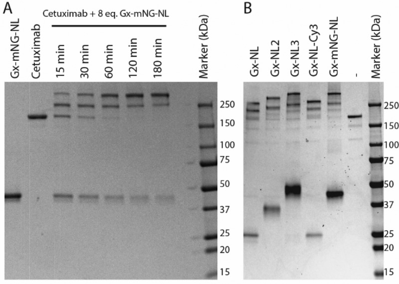
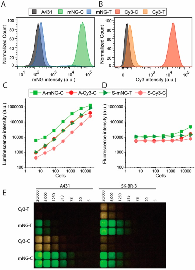
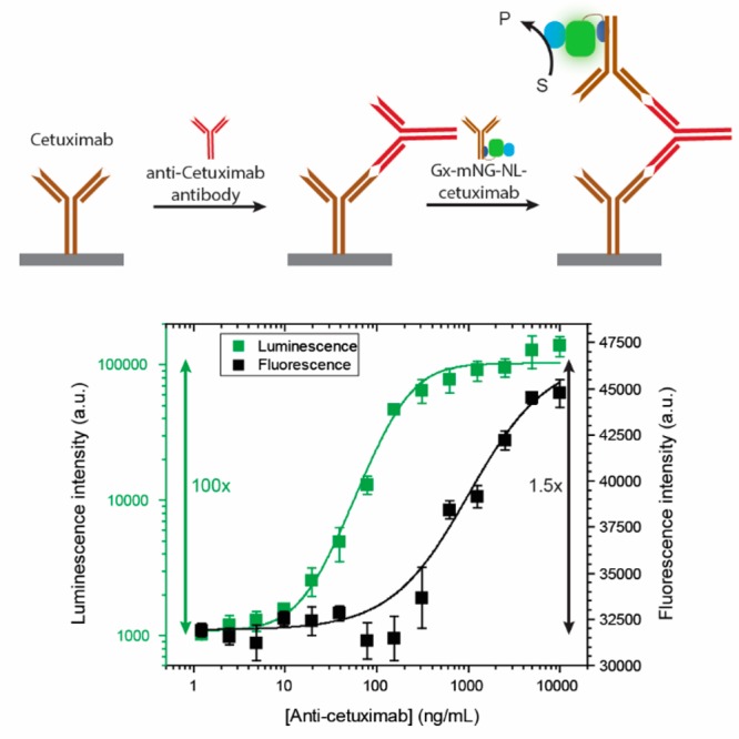
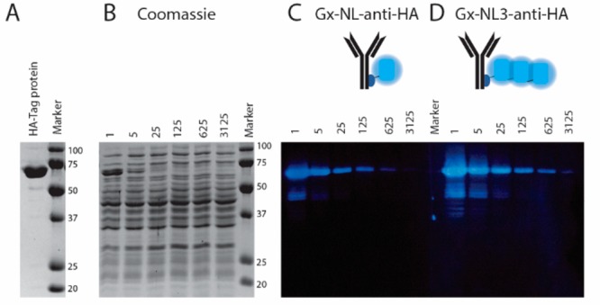
Bioluminescent antibodies represent attractive detection agents in both bioanalytical assays and imaging. Currently, their preparation relies on genetic fusion of luciferases to antibodies or nonspecific chemical conjugation strategies. Here, we report a generic method to generate well-defined covalent antibody-luciferase conjugates starting from commercially available monoclonal antibodies. Our approach uses fusion proteins consisting of the bright blue light-emitting luciferase NanoLuc (NL) and an Fc-binding protein domain (Gx) that can be photo-cross-linked to the antibody using UV light illumination. Green and red color variants were constructed by tight fusion of the NanoLuc with a green fluorescent acceptor domain and introduction of Cy3, respectively. To increase the already bright NanoLuc emission, tandem fusions were successfully developed in which the Gx domain is fused to two or three copies of the NanoLuc domain. The Gx-NL fusion proteins can be efficiently photo-cross-linked to all human immunoglobulin G (IgG) isotypes and most mammalian IgG's using 365 nm light, yielding antibodies with either one or two luciferase domains. The bioluminescent antibodies were successfully used in cell immunostaining and bioanalytical assays such as enzyme-linked immunosorbent assay (ELISA) and Western blotting.


Conflict of interest statement

The authors declare no competing financial interest.

Figures

Figure 1
Figure 1
Development of bioluminescent antibodies using Gx-NL fusion proteins. (A) Gx-NL fusions contain a pBPA moiety (inset), which is able to conjugate to the backbone of the Fc domain of an antibody under 365 nm illumination. (B) Schematic representation of protein sequence of Gx-NL fusion proteins. (C) Sodium dodecyl sulfate–polyacrylamide gel electrophoresis (SDS-PAGE) analysis of purified adapter proteins. (D) Normalized luminescence spectra of different colored adapter proteins. (E) Luminescence spectra of 100 pM fusion proteins with multiple tandem repeats of NanoLuc.
Figure 2
Figure 2
Photoconjugation of Gx-NL fusion proteins to cetuximab. (A) Nonreducing SDS-PAGE analysis of photo-cross-linking 8 equiv of Gx-mNG-NL (3.2 μM) to cetuximab (0.4 μM) with increasing illumination times. (B) Nonreducing SDS-PAGE analysis of 1 h photoconjugation of 0.4 μM cetuximab with 3.2 μM of various protein G–luciferase fusion proteins.
Figure 3
Figure 3
Immunostaining of cell surface receptors on A431 and SK-BR-3 cells using bioluminescent antibodies. Flow cytometry of A431 cells stained with 10 nM cetuximab (C) or trastuzumab (T) photo-cross-linked with (A) Gx-mNG-NL (mNG) or (B) Gx-NL-Cy3 (Cy3). (C, D) Plate reader read-out of stained cells (A, A431; S, SK-BR-3) with 1 nM bioluminescent antibody (C, cetuximab; T, trastuzumab) with either (C) luminescence or (D) fluorescence detected. (E) Analysis of the experiments described in (C) and (D) using a digital camera.
Figure 4
Figure 4
ELISA assay for the detection of the anti-cetuximab antibody using bioluminescently labeled cetuximab. A 1 μg/mL cetuximab-coated plate was incubated with anti-cetuximab antibody (1.2–10 000 ng/μL) and subsequently with 1.33 nM Gx-mNG-NL–cetuximab antibody. Both fluorescence and luminescence were recorded using a plate reader.
Figure 5
Figure 5
Bioluminescent antibody used as detection antibody in immunostaining of a Western blot. (A) Reducing SDS-PAGE gel showing the purified 65 kDa 2·HA-tag protein. (B) Coomassie-stained SDS-PAGE gel of cell lysate spiked with various dilutions (1–3 125×) of the 2·HA-tag protein. (C, D) Western blot of (B) stained with 3.32 nM Gx-NL-anti-HA (C) or Gx-NL3-anti-HA (D).

References

    1. Kaskova Z. M.; Tsarkova A. S.; Yampolsky I. V. (2016) 1001 Lights: luciferins, luciferases, their mechanisms of action and applications in chemical analysis, biology and medicine. Chem. Soc. Rev. 45, 6048–6077. 10.1039/C6CS00296J. - DOI - PubMed
    1. Yeh H.-W.; Ai H.-W. (2019) Development and applications of bioluminescent and chemiluminescent reporters and biosensors. Annu. Rev. Anal. Chem. 12, 129–150. 10.1146/annurev-anchem-061318-115027. - DOI - PMC - PubMed
    1. England C. G.; Ehlerding E. B.; Cai W. (2016) NanoLuc: A small luciferase is brightening up the field of bioluminescence. Bioconjugate Chem. 27, 1175–1187. 10.1021/acs.bioconjchem.6b00112. - DOI - PMC - PubMed
    1. Billiald P.; Mousli M.; Goyffon M.; Vaux D. (1997) Engineering of a bioluminescent antigen-binding protein. Biotechnol. Lett. 19, 1037–1041. 10.1023/A:1018463804651. - DOI
    1. Venisnik K. M.; Olafsen T.; Loening A. M.; Iyer M.; Gambhir S. S.; Wu A. M. (2006) Bifunctional antibody-renilla luciferase fusion protein for in vivo optical detection of tumors. Protein Eng., Des. Sel. 19, 453–460. 10.1093/protein/gzl030. - DOI - PubMed

Publication types