

doi: 10.1161/CIRCIMAGING.119.009791.
Epub 2020 Jan 8.
64Cu-ATSM Positron Emission Tomography/Magnetic Resonance Imaging of Hypoxia in Human Atherosclerosis
Affiliations
- PMID: 31910670
- PMCID: PMC7328725
- DOI: 10.1161/CIRCIMAGING.119.009791
Item in Clipboard
64Cu-ATSM Positron Emission Tomography/Magnetic Resonance Imaging of Hypoxia in Human Atherosclerosis
Circ Cardiovasc Imaging.
2020 Jan.
No abstract available
Keywords: atherosclerosis; carotid arteries; hypoxia; jugular veins; macrophage.
Conflict of interest statement
DISCLOSURES
There is no conflict of interest to disclose.
Figures
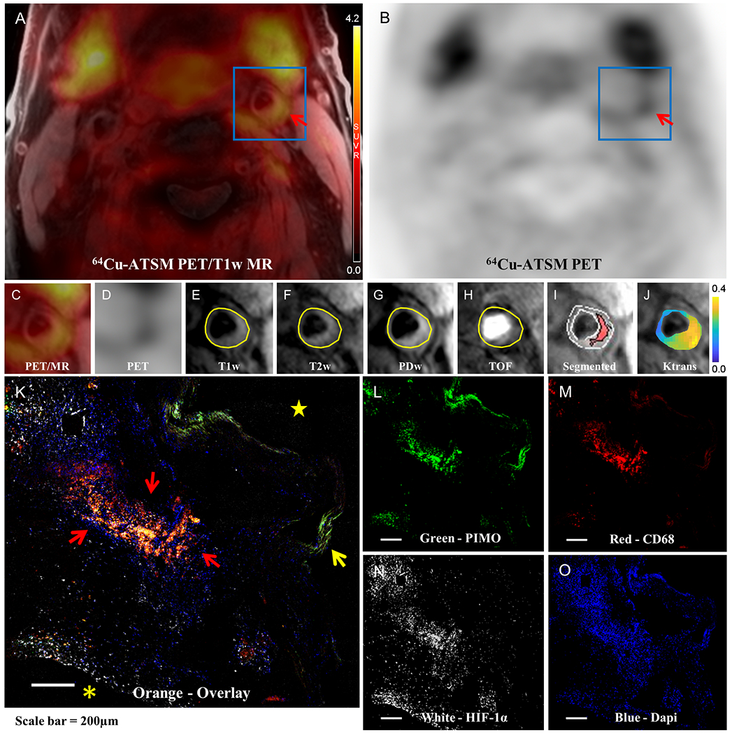
Focal 64Cu-ATSM uptake was detected in the plaque (red arrow) of the left carotid artery of a representative 69-year-old male patient as displayed in (A) 64Cu-ATSM PET/T1-weighted MR fused image and (B) 64Cu-ATSM PET only image. 64Cu-ATSM retention was also identified in the adjacent salivary glands, and a small amount of uptake is seen in muscle. (C) and (D) are the zoomed-in view of (A) and (B) with the region of interest (ROI) placed on the carotid artery (blue box). Plaque components of intraplaque hemorrhage and lipid-rich necrotic core (LRNC) are outlined in the corresponding (E) T1-weighted MRI, (F) T2-weighted MRI, (G) PD-weighted MRI, (H) TOF MRI. (I) Segmented T1-weighted MR image shows that there was one large region of hemorrhage & LRNC core comprising 25.6% of the entire plaque area. (J) Parametric map of Ktrans values color coded from 0 to 0.4 min−1 and overlaid on anatomic MR image suggest that the highly vascularized adventitia at the outer rim has the highest Ktrans values. The IHC staining of (L) PIMO for hypoxic cells, (M) CD68 for macrophages, (N) HIF-1α for hypoxia-inducible factor-1α and (O) DAPI (4′,6-diamidino-2-phenylindole) for nuclei on adjacent slices of CEA specimen slices from this patient were co-localized as shown in orange areas (red arrow) of (K) the overlaid image. Internal elastic lamina is denoted by the yellow arrow. Asterisk and star refer to the extracellular space and carotid artery lumen, respectively.
References
-
- Sluimer JC, Gasc JM, van Wanroij JL, Kisters N, Groeneweg M, Sollewijn Gelpke MD, Cleutjens JP, van den Akker LH, Corvol P, Wouters BG, et al. Hypoxia, hypoxia-inducible transcription factor, and macrophages in human atherosclerotic plaques are correlated with intraplaque angiogenesis. J Am Coll Cardiol. 2008;51:1258–65. - PubMed
-
- Fujibayashi Y, Taniuchi H, Yonekura Y, Ohtani H, Konishi J and Yokoyama A. Copper-62-ATSM: a new hypoxia imaging agent with high membrane permeability and low redox potential. J Nucl Med. 1997;38:1155–60. - PubMed
-
- Gaens ME, Backes WH, Rozel S, Lipperts M, Sanders SN, Jaspers K, Cleutjens JP, Sluimer JC, Heeneman S, Daemen MJ, et al. Dynamic contrast-enhanced MR imaging of carotid atherosclerotic plaque: model selection, reproducibility, and validation. Radiology. 2013;266:271–9. - PubMed
Publication types
MeSH terms
Substances
Grants and funding
LinkOut - more resources
Full Text Sources
