. 2020 Mar:136:104725.
doi: 10.1016/j.nbd.2019.104725. Epub 2020 Jan 3.

The drug adaptaquin blocks ATF4/CHOP-dependent pro-death Trib3 induction and protects in cellular and mouse models of Parkinson's disease

Affiliations

The drug adaptaquin blocks ATF4/CHOP-dependent pro-death Trib3 induction and protects in cellular and mouse models of Parkinson's disease

Pascaline Aimé et al. Neurobiol Dis. 2020 Mar.

Abstract

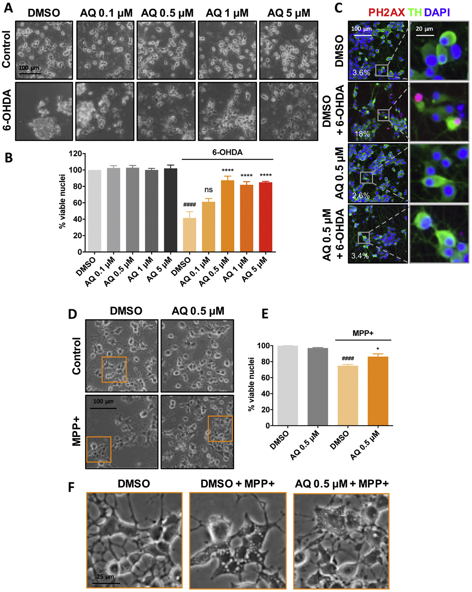
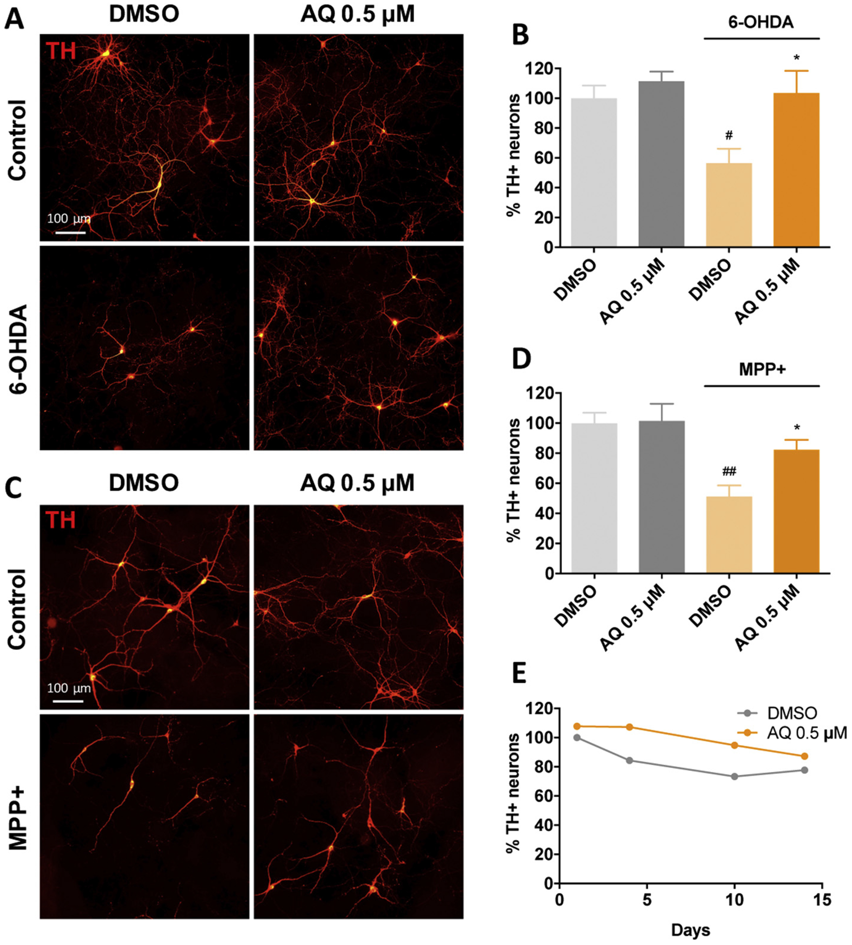
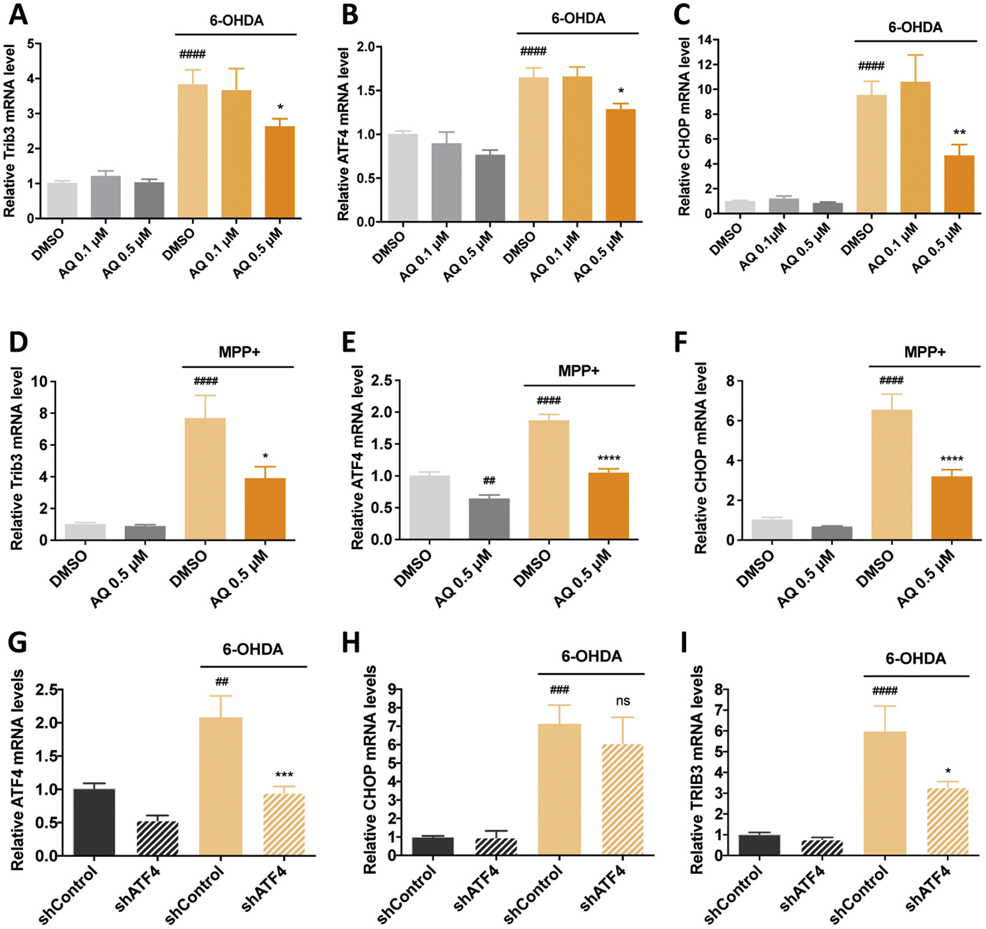
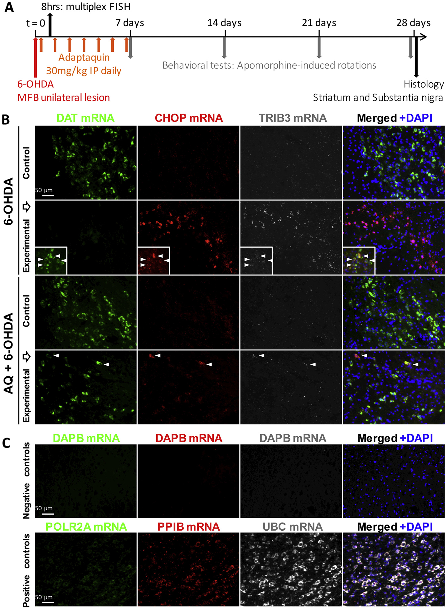
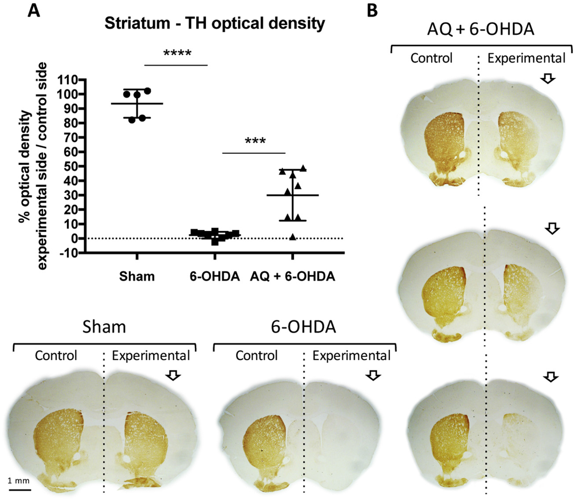
Identifying disease-causing pathways and drugs that target them in Parkinson's disease (PD) has remained challenging. We uncovered a PD-relevant pathway in which the stress-regulated heterodimeric transcription complex CHOP/ATF4 induces the neuron prodeath protein Trib3 that in turn depletes the neuronal survival protein Parkin. Here we sought to determine whether the drug adaptaquin, which inhibits ATF4-dependent transcription, could suppress Trib3 induction and neuronal death in cellular and animal models of PD. Neuronal PC12 cells and ventral midbrain dopaminergic neurons were assessed in vitro for survival, transcription factor levels and Trib3 or Parkin expression after exposure to 6-hydroxydopamine or 1-methyl-4-phenylpyridinium with or without adaptaquin co-treatment. 6-hydroxydopamine injection into the medial forebrain bundle was used to examine the effects of systemic adaptaquin on signaling, substantia nigra dopaminergic neuron survival and striatal projections as well as motor behavior. In both culture and animal models, adaptaquin suppressed elevation of ATF4 and/or CHOP and induction of Trib3 in response to 1-methyl-4-phenylpyridinium and/or 6-hydroxydopamine. In culture, adaptaquin preserved Parkin levels, provided neuroprotection and preserved morphology. In the mouse model, adaptaquin treatment enhanced survival of dopaminergic neurons and substantially protected their striatal projections. It also significantly enhanced retention of nigrostriatal function. These findings define a novel pharmacological approach involving the drug adaptaquin, a selective modulator of hypoxic adaptation, for suppressing Parkin loss and neurodegeneration in toxin models of PD. As adaptaquin possesses an oxyquinoline backbone with known safety in humans, these findings provide a firm rationale for advancing it towards clinical evaluation in PD.

Keywords: ATF4; Adaptaquin; CHOP; Neuroprotection; Oxyquinoline; Parkin; Parkinson's disease; Trib3.


Figures

Fig. 1.
Fig. 1.
Adaptaquin protects neuronal PC12 cells against 6-OHDA and MPP+ − induced cell death. Phase contrast images (A) and corresponding quantifications (B) of survival assay showing the remaining viable nuclei in neuronal PC12 cell cultures either untreated (control, DMSO) or treated with 150 μM 6-OHDA and indicated AQ concentrations for 24 h. 6-OHDA induces significant cell death. ANOVA with Tukey’s post-hoc test ####p < .0001; ****p < .0001; ns = not significant. Values are mean +/− SEM of 3 independent experiments. Representative immunofluorescence images (C) showing that AQ decreases the phosphorylation of histone 2A.X (PH2AX), a marker of apoptosis. Neuronal PC12 cells were treated with DMSO or 0.5 μM AQ and 150 μM 6-OHDA for 24 h and stained for the catecholaminergic marker tyrosine hydroxylase (TH, green), nuclei (DAPI, blue) and PH2AX (red). The percentage of PH2AX+ cells for each condition is annotated on the images. Phase contrast images (D) and corresponding quantifications (E) of survival assay showing the remaining viable nuclei in neuronal PC12 cell cultures either untreated (control, DMSO) or treated with 1 mM MPP+ and 0.5 μM AQ for 48 h. ANOVA with Tukey’s post-hoc test: ####p < .0001) and *p < .05). Values are mean +/− SEM of 3 independent experiments. Higher magnification images (F) showing that MPP+ elicits the formation of large intracellular vacuoles and that treatment with 0.5 μM AQ reduces their size.
Fig. 2.
Fig. 2.
Adaptaquin protects ventral midbrain dopaminergic neurons against 6-OHDA and MPP+-induced cell death. Representative immunofluorescence images (A, C) of untreated ventral midbrain primary cultures (Control, DMSO) or treated with 0.5 μM AQ and 40 μM 6-OHDA (A) or 40 μM MPP+ (C) for 24 h and immunostained for tyrosine hydroxylase (TH, red). Corresponding quantifications (B, D) of 3 independent experiments showing the percentage of remaining viable TH+ neurons in control cultures (DMSO) or ventral midbrain cultures treated with 0.5 μM AQ and 40 μM 6-OHDA (B) or 0.5 μM AQ and 40 μM MPP+ (D) for 24 h. Both 6-OHDA and MPP+ induce significant death of dopaminergic neurons (ANOVA with Tukey’s post-hoc tests, #p < .05, ##p < .005) while AQ restores cell viability of both 6-OHDA and MPP+ treated dopaminergic neurons (ANOVA with Tukey’s post-hoc tests *p < .05). Time-course (E) showing the percentage of remaining viable dopaminergic neurons in ventral midbrain primary cultures treated with DMSO or 0.5 μM AQ for up to 14 days and immunostained for TH.
Fig. 3.
Fig. 3.
A protective dose of adaptaquin blocks the induction of ATF4, CHOP and Trib3 mRNAs and ATF4 knockdown reduces Trib3 mRNA but not CHOP mRNA induction in response to 6-OHDA in neuronal PC12 cells. qPCR analysis of Trib3 (A, D), ATF4 (B, E) and CHOP (C, F) mRNA levels in neuronal PC12 cells treated with DMSO, 0.1 μM or 0.5 μM AQ and 150 μM 6-OHDA for 8 h (A–C); DMSO, 0.5 μM AQ and 1 mM MPP+ for 16 h (D–F). In DMSO-treated cultures, 6-OHDA induces an increase in Trib3, ATF4 and CHOP mRNA (ANOVA with Tukey’s post-hoc tests, ####p < .0001). For cultures treated with 6-OHDA and a 0.5 μM AQ significant decreased Trib3, ATF4 and CHOP mRNA levels (ANOVA with Tukey’s post-hoc tests, *p < .05, **p < .005). In DMSO-treated cultures, MPP+ increases Trib3, ATF4 and CHOP mRNA (ANOVA with Tukey’s post-hoc tests, ####p < .0001). In MPP+ treated cultures, 0.5 μM AQ significantly decreases Trib3, ATF4 and CHOP mRNA levels (ANOVA with Tukey’s post-hoc tests, *p < .05, ****p < .0001). In absence of MPP+, 0.5 μM AQ decreases ATF4 mRNA levels at 16 h (ANOVA with Tukey’s post-hoc tests, ##p < .005). All mRNA levels are expressed as mean +/− SEM of 3 independent experiments. qPCR analysis shows the effects of lentivirally-delivered ATF4 shRNA (shATF4) on levels of ATF4 (G), CHOP (H) and Trib3 (I) mRNA after treatment with 150 μM 6-OHDA for 8 h. Control cultures were infected with a control shRNA (shControl). In control cultures, 6-OHDA induces Trib3, ATF4 and CHOP mRNA (ANOVA with Tukey’s post-hoc tests, ##p < .005, ###p < .0005, ####p < .0001). ATF4 knockdown reduces the induction of ATF4 and Trib3 mRNA in response to 6-OHDA (ANOVA with Tukey’s post-hoc tests, *p < .05, ***p < .001). By contrast ATF4 knockdown does not reduce the induction of CHOP mRNA in response to 6-OHDA (ns). All mRNA levels are expressed as mean +/− SEM of 3 independent experiments.
Fig. 4.
Fig. 4.
A protective dose of adaptaquin block the induction of ATF4, CHOP and Trib3 proteins in neuronal PC12 cells treated with 6-OHDA and maintains Parkin protein levels. Western blot images (A) and corresponding quantifications (B–E) of 5 independent experiments showing the protein levels of ATF4 (B), CHOP (C), Trib3 (D) and Parkin (E) in neuronal PC12 cells treated with DMSO, 0.1 μM or 0.5 μM AQ and 150 μM 6-OHDA for 8 h. In DMSO-treated cultures, 6-OHDA induces ATF4, CHOP and Trib3 proteins and decreases Parkin protein (ANOVA with Tukey’s post-hoc tests, #p ≤ .05). By contrast, cultures treated with 6-OHDA and 0.5 μM displayed a significant decrease in Trib3, ATF4 and CHOP protein levels (ANOVA with Tukey’s post-hoc tests, *p < .05, **p < .001). In addition, a 0.5 μM dose of AQ significantly increased Parkin levels (ANOVA with Tukey’s post-hoc tests, *p = .05). Data are normalized to ERK. Non specific bands are marked with asterisks. Survival assay (F) showing the percentage of remaining dopaminergic neurons in ventral midbrain cultures infected with a lentivirus carrying a control shRNA (shControl) or an shRNA against Parkin (shParkin) for 14 days and immunostained for TH and GFP. Parkin knockdown induces the death of 31% of TH/GFP+ neurons (t-test, ***p < .001).
Fig. 5.
Fig. 5.
Adaptaquin blocks Trib3 and Ddit3/CHOP mRNA induction in the substantia nigra of 6-OHDA-injected mice. (A) Timeline of in vivo experiments: Stereotaxic surgery was performed and mice received a unilateral injection 6-OHDA in the right median forebrain bundle (MFB) at t = 0. After 2 h, a sub-group of mice received a single intraperitoneal (IP) injection of adaptaquin (AQ). Mice were sacrificed 8 h after the 6-OHDA lesion and their brains were processed for multiplex fluorescent in situ hybridization (FISH) and Trib3, Ddit3/CHOP and Slc6a3/DAT mRNA levels were analyzed. Another sub-group of mice started receiving daily IP AQ injections 2 h after the 6-OHDA lesion and for 7 consecutive days. At 7, 14, 21 and 28 days, mice were challenged by subcutaneous injections of the dopamine agonist apomorphine and contralateral rotations were measured to assess the functional integrity of the nigrostriatal system. Twenty-eight days after the 6-OHDA lesion, mice were sacrificed and their brains were processed for histology and tyrosine hydroxylase immunoreactivity was analyzed in the substantia nigra and the striatum. (B) Representative images of multiplex fluorescent in situ hybridization (FISH) showing Slc6a3/DAT mRNA expressed by midbrain dopaminergic neurons (green); Ddit3/CHOP (red) and Trib3 (white) mRNA signals in the unlesioned (control) or 6-OHDA lesioned (experimental) side of the substantia nigra of mice receiving either a vehicle IP injection 2 h after 6-OHDA treatment (6-OHDA) or a 30 mg/kg IP injection of AQ 2 h after 6-OHDA treatment (AQ + 6-OHDA). Eight hour after the 6-OHDA lesion, Slc6a3/DAT mRNA expression is decreased and only small subsets of dopaminergic neurons retain Slc6a3/DAT mRNA expression (inserts). Strong Ddit3/CHOP and Trib3 mRNA signals are detected in the experimental side of the substantia nigra of vehicle-treated mice. A single IP injection of AQ 2 h after the 6-OHDA lesion maintains Slc6a3/DAT mRNA expression and decreases Ddit3/CHOP and Trib3 mRNA expression in the substantia nigra. Arrow heads indicate neurons showing a colocalization of Slc6a3/DAT, Ddit3/CHOP and Trib3 mRNAs. (C) Representative images of control multiplex FISH experimental procedures using probes targeting the bacterial gene dapb (negative control) and the ubiquitous genes Polr2a, Pp1b and Ubc showing low, moderate and high mRNA expression levels, respectively (positive controls).
Fig. 6.
Fig. 6.
Adaptaquin partially prevents the loss of dopaminergic neurons in the substantia nigra of 6-OHDA-injected mice. Representative images (A) of TH immunohistochemistry (brown) with thionin counterstain (blue) showing dopaminergic neurons in the unlesioned (control) or 6-OHDA lesioned (experimental) side of the posterior and anterior regions of the substantia nigra of mice who received a sham surgery or a 6-OHDA lesion and either a control IP injection (6-OHDA) or a daily 30 mg/kg IP injection of AQ for 7 consecutive days (AQ + 6-OHDA). Higher magnification images (B) showing the dopaminergic neurons in the experimental side of the anterior substantia nigra of sham, 6-OHDA and AQ + 6-OHDA mice. Unbiased stereology counts (C) and proportions between the experimental and the control side (D) of the total number of dopaminergic TH+ neurons in all three groups showing that 6-OHDA induces a drastic reduction of the counts (ANOVA with Sidak’s multiple comparison test ****p < .0001) and the proportion (ANOVA with Tukey’s post-hoc test ****p < .0001) of dopaminergic neurons in the substantia nigra, compared to sham mice. AQ post-treatment for 7 days significantly increases both the counts (ANOVA with Sidak’s multiple comparison test ***p < .0005) and the proportion (ANOVA with Tukey’s post-hoc test ***p < .0005) of nigral dopaminergic neurons.
Fig. 7.
Fig. 7.
Adaptaquin partially restores the loss of TH immunoreactivity in the striatum of 6-OHDA-injected mice. Quantification (A) and representative images (B) of TH immunohistochemistry (brown) showing the optical density of the TH signal from dopaminergic fibers in the unlesioned (control) or 6-OHDA lesioned (experimental) side of the striatum of mice who received a sham surgery or a 6-OHDA lesion and either a control IP injection (6-OHDA) or a daily 30 mg/kg IP injection of AQ for 7 consecutive days (AQ + 6-OHDA). 6-OHDA induces a drastic reduction of TH immunostaining in experimental side of the striatum, relative to the control side and compared to sham mice (ANOVA with Tukey’s post-hoc test ****p < .0001). AQ post-treatment for 7 days significantly increases the proportion of TH immunoreactivity in the striatum (ANOVA with Tukey’s post-hoc test ***p < .001). Given the variability in the AQ + 6-OHDA group, striatum images of 3 individual mice are shown in B.
Fig. 8.
Fig. 8.
Adaptaquin partially restores movement control in 6-OHDA-injected mice. (A–D) Behavioral analysis by quantification of the net contralateral rotations induced by a subcutaneous injections of apomorphine at 7 days (A), 14 days (B), 21 days (C) and 28 days after mice received a sham surgery or a 6-OHDA lesion and either a control IP injection (6-OHDA) or a daily 30 mg/kg IP injection of AQ for 7 consecutive days (AQ + 6-OHDA). n = 5 for sham, n = 13 for 6-OHDA alone and n = 13 for AQ + 6-OHDA group. 6-OHDA induces a drastic increase in the number of contralateral rotations at all time points measured compared to sham mice (ANOVA with Tukey’s post hoc test; *p < .05, ***p < .0005). AQ post-treatment for 7 days significantly decreases the number of contralateral rotations at 14 days, 21 days and 28 days (ANOVA with Tukey’s post hoc test; ***p < .0005, ****p < .0001). (E–G) Correlation analysis of the results of the AQ-treated group showing that the number of dopaminergic neurons in the substantia nigra is highly correlated to the density of TH immunoreactivity in the striatum (E, Pearson correlation coefficient r = 0.968, p < .0001), the density TH immunoreactivity in the striatum is highly correlated to the number of apomorphine-induced rotations (F, Pearson correlation coefficient r = −0.966, p < .0001) and the number of dopaminergic neurons in the substantia nigra is highly correlated to the number of apomorphine-induced rotations (G, Pearson correlation coefficient r = −0.971, p < .0001).

References

    1. Aimé P, et al., 2015. Trib3 is elevated in Parkinson’s disease and mediates death in Parkinson’s disease models. J. Neurosci 35, 10731–10749. 10.1523/JNEUROSCI.0614-15.2015. - DOI - PMC - PubMed
    1. Aimé P, Sun X, Greene LA, 2018. Manipulation and study of gene expression in neurotoxin-treated PC12 and SH-SY5Y cells for in vitro studies of Parkinson’s disease gene expression and regulation in mammalian cells. In: Transcription Toward the Establishment of Novel Therapeutics. IntechOpen 2018. 10.5772/intechopen.71811. - DOI
    1. Bowenkamp KE, et al., 1996. 6-hydroxydopamine induces the loss of the dopaminergic phenotype in substantia nigra neurons of the rat. A possible mechanism for restoration of the nigrostriatal circuit mediated by glial cell line-derived neurotrophic factor. Exp. Brain Res 111, 1–7. 10.1007/bf00229549. - DOI - PubMed
    1. Chen X, et al., 2012. Neurotrophic effects of serum- and glucocorticoid-inducible kinase on adult murine mesencephalic dopamine neurons. J. Neurosci 32, 11299–11308. 10.1523/JNEUROSCI.5910-11.2012. - DOI - PMC - PubMed
    1. Dawson TM, Dawson VL, 2010. The role of parkin in familial and sporadic Parkinson’s disease. Mov. Disord 25 (Suppl. 1), S32–S39. 10.1002/mds.22798. - DOI - PMC - PubMed

Publication types

MeSH terms