Review
. 2020 Mar;32(3):573-594.
doi: 10.1105/tpc.19.00535. Epub 2020 Jan 6.

Matrix Redox Physiology Governs the Regulation of Plant Mitochondrial Metabolism through Posttranslational Protein Modifications

Affiliations
Review

Matrix Redox Physiology Governs the Regulation of Plant Mitochondrial Metabolism through Posttranslational Protein Modifications

Ian Max Møller et al. Plant Cell. 2020 Mar.

Abstract

Mitochondria function as hubs of plant metabolism. Oxidative phosphorylation produces ATP, but it is also a central high-capacity electron sink required by many metabolic pathways that must be flexibly coordinated and integrated. Here, we review the crucial roles of redox-associated posttranslational protein modifications (PTMs) in mitochondrial metabolic regulation. We discuss several major concepts. First, the major redox couples in the mitochondrial matrix (NAD, NADP, thioredoxin, glutathione, and ascorbate) are in kinetic steady state rather than thermodynamic equilibrium. Second, targeted proteomics have produced long lists of proteins potentially regulated by Cys oxidation/thioredoxin, Met-SO formation, phosphorylation, or Lys acetylation, but we currently only understand the functional importance of a few of these PTMs. Some site modifications may represent molecular noise caused by spurious reactions. Third, different PTMs on the same protein or on different proteins in the same metabolic pathway can interact to fine-tune metabolic regulation. Fourth, PTMs take part in the repair of stress-induced damage (e.g., by reducing Met and Cys oxidation products) as well as adjusting metabolic functions in response to environmental variation, such as changes in light irradiance or oxygen availability. Finally, PTMs form a multidimensional regulatory system that provides the speed and flexibility needed for mitochondrial coordination far beyond that provided by changes in nuclear gene expression alone.


Figures

Figure 1.
Figure 1.
PTMs of Protein Side Chains Discussed in This Review. Protein phosphorylation of Ser, Thr, and Tyr is catalyzed by protein kinases (PK) and is reversible via a reaction catalyzed by protein phosphatases (PP). Oxidation of Cys can occur via the action of ROS. These nonenzymatic redox modifications form mixed disulfides (e.g., glutathionylation) and Cys derivatives with sulfenic, sulfinic, and cysteic acid groups. The reduction of mixed disulfides, like glutathionylation products, occurs via the action of Trx, while at least in peroxiredoxins, Cys sulfinic acid groups can be reduced by sulfiredoxin (Srx) in the presence of ATP (Biteau et al., 2003). Lys acetylation is catalyzed by acetyltransferases (AT) and can be reversed by deacetylases (DAC). Met oxidation to Met-sulfoxide and Met-sulfone is nonenzymatic, and the first step can be reversed by Met sulfoxide reductase (MSR). Trp oxidation is also nonenzymatic and occurs due to the interaction with ROS. Carbonylation of Arg, Lys, Pro, and Thr occurs via interaction with ROS nonenzymatically and irreversibly. Protein S-nitrosylation of Cys and nitration of Tyr are nonenzymatic and take place due to the interaction with reactive nitrogen species (RNS). The dominant charged state of the side chains at pH 7.0 is shown only for the phosphorylated Ser, Thr, and Tyr.
Figure 2.
Figure 2.
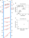
Regulation of the Redox Characteristics in the Matrix of Plant Mitochondria. (A) The redox potentials of the major redox pairs at pH 6.8 and 8.0. The further the redox potentials are from each other, the more different their reduction levels will be at thermodynamic equilibrium. The values of redox potentials at pH 6.8 and 8.0 for the pairs of oxidized/reduced Trx, peroxiredoxin (Prx), and Grx are shown by arrows pointing to the graph from the left and right to distinguish them from the low-molecular-weight redox pairs. Note that redox potentials of Trx, Prx and Grx proteins can differ strongly within the respective protein families. Specific half reactions under consideration are shown in Supplemental Figure 1. (B) The pool sizes of NAD and NADP and their redox levels in pea leaf mitochondria depending on light and CO2 supply (Igamberdiev and Gardeström, 2003). (C) The accumulation of nitric oxide (NO), depending on the NADH/NAD+ ratios in anoxic alfalfa (Medicago sativa) roots (Dordas et al., 2003; Igamberdiev et al., 2004). FW, fresh weight.
Figure 3.
Figure 3.
The Link between the Potentials of Matrix Redox Couples and PTMs. Interactions between pools of the soluble redox couples Cys-SH/Cys-S-S-Cys, NAD(P)H, Trx, glutathione, nitrite, NO, O2, superoxide, and H2O2 are shown. Cys-S-S-Cys to Cys-SH conversions are promoted when the redox potential is very negative [high NAD(P)H/NAD(P)+]. Cys and Met oxidations are promoted when the redox potential is very negative and ROS production is high. Protein nitrosylation occurs when the redox potential is very negative with the concomitant NO and ROS accumulation (e.g., during hypoxia). The formation of highly negative redox potential can also result in protein carbonylations (upon high ROS production). Protein phosphorylation is promoted in state 4 (high ATP level), accompanied by very negative redox potential and ROS accumulation. Protein acetylation is observed upon moderately negative redox potential (allowing PDH to resume acetyl-CoA production). Protein deacetylation by sirtuins takes place when the concentration of NAD+ is high (irrespective of the reduction level). The figure layout was adopted and significantly modified from Noctor (2006). Abbreviations not defined in the text are as follows: APX, ascorbate peroxidase; DHAR, dehydroascorbate reductase; GR, glutathione reductase; MDH, malate dehydrogenase; MDHAR, monodehydroascorbate reductase; NDA, internal rotenone-insensitive NADH dehydrogenase; NDB, external rotenone-insensitive NAD(P)H dehydrogenase; NDC, internal rotenone-insensitive NADPH dehydrogenase; NDPK, nucleoside diphosphate kinase; OX, oxidation; P5C DH, pyrroline-5-carboxylate dehydrogenase; RED, reduction; TH-R, transhydrogenase-like reaction.
Figure 4.
Figure 4.
Posttranslational Modifications of TCA Enzymes and ETC Components. Modifications are denoted as follows: A, acetylation; N, S-nitrosylation; P, phosphorylation; T, Trx-dependent reduction/oxidation. The mitochondrial matrix enzymes and ETC components shown are as follows: ACO, aconitase; AOX; bc1, complex III; c, cytochrome c; COX, complex IV (cytochrome c oxidase); CS, citrate synthase; FDH, formate dehydrogenase; FUM, fumarase; GDC; I, complex I; II, complex II (succinate dehydrogenase); ICDH; IMM; IMS, intermembrane space; MDH, malate dehydrogenase; NDA, internal rotenone-insensitive NADH dehydrogenase; NDB1 and NDB2, external rotenone-insensitive NADPH and NADH dehydrogenases; NDC, internal rotenone-insensitive NADPH dehydrogenase; OGDH, 2-oxoglutarate dehydrogenase complex; PDC; SCS, succinyl-CoA synthetase; SHMT, Ser hydroxymethyltransferase. The figure is based on data from (Bykova et al. (2003a), Balmer et al. (2004), Gelhaye et al. (2004), Kristensen et al. (2004), Umbach et al. (2006), Havelund et al. (2013), Yoshida et al. (2013), König et al. (2014a), Salvato et al. (2014), Nietzel et al. (2017), and Millar et al. (2019). The TCA cycle is fed in plant mitochondria not only by acetyl-CoA formed from glycolytic pyruvate but also from malate that can be converted to pyruvate by NAD-malic enzyme (ME) and oxaloacetate (OAA) that can be converted to pyruvate by MDH and ME. Plant-specific proteins of the mitochondrial ETC and the enzymes that participate in photorespiration are shown in green. A more complete overview of mitochondrial proteins with multiple PTMs is provided in Supplemental Table 1.
Figure 5.
Figure 5.
Regulation of AOX by Reduction and Metabolites. AOX biosynthesis is regulated transcriptionally by citrate and by elevated levels of ROS. AOX is modified posttranslationally by the mitochondrial Trx o, which mediates the conversion between an inactive form with a disulfide bridge between the monomers and an active form with free thiol groups (Gelhaye et al., 2004; Umbach et al., 2006). Thioredoxin reductase (Trx-R) regenerates Trx o to the reduced form; NADPH for this reaction is supplied by NADP-ICDH or other dehydrogenases. AOX is activated by oxo-acids (R-CO-COO-) including pyruvate (coming from glycolysis and malic enzyme reactions), 2-oxoglutarate (2-OG) and oxaloacetate (OAA; formed in the TCA cycle), and glyoxylate (from photorespiration). The posttranslational binding of pyruvate and other oxo-acids to the reduced form further activates the enzyme. The increase in the concentration of the AOX substrate ubiquinol (UQH2) inside the inner membrane stimulates AOX activity. The figure was significantly modified from Web Figure 12.3C of Taiz et al. (2015): http://6e.plantphys.net/topic12.03.html. The regulation of AOX at different levels is reviewed by Selinski et al. (2018b).
Figure 6.
Figure 6.
Acetylation of TCA Cycle Proteins and Proposed Redox Regulation via Depletion of the NAD Pool during Sirtuin-Dependent Deacetylation and Poly-ADP-Ribosylation. The figure is based on data from König et al. (2014a). The proposed joint operation of the TCA cycle and reversible acetylation-deacetylation results in the regulation of NAD pool size and reduction level (top box). Protein Lys acetylation of TCA cycle enzymes can take place via KAT activity or nonenzymatically (non-Enz.) at pH 8.0 and higher in the presence of acetyl-CoA, while NAD+-dependent KDAC of the sirtuin family consumes NAD+ during the process of deacetylation. Another process stimulated by high NAD+ and depleting the NAD pool is poly-ADP-ribosylation of proteins (bottom box). PARP, whose presence in plant mitochondria is still controversial, cleaves NAD+ and attaches the ADP-ribose moiety to acceptor proteins, whereas poly(ADP-ribose) glycohydrolase (PARG) cleaves the ribose-ribose backbone bond of poly(ADP-ribose), releasing free ADP-ribose (Briggs and Bent, 2011). The inhibition of TCA cycle enzymes by Lys acetylation that affects the metabolic flux, so that only parts of it are used (Sweetlove et al., 2010), is still hypothetical. Pyruvate comes from glycolysis, and malate and oxaloacetate (OAA) can come from glycolysis or be formed in the cycle. Acetyl-CoA is abbreviated as Ac-CoA, and acetylation is denoted by the letter A in a circle. Other abbreviations are as follows: ACO, aconitase; CS, citrate synthase; FUM, fumarase; MDH, malate dehydrogenase; ME, NAD-malic enzyme; NAM, nicotinamide; 2′-OAADP-ribose, 2′-O-acetyl-ADP-ribose; OGDH, 2-oxoglutarate dehydrogenase complex; PAR, poly(ADP-ribose); SCS, succinyl-CoA synthetase; SDH, succinate dehydrogenase; SHMT, Ser hydroxymethyltransferase.
Figure 7.
Figure 7.
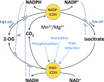
Regulation of NAD- and NADP-Dependent ICDH in Plant Mitochondria. The NAD-ICDH reaction is essentially irreversible, yet the forward reaction is regulated by phosphorylation (Bykova et al., 2003a) and Trx-mediated thiol reduction (Salvato et al., 2014). The NADP-ICDH reaction is reversible, and the protein is not known to be regulated by PTMs. However, the activity of this reaction is stimulated by PTM-induced inhibition of NAD-ICDH, which functions as the master regulator of this step in the TCA cycle and induces isocitrate accumulation. Likewise, the forward reaction of mitochondrial NADP-ICDH could be stimulated at the substrate level by glutathionylation, as observed for cytosolic NADP-ICDH (Niazi et al., 2019). The regulation of this reaction by metabolites and pH is shown as described by Igamberdiev and Gardeström (2003). nc, noncompetitive inhibition; in all other cases, the inhibition is competitive.
Figure 8.
Figure 8.
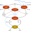
Regulation of Individual GDC Proteins and Ser hydroxymethyltransferase by Trx and Other Posttranslational Modifications. The P-protein of the GDC is phosphorylated, S-nitrosylated, and S-glutathionylated, and the latter two processes inhibit GDC activity (Palmieri et al., 2010). All four GDC subunits (P, L, H, and T) as well as Ser hydroxymethyltransferase (SHMT) are potentially regulated by Trx (Balmer et al., 2004) and Lys acetylation (Supplemental Table 1). The figure is based on the data of Balmer et al. (2004), Palmieri et al. (2010), Salvato et al. (2014), and Millar et al. (2019).

References

    1. Agius S.C., Rasmusson A.G., Møller I.M. (2001). NAD(P) turnover in plant mitochondria. Aust. J. Plant Physiol. 28: 461–470.
    1. Albrecht S.C., Sobotta M.C., Bausewein D., Aller I., Hell R., Dick T.P., Meyer A.J. (2014). Redesign of genetically encoded biosensors for monitoring mitochondrial redox status in a broad range of model eukaryotes. J. Biomol. Screen. 19: 379–386. - PubMed
    1. Anderson K.A., et al. (2017).. SIRT4 is a lysine deacylase that controls leucine metabolism and insulin secretion. Cell Metab. 25: 838–855.e15. - PMC - PubMed
    1. Atkin O.K., Evans J.R., Siebke K. (1998). Relationship between the inhibition of leaf respiration by light and enhancement of leaf dark respiration following light treatment. Aust. J. Plant Physiol. 25: 437–443.
    1. Baeza J., Smallegan M.J., Denu J.M. (2016). Mechanisms and dynamics of protein acetylation in mitochondria. Trends Biochem. Sci. 41: 231–244. - PMC - PubMed

Publication types

MeSH terms

Substances

LinkOut - more resources