Comment
. 2020 Jun 1;26(11):2654-2663.
doi: 10.1158/1078-0432.CCR-19-3563. Epub 2020 Jan 7.

Tumor Analyses Reveal Squamous Transformation and Off-Target Alterations As Early Resistance Mechanisms to First-line Osimertinib in EGFR-Mutant Lung Cancer

Affiliations
Comment

Tumor Analyses Reveal Squamous Transformation and Off-Target Alterations As Early Resistance Mechanisms to First-line Osimertinib in EGFR-Mutant Lung Cancer

Adam J Schoenfeld et al. Clin Cancer Res. .

Abstract

Purpose: Patterns of resistance to first-line osimertinib are not well-established and have primarily been evaluated using plasma assays, which cannot detect histologic transformation and have differential sensitivity for copy number changes and chromosomal rearrangements.

Experimental design: To characterize mechanisms of resistance to osimertinib, patients with metastatic EGFR-mutant lung cancers who received osimertinib at Memorial Sloan Kettering Cancer Center and had next-generation sequencing performed on tumor tissue before osimertinib initiation and after progression were identified.

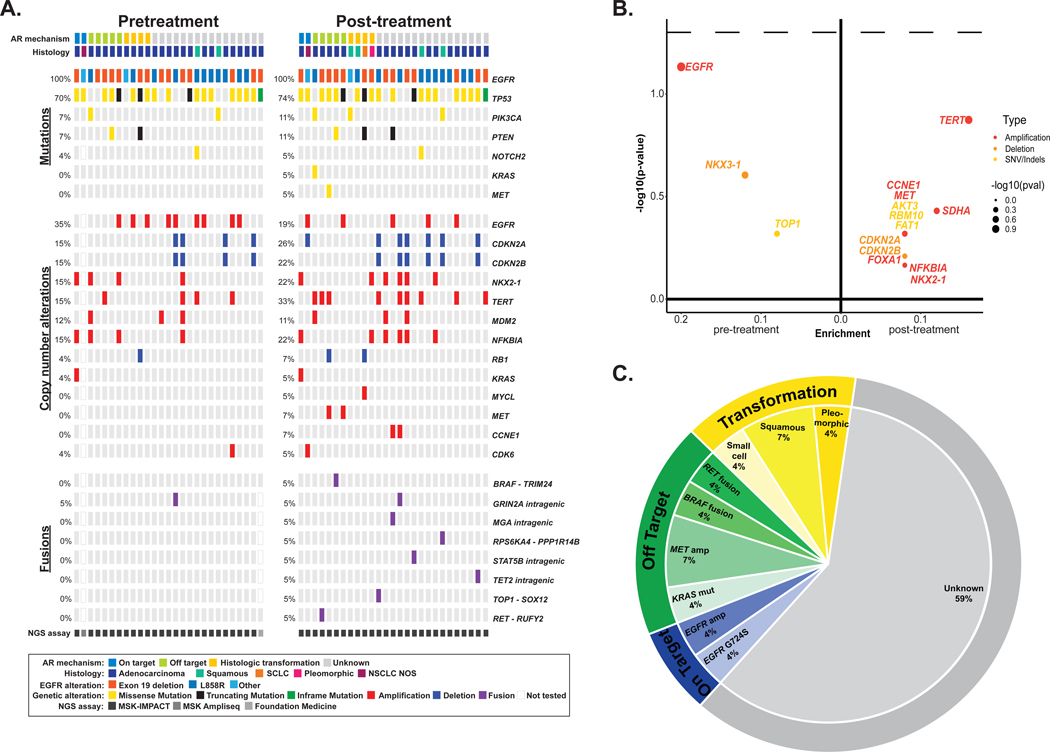
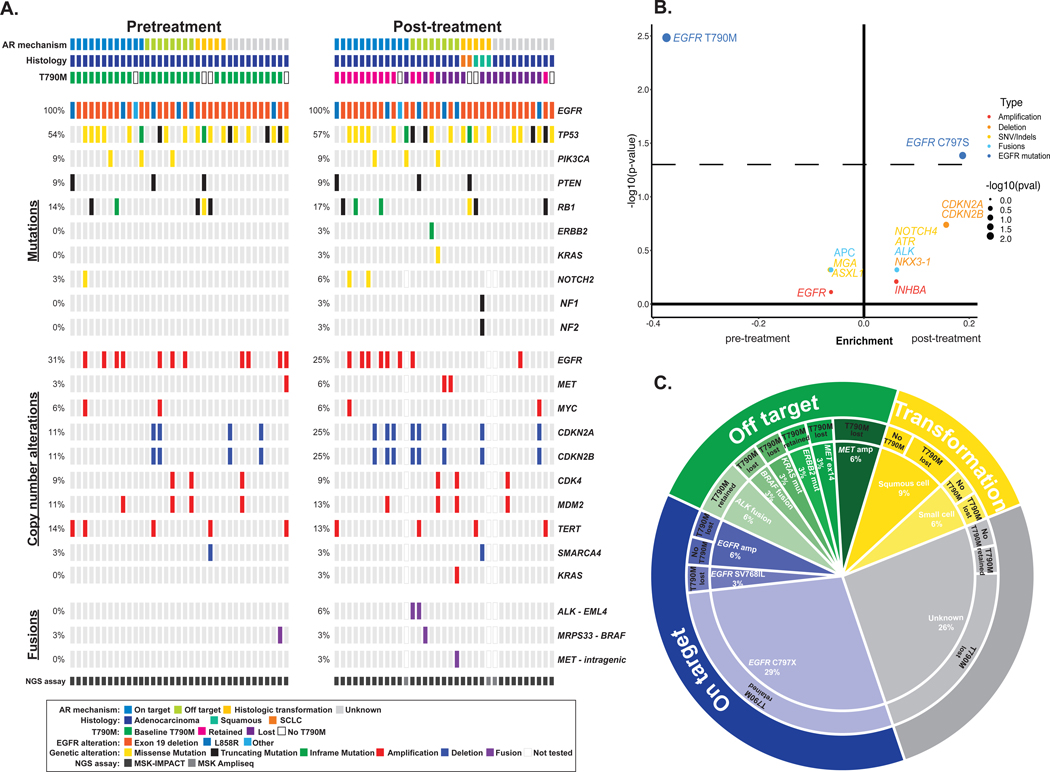
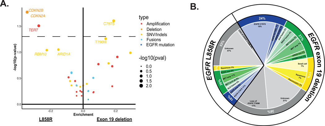
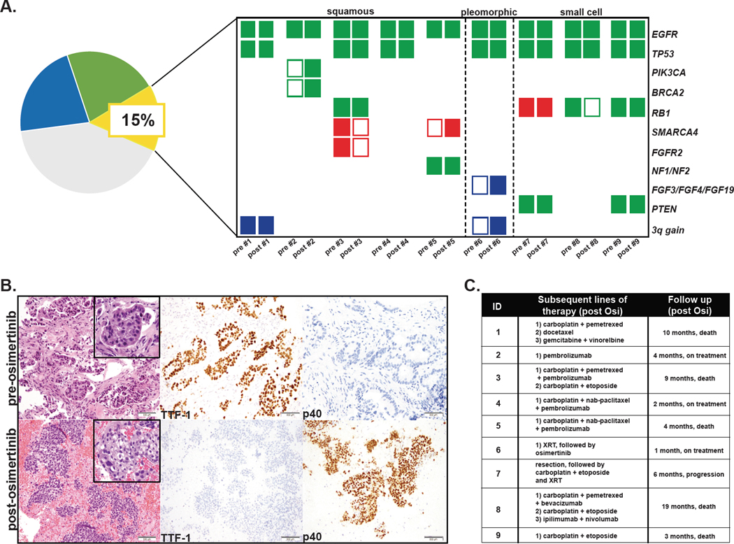
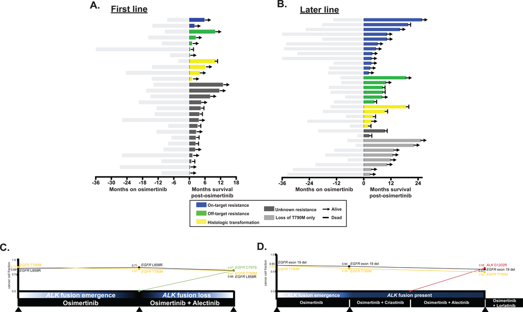
Results: Among 62 patients who met eligibility criteria, histologic transformation, primarily squamous transformation, was identified in 15% of first-line osimertinib cases and 14% of later-line cases. Nineteen percent (5/27) of patients treated with first-line osimertinib had off-target genetic resistance (2 MET amplification, 1 KRAS mutation, 1 RET fusion, and 1 BRAF fusion) whereas 4% (1/27) had an acquired EGFR mutation (EGFR G724S). Patients with squamous transformation exhibited considerable genomic complexity; acquired PIK3CA mutation, chromosome 3q amplification, and FGF amplification were all seen. Patients with transformation had shorter time on osimertinib and shorter survival compared with patients with on-target resistance. Initial EGFR sensitizing mutation, time on osimertinib treatment, and line of therapy also influenced resistance mechanism that emerged. The compound mutation EGFR S768 + V769L and the mutation MET H1094Y were identified and validated as resistance mechanisms with potential treatment options.

Conclusions: Histologic transformation and other off-target molecular alterations are frequent early emerging resistance mechanisms to osimertinib and are associated with poor clinical outcomes.See related commentary by Piotrowska and Hata, p. 2441.


Figures

Figure 1.
Figure 1.. Genomic alterations identified with first-line osimertinib.
A, Frequency of alterations pre- and post-osimertinib in patients treated with first-line osimertinib. The in-figure legend specifies details on acquired resistance mechanism, histology, initial EGFR mutation, alteration type, and next-generation sequencing (NGS) assay. B, Enrichment of individual altered genes pre-osimertinib (left) and post-osimertinib (right). Alteration type is indicated in the in-figure legend. The dashed line represents a P-value of 0.05. The frequency difference between the two sample sets is plotted on the x-axis and its significance [-log10(p-value)] on the y-axis. C, The distribution of established mechanisms of resistance by type of alteration in patients treated with first-line osimertinib. SCLC, small-cell lung cancer; NSCLC, non-small cell lung cancer; AR, acquired resistance.
Figure 2.
Figure 2.. Genomic alterations identified with later-line osimertinib.
A, Frequency of alterations pre- and post-osimertinib in patients treated with later-line osimertinib. The in-figure legend specifies details on acquired resistance mechanism, histology, initial EGFR mutation, alteration type, and next-generation sequencing (NGS) assay. B, Enrichment of individual altered genes pre-osimertinib (left) and post-osimertinib (right). The dashed line represents a P-value of 0.05. the frequency difference between the two sample sets is plotted on the x-axis and its significance [-log10(P-value)] on the y-axis. C, The distribution of established mechanisms of resistance by type of alteration in patients treated with later-line osimertinib. SCLC, small-cell lung cancer; NSCLC, non-small cell lung cancer; AR, acquired resistance
Figure 3.
Figure 3.. Molecular alterations by sensitizing EGFR mutation.
A, Enrichment of genomic alterations by sensitizing EGFR mutation. The dotted line represents a P-value of 0.05. Enrichment of mutations in patients with EGFR exon 19 deletions (right) versus EGFR L858R (left). For each mutation, the frequency difference between the two cohorts is plotted on the x-axis and its significance -log10(P-value) on the y-axis. B, The distribution of mechanisms of resistance organized by sensitizing EGFR mutation
Figure 4.
Figure 4.. The genomic profile and clinical outcomes of patient with histologic transformation.
A, Genomic patterns pre- and post-osimertinib by histologic transformation subtype. Green shaded rectangle represents mutation. Blue shaded rectangle represents copy number gain. Red shaded rectangle represents deletion. B, Pre-treatment biopsy shows adenocarcinoma, which is positive for TTF-1 and negative for p40 by immunohistochemistry. Post-treatment sample show squamous cell carcinoma, which is negative for TTF-1 and positive for p40 by immunohistochemistry. C, Treatment regimens received by patients with histologic transformation and overall survival after osimertinib.
Figure 5.
Figure 5.. Clinical outcomes pre/post osimertinib progression by resistance mechanism.
A,B Time on osimertinib (months) is depicted to the left (light grey) and overall survival after osimertinib (months) is depicted on the right for patients who A, received first-line osimertinib. B, received later-line osimertinib. C,D Longitudinal analysis of 2 patients who received later-line osimertinib. C, One patient acquired an EGFR C797S mutation and lost an ALK fusion after treatment with osimertinib and alectinib. D, The other patient acquired an ALK G1202R mutation after treatment with osimertinib and alectinib. Treatment summaries from osimertinib onwards are noted in black bars with time (months) along the x-axis. Summary of molecular alterations (MSK-IMPACT) prior to starting osimertinib and at 2 resistance timepoints are shown for each patient (colored lines). Cancer cell fractions (CCF) of driver and resistance alterations based on FACETS analysis (see supplementary methods) at each biopsy timepoint.
Figure 6.
Figure 6.. Combined inhibition of MET and EGFR overcomes MET-H1094-mediated osimertinib resistance.
A, MET-H1094 mutations were introduced by site-directed mutagenesis and confirmed by Sanger sequencing. B, Plasmids containing the MET mutants were stably introduced into PC9 cells via lentiviral transduction and then expression of MET confirmed by Western blotting. C and D, Isogenic PC9 cells were treated with osimertinib, C, or crizotinib, D, for 96 h and then growth determined using alamarBlue viability dye (top panel). IC50 values were determined by non-linear regression analysis using GraphPad Prism (bottom panel). E-G, Cells were treated with the indicated combined concentrations of osimertinib and crizotinib for 96 h and then growth determined. E and F, The percent inhibition of growth at each drug combination. G, The Chou-Talalay method was used to examine whether crizotinib and osimertinib inhibited growth in a synergistic manner. The dot plot shows the combination index (CI) as a function of the fraction affected. CI<1 indicates synergy between the two inhibitors. H, Caspase 3/7 activity was determined in cells treated for 48 h with the indicated inhibitors for 48 h and then caspase 3/7 activity determined. Results represent the fold change in caspase 3/7 enzymatic activity above the corresponding untreated cells. All experiments were repeated 3 times and included triplicate determinations of each condition. EV: empty vector. The K110A mutation results in an inactive kinase. Results represent the mean ± SD. *p<0.05 **p<0.01, ***p<0.001.

Comment in

Comment on

References

    1. Yu HA, Arcila ME, Rekhtman N, Sima CS, Zakowski MF, Pao W, et al. Analysis of tumor specimens at the time of acquired resistance to EGFR-TKI therapy in 155 patients with EGFR-mutant lung cancers. Clinical cancer research : an official journal of the American Association for Cancer Research 2013;19(8):2240–7 doi 10.1158/1078-0432.ccr-12-2246. - DOI - PMC - PubMed
    1. Sequist LV, Waltman BA, Dias-Santagata D, Digumarthy S, Turke AB, Fidias P, et al. Genotypic and histological evolution of lung cancers acquiring resistance to EGFR inhibitors. Science translational medicine 2011;3(75):75ra26 doi 10.1126/scitranslmed.3002003. - DOI - PMC - PubMed
    1. Mok TS, Wu YL, Ahn MJ, Garassino MC, Kim HR, Ramalingam SS, et al. Osimertinib or Platinum-Pemetrexed in EGFR T790M-Positive Lung Cancer. The New England journal of medicine 2017;376(7):629–40 doi 10.1056/NEJMoa1612674. - DOI - PMC - PubMed
    1. Soria JC, Ohe Y, Vansteenkiste J, Reungwetwattana T, Chewaskulyong B, Lee KH, et al. Osimertinib in Untreated EGFR-Mutated Advanced Non-Small-Cell Lung Cancer. The New England journal of medicine 2018;378(2):113–25 doi 10.1056/NEJMoa1713137. - DOI - PubMed
    1. Piotrowska Z, Isozaki H, Lennerz JK, Gainor JF, Lennes IT, Zhu VW, et al. Landscape of acquired resistance to osimertinib in EGFR-mutant NSCLC and clinical validation of combined EGFR and RET inhibition with osimertinib and BLU-667 for acquired RET fusion. Cancer discovery 2018;8(12):1529–39 doi 10.1158/2159-8290.Cd-18-1022. - DOI - PMC - PubMed

Publication types

MeSH terms