Comparative Study
. 2020 Jan 7;11(1):16.
doi: 10.1038/s41467-019-13845-4.

Different ways to transport ammonia in human and Mycobacterium tuberculosis NAD+ synthetases

Affiliations
Comparative Study

Different ways to transport ammonia in human and Mycobacterium tuberculosis NAD+ synthetases

Watchalee Chuenchor et al. Nat Commun. .

Abstract

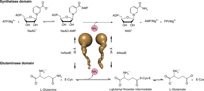
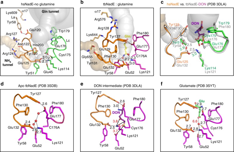
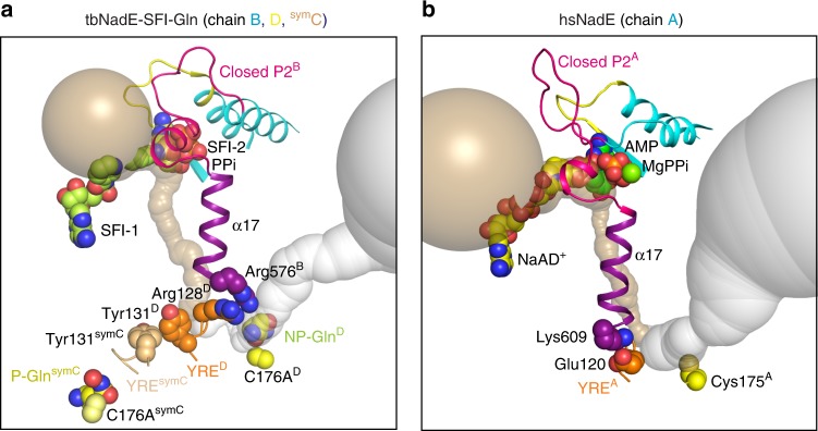
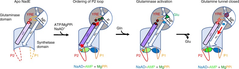
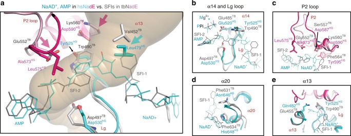
NAD+ synthetase is an essential enzyme of de novo and recycling pathways of NAD+ biosynthesis in Mycobacterium tuberculosis but not in humans. This bifunctional enzyme couples the NAD+ synthetase and glutaminase activities through an ammonia tunnel but free ammonia is also a substrate. Here we show that the Homo sapiens NAD+ synthetase (hsNadE) lacks substrate specificity for glutamine over ammonia and displays a modest activation of the glutaminase domain compared to tbNadE. We report the crystal structures of hsNadE and NAD+ synthetase from M. tuberculosis (tbNadE) with synthetase intermediate analogues. Based on the observed exclusive arrangements of the domains and of the intra- or inter-subunit tunnels we propose a model for the inter-domain communication mechanism for the regulation of glutamine-dependent activity and NH3 transport. The structural and mechanistic comparison herein reported between hsNadE and tbNadE provides also a starting point for future efforts in the development of anti-TB drugs.


Conflict of interest statement

The authors declare no competing interests.

Figures

Fig. 1
Fig. 1. Reaction catalyzed by glutamine-dependent NAD+ synthetase.
Ammonia tunnels of hsNadE and tbNadE shown in gold indicate the ammonia transfer from the glutaminase to the synthetase active site.
Fig. 2
Fig. 2. Structures of hsNadE and tbNadE in the activated complexes.
ad Diagram and structures of the glutaminase and synthetase domains in one polypeptide chain of the superimposed structure (b) of hsNadE with NaAD+, AMP, and MgPPi (c) and tbNadE with the sulfonamide derivative 1 (SFI) and PPi (d) using the glutaminase domains alone for superimposition. Residue numbering in the schematic diagram of hsNadE and tbNadE is shown in black and red, respectively. The P1 and P2 loops at the synthetase active sites, the YRE at the glutaminase domain, and the linker connecting the glutaminase and synthetase domains are colored accordingly to the diagram. Ligands bound in the structures are shown in spheres. e, f Comparison of the molecules in the asymmetric unit and in the subunit interfaces in hsNadE (e) and tbNadE (f). The buried surfaces of both complexes are compared between subunits A and B in hsNadE, subunits B and D, and subunits B and C in tbNadE. The number was calculated by PISA and shown in Å.
Fig. 3
Fig. 3. Octameric assembly and molecular tunnels connecting the two active sites in hsNadE.
a The biological unit of hsNadE is generated by crystal symmetry as an octamer. The glutamine and ammonia tunnels in subunits A and B are in gray and golden, respectively. The red arrow indicates the direction of the substrate/product flows in/out the molecular tunnel. b, c Comparison of the constrictions located in the molecular tunnels found in hsNadE (b) and tbNadE-SFI complexes (c) within the single subunit A and two subunits Dglu–Bsyn, respectively. Key residues (glutamine-binding residue, catalytic cysteine, and last position of the ammonia transport) are shown as spheres. d Polar, nonpolar, and charged residues forming the ammonia tunnel and constrictions in the hsNadE complex. The residues lining the ammonia tunnel belong to the glutaminase domain (green), the YRE loop (orange), and the synthetase domain (cyan). A water molecule coordinated with the residues forming constriction 1-1* and the YRE loop residues is shown in gray sphere with a light blue contour and its position indicates the start of the ammonia transport. eg Ammonia tunnel radii calculated in subunit A of hsNadE complex (e), in subunits Dglu–Bsyn (f), and in subunits Cglu-symCsyn (g) of the tbNadE–SFI complex. The dashed line at constriction 1-1* shows the boundary of the glutamine and ammonia tunnels. 2.1 and 1.6 Å cut-off radii are used for the glutamine and ammonia tunnels, respectively. Location of the glutamine-binding site is defined by Tyr123HS/Phe130TB and Trp179HS/Phe180TB. The last position of ammonia in the ammonia tunnel is marked by Cys531HS/Asp497TB.
Fig. 4
Fig. 4. Structural basis of substrate specificity of the synthetase domain.
a, b Binding of the NaAD+ (yellow stick, a) and sulfonamide derivative 1 (light green stick, SFI, b) in NaAD+-binding site of hsNadE and tbNadE, respectively. c, d Binding of the AMP, MgPPi (green/orange stick, green sphere) in hsNadE (c) and SFI, PPi in tbNadE (d). Cl atom hydrogen bonds to Mg2+ is in red sphere. In the tbNadE–SFI complex, the first SFI molecule is bound in the NaAD+-binding site (b) and the second SFI together with PPi is bound in the ATP-binding site (d) in a similar manner than the synthetase intermediate analog. This results in the ordering of the P2 loop (pink residues).
Fig. 5
Fig. 5. Role of the YRE loop and arrangement of the glutaminase active site.
a This panel illustrates the hsNadE glutaminase domain with the glutaminase active site free of ligand in a complex with the synthetase active site fully occupied with the intermediate analog. b The glutaminase active site of tbNadE subunit D is occupied with glutamine in a complex with SFIs and PPi bound in the synthetase active site of the coupled subunit B. The YRE loop residues are shown in orange sticks in all complexes, the catalytic and substrate-binding residues are shown in green in hsNadE and purple in tbNadE. The glutamine substrate and water in the ammonia tunnel of the tbNadE complex are shown as a yellow stick and red sphere. c Overlay of the hsNadE and tbNadE–DON complexes (PDB code 3DLA). Distance of hydrogen bonding between (i) YRE loop’s glutamate and catalytic lysine and (ii) the glutamate and constriction 1’s tyrosine is shown as green (hsNadE) and gray (tbNadE) numbers. df The glutaminase active site of previously reported tbNadE structures bound to waters in the apo form (PDB code 3SDB) (d), with DON bound (dark purple stick, PDB code 3DLA) (e), and with glutamate bound (green stick, PDB code 3SYT (f).
Fig. 6
Fig. 6. Comparison of the regulatory elements between the tbNadE and hsNadE complexes.
a, b Cartoon structures of the closed P2 loop (cyan), the α-17 helix (purple), and the YRE loop (orange for the coupled subunit, and brown for the uncoupled subunit) in tbNadE–SFI complex bound to glutamine (a) and hsNadE (b). Panel a shows the α-17 helix connecting the closed P2 loop of Bsyn to the YRE loop of the coupled subunit Dglu. This YRE loop makes also contact with the YRE loop of the uncoupled subunit symC thus resulting in a productive binding of the glutamine substrate in symC of tbNadE. Panel b shows the α17 connects the closed P2 and YRE loops with the same subunit (chain A) in hsNadE. The pairs of Arg128TB/Arg576TB and Glu120HS/Lys609HS connecting the YRE and α-17 helix of the coupled subunits in tbNadE and hsNadE, respectively, are shown as orange and purple spheres. The Tyr131TB/symCTyr131TB in tbNadE are shown as orange and brown spheres, respectively. The catalytic cysteine and the C176A variant in the glutaminase active site are shown as yellow spheres. The Caver calculated ammonia and glutamine tunnels are shown in golden and gray, respectively.
Fig. 7
Fig. 7. Proposed shared steps in hsNadE and tbNadE for the glutaminase activation.
Dash and solid curved lines represents disorder and ordered loops, respectively. NP and P indicate a non-productive and a productive glutaminase active site, respectively. Light and dark purple coloring of α-17 indicate the absence or presence of activating interactions with the YRE loop. Similarly, white and color filled stars represent the absence or presence of interactions between α-17 and YRE loop (in red), and YRE and the catalytic triad (in green). Orange, green, dark blue, and light blue spheres represent PPi, the AMP, and NaAD+ moiteties of NaAD–AMP, and ammonia. The green filled triangle represents glutamate. The “no access” sign shows the gating mechanism that blocks access to the glutamine tunnel. The addition of Gln is shown in the model after formation of the NaAD–AMP for simplicity but the order of its addition remains unclear.
Fig. 8
Fig. 8. Implication for drug design specific to tbNadE.
a Overlay of tbNadE and hsNadE complexes. NaAD+ shown with AMP in hsNadE in cyan and SFIs in tbNadE in gray (PPis are omitted for clarity). The P2 loop residues are pink and light pink in hsNadE and tbNadE, respectively. Residues important for future anti-TB drug design are shown as sticks. Pink and cyan arrows highlight the residue with the biggest difference in size and/or character between homologs. be Comparison of the synthetase-binding site between homologs at α14 and the Lg loop (b), P2 loop (c), α20 (d), and α13 (e).

References

    1. Magni G, Orsomando G, Raffelli N, Ruggieri S. Enzymology of mammalian NAD+ metabolism in health and disease. Front. Biosci. 2008;13:6135–6154. doi: 10.2741/3143. - DOI - PubMed
    1. Yang Y, Sauve AA. NAD+ metabolism: bioenergetics, signaling and manipulation for therapy. BBA—Proteins Proteom. 2016;1864:1787–1800. doi: 10.1016/j.bbapap.2016.06.014. - DOI - PMC - PubMed
    1. Li J, et al. A conserved NAD+ binding pocket that regulates protein–protein interactions during aging. Science. 2017;355:1312–1317. doi: 10.1126/science.aad8242. - DOI - PMC - PubMed
    1. Moure VR, et al. Regulation of nitrogenase by reversible mono-ADP-ribosylation. Curr. Top. Microbiol. Immunol. 2015;384:89–106. - PubMed
    1. Aravind L, Zhang D, de Souza RF, Anand S, Iyer M. The natural history of ADP-ribosyltransferases and the ADP-ribosylation system. Curr. Top. Microbiol. Immunol. 2015;384:3–32. - PMC - PubMed

Publication types

MeSH terms