. 2020 Nov;77(21):4397-4411.
doi: 10.1007/s00018-019-03424-7. Epub 2020 Jan 7.

An intact keratin network is crucial for mechanical integrity and barrier function in keratinocyte cell sheets

Affiliations

An intact keratin network is crucial for mechanical integrity and barrier function in keratinocyte cell sheets

Susanne Karsch et al. Cell Mol Life Sci. 2020 Nov.

Abstract

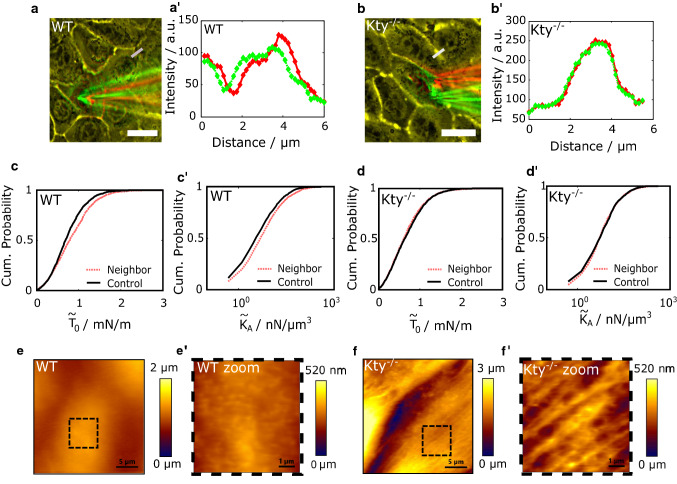
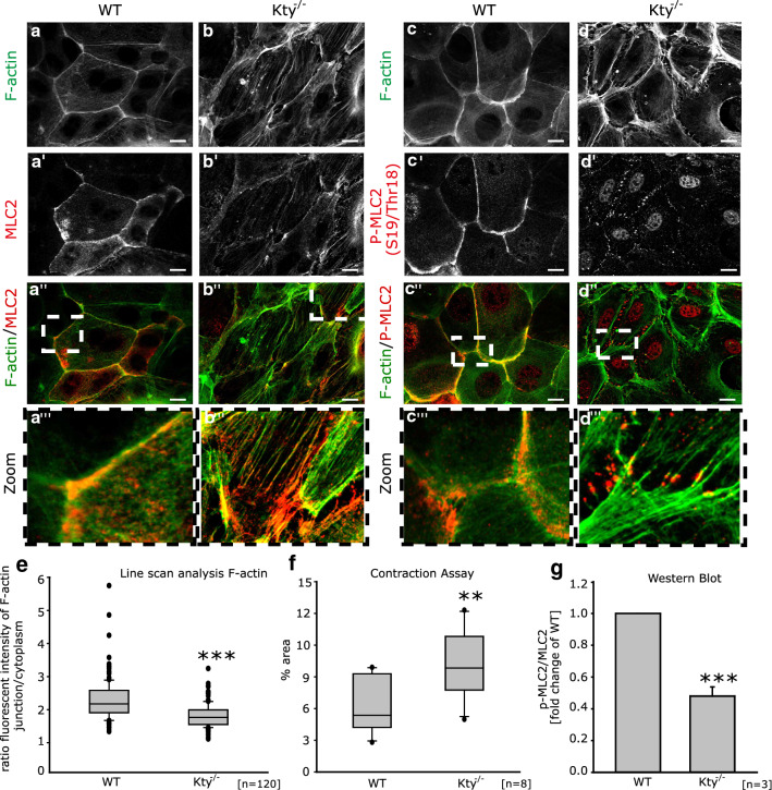
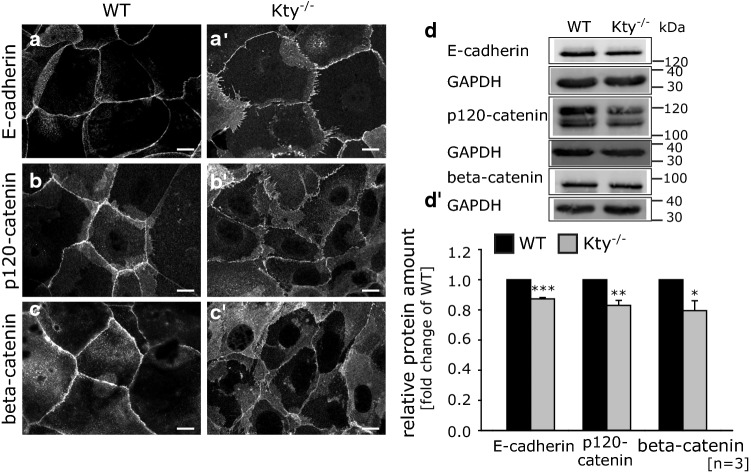
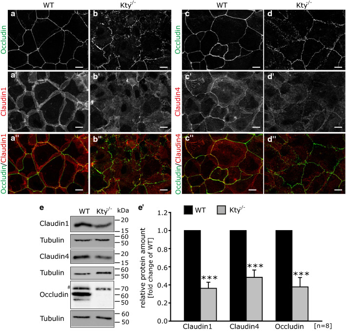
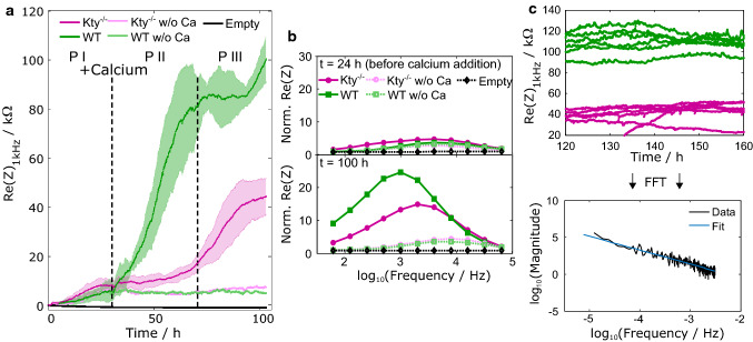
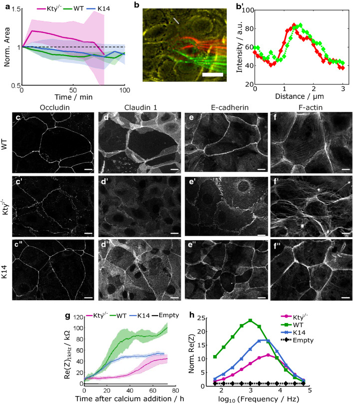
The isotype-specific composition of the keratin cytoskeleton is important for strong adhesion, force resilience, and barrier function of the epidermis. However, the mechanisms by which keratins regulate these functions are still incompletely understood. In this study, the role and significance of the keratin network for mechanical integrity, force transmission, and barrier formation were analyzed in murine keratinocytes. Following the time-course of single-cell wound closure, wild-type (WT) cells slowly closed the gap in a collective fashion involving tightly connected neighboring cells. In contrast, the mechanical response of neighboring cells was compromised in keratin-deficient cells, causing an increased wound area initially and an inefficient overall wound closure. Furthermore, the loss of the keratin network led to impaired, fragmented cell-cell junctions, and triggered a profound change in the overall cellular actomyosin architecture. Electric cell-substrate impedance sensing of cell junctions revealed a dysfunctional barrier in knockout (Kty-/-) cells compared to WT cells. These findings demonstrate that Kty-/- cells display a novel phenotype characterized by loss of mechanocoupling and failure to form a functional barrier. Re-expression of K5/K14 rescued the barrier defect to a significant extent and reestablished the mechanocoupling with remaining discrepancies likely due to the low abundance of keratins in that setting. Our study reveals the major role of the keratin network for mechanical homeostasis and barrier functionality in keratinocyte layers.

Keywords: Barrier functionality; Keratin cytoskeleton; Mechanical homeostasis; Wound closure.


Conflict of interest statement

The authors declare no competing interests.

Figures

Fig. 1
Fig. 1
Inefficient wound closure in Kty−/− cells. a A single cell in a confluent layer was scratched apically with a micropipette, leading to a single-cell wound. b Fluorescence micrographs including xz and yz sections of actin (red) and keratin 14 (green) in the wounded cell (white contour) after micropipette-mediated wounding. c Phase-contrast images of the indicated cell 1 min after wounding (blue contour) and 12 min after wounding (red contour). d Individual cell area changes of defect cells over time for WT (green) and Kty−/− cells (magenta). Normalization was done with respect to the area prior to wounding. Arrowhead marks time point of 12 min which is visualized in (c). e Cumulative histogram of the apparent pretension T0 capturing the resistance to external force at low indentation depth (p < 0.001) for homeostatic WT cells (green, solid line) and homeostatic Kty−/− cells (magenta, dashed line) The inset shows representative force-indentation curves recorded either on WT (green) or Kty−/− (magenta) cells. f Cumulative histogram of the apparent area compressibility modulus KA probing larger indentation depths (p < 0.001) for the same cells. Number of analyzed force curves n = 2441 for Kty−/− and n = 2563 for WT cells which correspond to N = 5 individual experiments/force maps. Scale bars: 20 µm
Fig. 2
Fig. 2
Impaired mechanical coupling in the absence of keratins. a Merged image of colored (red, green) phase-contrast images taken during wounding of WT cells. Yellow color indicates exact overlay of images; red-colored image was taken before micropipette movement, green after movement. a’ Line profiles of the two images at a position of a cell junction one cell away from the defect (grey bar in a) indicating junction movement during wounding. bb’ Same analysis for Kty−/− cells during wounding. c Cumulative histogram of the apparent pretension T0 and (c’) apparent area compressibility modulus KA of WT cells neighboring a defect (red, dashed) and control cells in an intact layer (black, solid) (both p < 0.001; n = 2659 (neighbor) and n = 2563 (control)). dd’ Cumulative histograms of the same mechanical parameters for Kty−/− cells [p = 0.112 and p = 0.288, respectively; n = 2225 (neighbor) and n = 2441 (control)]. e AFM height scan of WT cells revealing homogeneous junctions. e’ Zoom into the marked area in e showing homogeneous cortex structures. f AFM height scan of Kty−/− cells revealing fragmented junctions. f’ Zoom into marked area in f showing fibrous structures. Scale bars: a and b: 20 µm, e and f: 5 µm, and e’ and f’: 1 µm
Fig. 3
Fig. 3
Altered actomyosin organization in Kty−/− cells. ad’’’ Immunostaining for F-actin (Alexa Fluor 488-Phalloidin), MLC2, and phospho-myosin light chain (pMLC) in WT and Kty−/− cells 48 h after Ca2+ switch. Scale bars: 10 µm. e Quantification of F-actin staining using line scan analysis. For each cell line, 120 cell borders in three independent experiments were measured. f Epithelial sheet contraction assay of WT and Kty−/− cells. g Quantification of pMLC-levels relative to total MLC2 (mean ± SEM, n = 3)
Fig. 4
Fig. 4
Impaired formation of adherens junctions. ac’ Immunostaining for E-cadherin, p120-catenin, and β-catenin in WT and Kty−/− cells 48 h after Ca2+-switch. Scale bars: 10 µm. d Western blots of total protein lysates for E-cadherin, p120-catenin, β-catenin, and Tubulin. d’ Quantification of protein levels relative to tubulin as loading control (mean ± SEM, n = 4)
Fig. 5
Fig. 5
Altered organization of tight junctions in keratin-free keratinocytes. ad’’ Immunostaining for occludin, claudin 1, and claudin 4 in WT and Kty−/− cells 48 h after Ca2+ switch. Scale bars: 10 µm. e WB of total protein lysates for occludin, claudin 1, claudin 4, and α-tubulin (# indicates the band for quantification). e’ Quantification of protein levels relative to α-tubulin as loading control (mean ± SEM, n = 8)
Fig. 6
Fig. 6
Barrier formation is compromised in Kty−/− cells. a Real part of the complex impedance Z recorded with electric cell-substrate impedance sensing at 1 kHz as an indicator for junction functionality followed over time for WT (green, n = 4), Kty−/− (magenta, n = 4), ‘low calcium’ controls (light green, light magenta, respectively) and empty electrodes (black). b Normalized impedance spectra at t = 24 h (before calcium addition) and after impedance leveling in Kty−/− cells at t = 100 h. c For fluctuation analysis, each individual signal is Fourier transformed (black) and the resulting power spectrum is fitted with a linear slope (blue)
Fig. 7
Fig. 7
Re-expression of K14 partially restores TJ organization and barrier formation. a Averaged cell area changes of defect cells over time for WT (green, n = 4), Kty−/− cells (magenta, n = 9), and K14 cells (blue, n = 7) including standard deviations (shaded areas). Normalization was computed with respect to the individual area shortly before wounding. b Merge of colored phase-contrast images taken during wounding in K14 cells. Yellow color indicates exact overlay of images; red image was taken before micropipette movement, green after movement. Scale bar: 20 µm. b’ Line profiles of the two images at a position of a cell junction one cell away from the defect (grey bar in b) indicating displacement of junctions during wounding. ce’’ Immunostaining for occludin and claudin 1 in WT, Kty−/−, and K14 rescue cells 48 h after Ca2+ switch. Scale bars: 10 µm. ff’’ Immunostaining for F-actin (Phalloidin-Alexa 488) in WT, Kty−/−, and K14 rescue cells 48 h after Ca2+ switch. Scale bars: 10 µm. g Real part of the impedance Z recorded at 1 kHz as a representative indicator for junction functionality followed over time for WT (green, n = 4), Kty−/− (magenta, n = 4), K14 rescue cells (blue, n = 4), and the empty electrode (black). h Normalized impedance spectra (real part) 48 h after calcium addition
Fig. 8
Fig. 8
Schematic model of the contractile pattern inside Kty−/− (top) and WT cells (bottom). Actomyosin in Kty−/− cells is bundled into thick fibers spanning the cell (black springs). Only at distinct spots, namely the respective end of the fibers, cells are connected via cell–cell junctions (blue dots). In WT cells, a contractile cortex exists, lining the cell. Keratin filaments (green) are distributed inside cells acting as shock absorbers. Cell–cell junctions are distributed all over the cell’s periphery. Upon wounding and concomitant loss of contractile filaments inside one cell (grey X), adjacent contractile filaments are directly affected. In surrounding Kty−/− cells, forces are not balanced anymore, so that contractile filaments shrink and pull cell borders (indicated by red springs and borders), while in WT cells, forces are distributed more evenly and stable junction positions and resting lengths of the contractile filaments are possible

References

    1. Simpson CL, Patel DM, Green KJ. Deconstructing the skin: cytoarchitectural determinants of epidermal morphogenesis. Nat Rev Mol Cell Biol. 2011;12(9):565–580. doi: 10.1038/nrm3175. - DOI - PMC - PubMed
    1. Hoffman BD, Yap AS. Towards a dynamic understanding of cadherin-based mechanobiology. Trends Cell Biol. 2015;25(12):803–814. doi: 10.1016/j.tcb.2015.09.008. - DOI - PubMed
    1. Tunggal JA, Helfrich I, Schmitz A, Schwarz H, Günzel D, Fromm M, Kemler R, Krieg T, Niessen CM. E-cadherin is essential for in vivo epidermal barrier function by regulating tight junctions. EMBO J. 2005;24(6):1146–1156. doi: 10.1038/sj.emboj.7600605. - DOI - PMC - PubMed
    1. Loschke F, Seltmann K, Bouameur J-E, Magin TM. Regulation of keratin network organization. Curr Opin Cell Biol. 2015;32:56–64. doi: 10.1016/j.ceb.2014.12.006. - DOI - PubMed
    1. Shaw TJ, Martin P. Wound repair at a glance. J Cell Sci. 2009;122(18):3209–3213. doi: 10.1242/jcs.031187. - DOI - PMC - PubMed

Grants and funding