. 2020 Mar;21(3):415-428.
doi: 10.1111/mpp.12905. Epub 2020 Jan 7.

An RXLR effector PlAvh142 from Peronophythora litchii triggers plant cell death and contributes to virulence

Affiliations

An RXLR effector PlAvh142 from Peronophythora litchii triggers plant cell death and contributes to virulence

Junjian Situ et al. Mol Plant Pathol. 2020 Mar.

Abstract

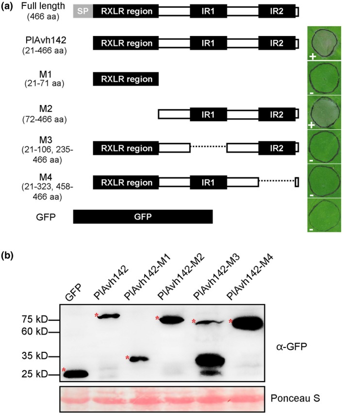
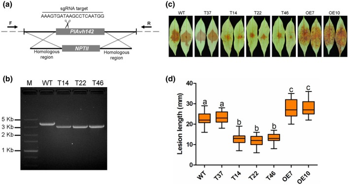
Litchi downy blight, caused by the phytopathogenic oomycete Peronophythora litchii, results in tremendous economic loss in litchi production every year. To successfully colonize the host cell, Phytophthora species secret hundreds of RXLR effectors that interfere with plant immunity and facilitate the infection process. Previous work has already predicted 245 candidate RXLR effector-encoding genes in P. litchii, 212 of which have been cloned and tested for plant cell death-inducing activity in this study. We found three such RXLR effectors could trigger plant cell death through transient expression in Nicotiana benthamiana. Further experiments demonstrated that PlAvh142 could induce cell death and immune responses in several plants. We also found that PlAvh142 localized in both the cytoplasm and nucleus of plant cells. The cytoplasmic localization was critical for its cell death-inducing activity. Moreover, deletion either of the two internal repeats in PlAvh142 abolished the cell death-inducing activity. Virus-induced gene silencing assays showed that cell death triggered by PlAvh142 was dependent on the plant transduction components RAR1 (require for Mla12 resistance), SGT1 (suppressor of the G2 allele of skp1) and HSP90 (heat shock protein 90). Finally, knockout of PlAvh142 resulted in significantly attenuated P. litchii virulence on litchi plants, whereas the PlAvh142-overexpressed mutants were more aggressive. These data indicated that PlAvh142 could be recognized in plant cytoplasm and is an important virulence RXLR effector of P. litchii.

Keywords: Peronophythora litchii; RXLR effector; cell death; plant immunity.


Figures

Figure 1
Figure 1
Analysis of the cell death phenotype of RXLR effectors from Peronophythora litchii. (a) Cell death triggered by P. litchii RXLR effectors in Nicotiana benthamiana leaves. Leaves of N. benthamiana were infiltrated with Agrobacterium tumefaciens carrying pBIN::GFP‐PlAvh142. Photographs were taken at 5 days post‐agroinfiltration (dpa). Green fluorescent protein (GFP) and INF1 were used as negative and positive control, respectively. (b) Cell death symptoms induced by PlAvh142 in different plants. A. tumefaciens carrying PlAvh142 was infiltrated into the leaves of N. benthamiana, Solanum melongena, and Solanum lycopersicum. Photographs were taken at 5 dpa. GFP was used as negative control. (c) Confirmation of proteins accumulation. Total proteins were extracted from N. benthamiana leaves at 2 dpa. Immunoblot analyses were performed using anti‐GFP (top panel) antibody. Ponceau S staining of total protein serves as loading control (bottom panel). Representative images were chosen for the results obtained from three independent experiments
Figure 2
Figure 2
Transient expression of PlAvh142‐activated defence responses in Nicotiana benthamiana. (a) Reactive oxygen species (ROS) accumulation and callose deposition were observed in PlAvh142‐expressing plants at 36 hr post‐agroinfiltration (hpa). Representative images present the results obtained from three independent experiments. (b) Quantification of the ROS using ImageJ software. Mean ± SE was derived from three independent biological repeats (n ≥ 10), and *** denotes significant differences from the green fluorescent protein (GFP) (Student's t test: p < .01). (c) Quantification of the callose deposits per microscopic field using ImageJ software. Mean ± SE was derived from three independent biological repeats (n ≥ 10), and *** denotes significant differences from the GFP (Student's t test: p < .01) (D) Relative transcript levels of defence‐related genes in N. benthamiana. Leaves were harvested for RNA extraction of PlAvh142 or GFP control at 0, 12, 24, and 36 hpa. Transcript levels of candidate genes in PlAvh142‐treated leaves were normalized to that of GFP control, which was arbitrarily set as 1. The constitutively expressed gene NbEF1α was used as internal reference, according to the 2−ΔΔ C t method. Data represent means ± SD from three independent biological repeats, asterisks denote significant differences from the control group (Student's t test: *p < .05; ***p < .01)
Figure 3
Figure 3
Deleting either of the internal repeats in PlAvh142 abolished the ability to trigger cell death. (a) Schematic diagrams of the protein structures of the PlAvh142 deletion mutants are shown in the left. Cell death symptoms in Nicotiana benthamiana leaves expressing PlAvh142 deletion mutants are shown in the right. Photographs were taken at 5 days post‐agroinfiltration. (b) Western blot confirmation of expression of PlAvh142 mutants using anti‐GFP (green fluorescent protein) antibody. The red asterisks indicate protein bands of the correct size. Protein loading was indicated by Ponceau S staining. Similar results were obtained from three independent experiments
Figure 4
Figure 4
Cytoplasmic localization is critical for PlAvh142‐induced cell death. (a) Confocal microscopy imaging shows that green fluorescent protein (GFP)‐tagged PlAvh142 localizes to both the nucleus and the cytoplasm. GFP‐tagged PlAvh142 was transiently coexpressed with red fluorescent protein (RFP) via agroinfiltration in Nicotiana benthamiana with an OD600 of 0.1. Images are from GFP channel (left panel), RFP channel (middle panel), and the overlay (right panel) in N. benthamiana leaf cells. Scale bar, 20 μm. Photographs were taken at 36 hr post‐agroinfiltration (hpa). (b) Confocal microscopy images showing the subcellular localization of PlAvh142 attached with the nuclear localization signal (NLS) and nuclear export signal (NES), and the mutant forms nes and nls. The fusion constructs were agroinfiltrated at a final OD600 of 0.1. Photographs were taken at 36 hpa. Scale bars, 20 μm. (c) PlAvh142 could still induce cell death when localized to the cytoplasm. Strains with NLS‐, NES‐, nls‐ or nes‐tagged PlAvh142 were agroinfiltrated at a final OD600 of 0.1. Cell death triggered by NLS‐targeted PlAvh142 is delayed and weak. Photographs were taken at 5 days post‐agroinfiltration. Representative images for each construct were selected from three biological repeats, each of which contained at least five leaves for agroinfiltration
Figure 5
Figure 5
RAR1, SGT1, and HSP90 were required for cell death induced by PlAvh142. (a) Representative photographs of PlAvh142‐induced cell death in silenced Nicotiana benthamiana leaves at 5 days post‐agroinfiltration (dpa). Agrobacterium tumefaciens carrying PlAvh142 was infiltrated into the upper leaves of silenced plants at 16–20 dpa of tobacco rattle virus (TRV) constructs. CK, unsilenced control. (b) Quantification of cell death in N. benthamiana leaves scored at 5 dpa. The degree of cell death was divided into three levels: no cell death, weak cell death, and strong cell death. Asterisks indicate significant differences from green fluorescent protein (GFP)‐silenced plants (Wilcoxon rank‐sum test: ***p < .001). (c) The transcript abundance of RAR1, SGT1, and HSP90 in corresponding silenced plants was analysed by quantitative reverse transcription PCR (RT‐qPCR). The constitutively expressed gene NbEF1α was used as internal reference. Error bars represent the SD of three biological replicates. (d) Western blot of PlAvh142 in silenced leaves using the anti‐GFP antibody. Protein loading is indicated by Ponceau S staining. Similar results were obtained from three independent experiments
Figure 6
Figure 6
Expression profile of Peronophythora litchii RXLR effector gene PlAvh142. The relative transcript levels of PlAvh142 during different development and infection stages of P. litchii were assessed by quantitative reverse transcription PCR (RT‐qPCR). MY, P. litchii mycelia grown in CJA medium; ZO, zoospores. Litchi leaves inoculated with P. litchii zoospores were harvested at 1.5, 3, 6, 12, and 24 hr post‐inoculation (hpi). The relative expression level was calibrated to the levels for the MY set as 1. The constitutively expressed gene PlActin was used as internal reference. Error bars represent the SD of three biological replicates
Figure 7
Figure 7
Knockout of PlAvh142 in Peronophythora litchii attenuated the virulence to litchi. (a) Schematic diagram of the gene replacement at the PlAvh142 locus by CRISPR/Cas9. The primers (F and R) used for PCR analysis are indicated by the horizontal arrows. (b) PCR analysis of the PlAvh142 knockout mutants. Wild‐type (WT): 1,401 bp (PlAvh142) + 2,134 bp; knockout mutants (T14, T22, and T46): 795 bp (NPTII) + 2,134 bp. (c) Pathogenicity assays of WT and PlAvh142 mutants on litchi leaves. Tender leaves were inoculated with 100 zoospores from WT, T37 (negative control), three PlAvh142 knockout mutants T14, T22, and T46, and two overexpression mutants OE7 and OE10. Disease symptoms were visually monitored over a period of 48 hr and photographs were taken at 48 hr post‐inoculation (hpi). (d) Lesion length was measured after 48 hpi. Different letters represent significant differences (p < .01; Duncan's multiple range test). Bars represent medians and boxes the 25th and 75th percentiles. There were 30 leaves used in each of the three biological replicates

References

    1. Andrade, M.A. , Perez‐Iratxeta, C. and Ponting, C.P. (2001) Protein repeats: structures, functions, and evolution. Journal of Structural Biology, 134, 117–131. - PubMed
    1. Asai, S. and Yoshioka, H. (2009) Nitric oxide as a partner of reactive oxygen species participates in disease resistance to necrotrophic pathogen Botrytis cinerea in Nicotiana benthamiana . Molecular Plant‐Microbe Interactions, 22, 619–629. - PubMed
    1. Asai, S. , Furzer, O.J. , Cevik, V. , Kim, D.S. , Ishaque, N. , Goritschnig, S. et al (2018) A downy mildew effector evades recognition by polymorphism of expression and subcellular localization. Nature Communications, 9, 5159. - PMC - PubMed
    1. Azevedo, C. , Sadanandom, A. , Kitagawa, K. , Freialdenhoven, A. , Shirasu, K. and Schulze‐Lefert, P. (2002) The RAR1 interactor SGT1, an essential component of R gene‐triggered disease resistance. Science, 295, 2073–3016. - PubMed
    1. Balint‐Kurti, P. (2019) The plant hypersensitive response: concepts, control and consequences. Molecular Plant Pathology, 20, 1163–1178. - PMC - PubMed

Publication types

LinkOut - more resources