. 2020 Jan 8;10(1):3.
doi: 10.1038/s41408-019-0270-0.

A new regulatory mechanism of protein phosphatase 2A activity via SET in acute myeloid leukemia

Affiliations

A new regulatory mechanism of protein phosphatase 2A activity via SET in acute myeloid leukemia

Elena Arriazu et al. Blood Cancer J. .

Abstract

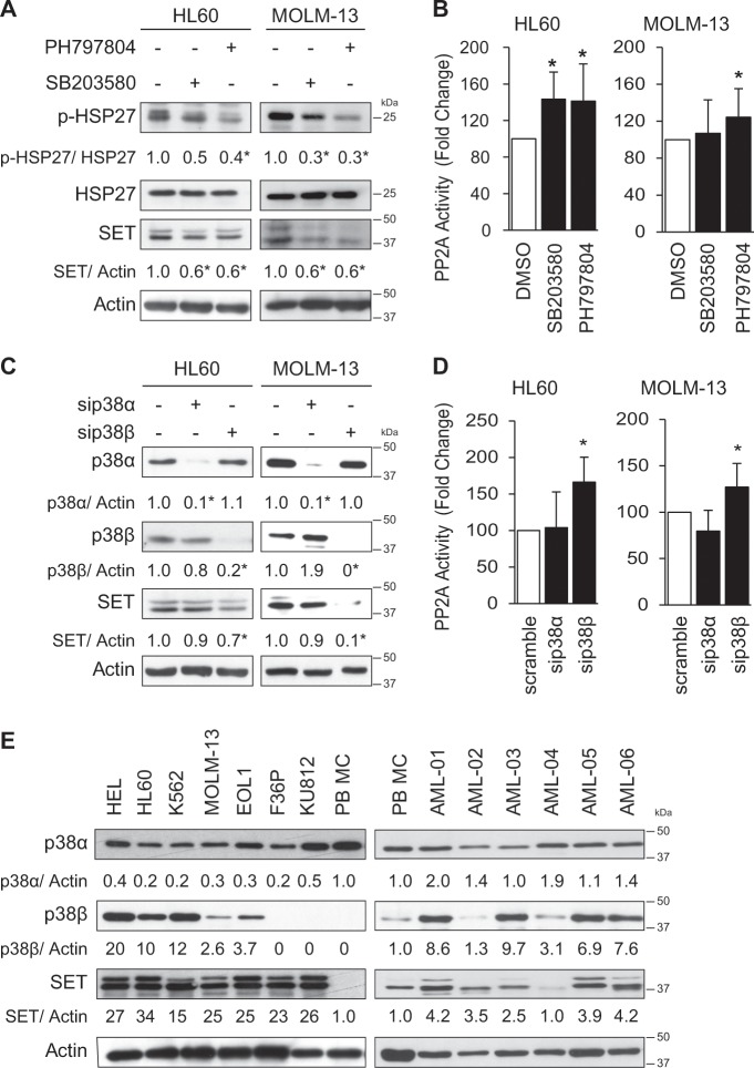
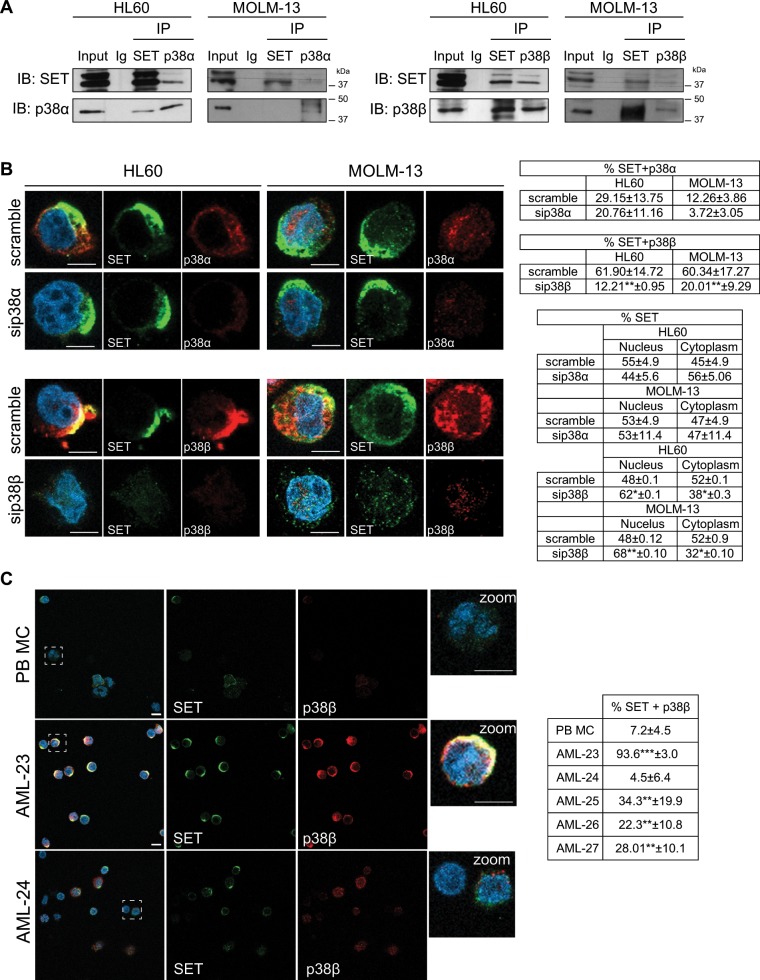
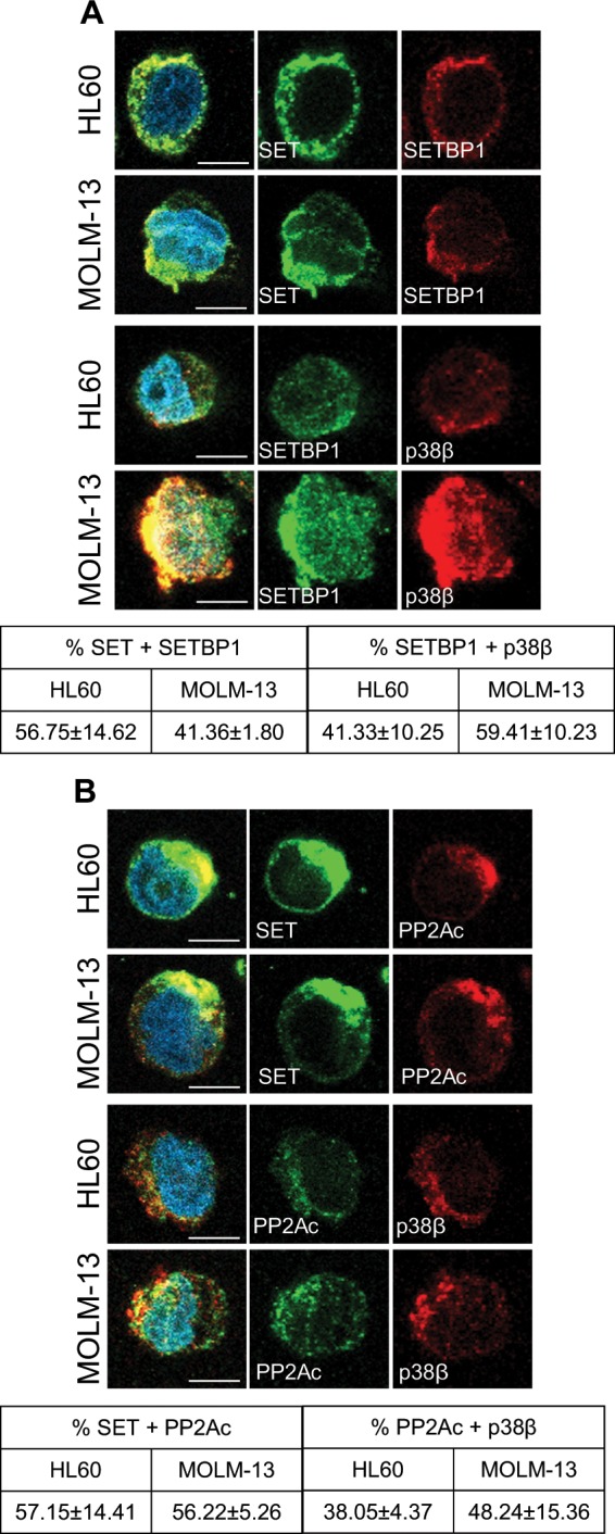
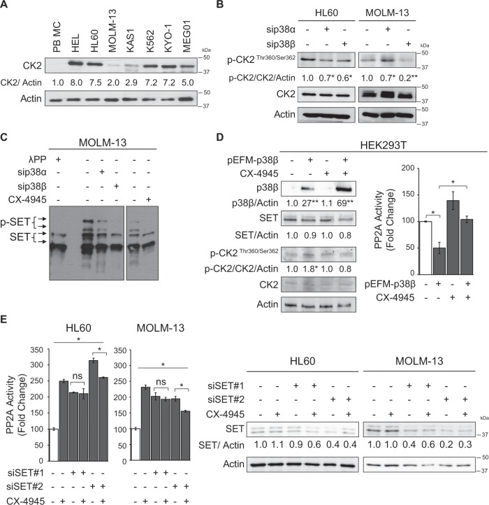
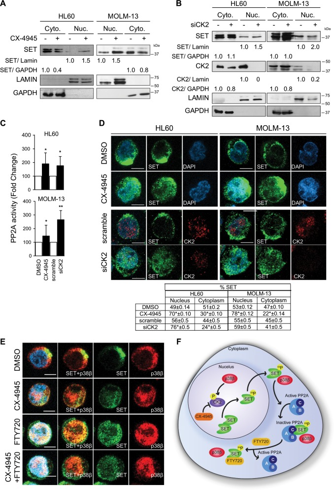
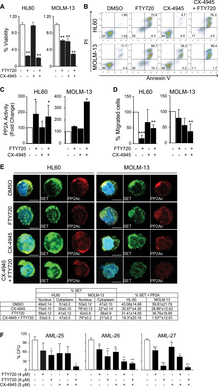
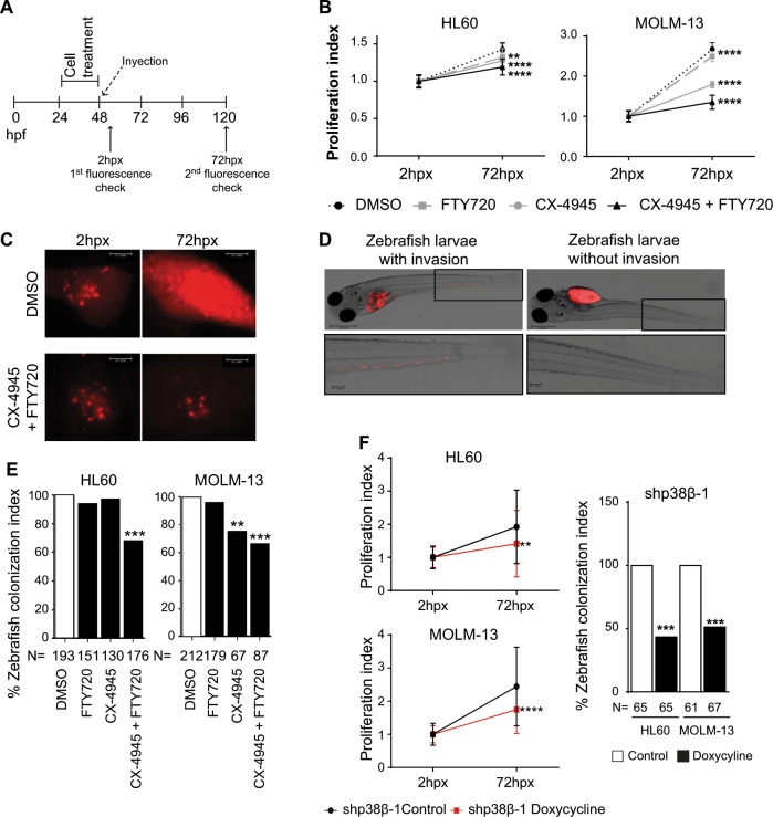
Acute myeloid leukemia (AML) is an aggressive hematologic malignancy. Although novel emerging drugs are available, the overall prognosis remains poor and new therapeutic approaches are required. PP2A phosphatase is a key regulator of cell homeostasis and is recurrently inactivated in AML. The anticancer activity of several PP2A-activating drugs (e.g., FTY720) depends on their interaction with the SET oncoprotein, an endogenous PP2A inhibitor that is overexpressed in 30% of AML cases. Elucidation of SET regulatory mechanisms may therefore provide novel targeted therapies for SET-overexpressing AMLs. Here, we show that upregulation of protein kinase p38β is a common event in AML. We provide evidence that p38β potentiates SET-mediated PP2A inactivation by two mechanisms: facilitating SET cytoplasmic translocation through CK2 phosphorylation, and directly binding to and stabilizing the SET protein. We demonstrate the importance of this new regulatory mechanism in primary AML cells from patients and in zebrafish xenograft models. Accordingly, combination of the CK2 inhibitor CX-4945, which retains SET in the nucleus, and FTY720, which disrupts the SET-PP2A binding in the cytoplasm, significantly reduces the viability and migration of AML cells. In conclusion, we show that the p38β/CK2/SET axis represents a new potential therapeutic pathway in AML patients with SET-dependent PP2A inactivation.


Conflict of interest statement

The authors declare that they have no conflict of interest.

Figures

Fig. 1
Fig. 1. p38β is overexpressed in AML and its inhibition decreases SET protein levels, increasing PP2A activity.
a HL60 and MOLM-13 cell lines were treated with the p38 inhibitors SB203580 (2.5 µM) and PH797804 (250 nM) for 24 h. Protein expression for p-HSP27/HSP27 (p38 substrate) and SET was analyzed by western blot. b Measurement of PP2A activity after p38 inhibition by immunoprecipitation and phosphatase assay. c Silencing of p38α and p38β with specific siRNA (50 nM for 48 h), and analysis of total protein by western blot. d Measurement of PP2A activity by immunoprecipitation and phosphatase assay. e Western blot analysis of total protein in AML cell lines and AML patient samples, compared to peripheral blood mononuclear cells (PB-MC). The results are corrected by the specific loading control and are expressed as fold-change of the control, which are assigned a value of 1 and are mean values. Experiments were performed in triplicate four times. *p < 0.05.
Fig. 2
Fig. 2. p38β co-localizes with SET in AML cells.
a Immunoprecipitation of SET, p38α and p38β with specific antibodies in HL60 and MOLM-13 cells. Normal goat Ig was used as negative control b Knockdown of p38α and p38β with siRNA (50 nM for 48 h), using scramble siRNA as control, in HL60 and MOLM-13 cells. Immunofluorescence analysis of either p38α or p38β (red) and SET (green). Nuclei were stained with DAPI. Immunofluorescences were visualized by confocal microscopy. Quantification table of colocalization fluorescence and green fluorescence (SET) in nucleus and cytoplasm. Quantification analysis showed ~60% of SET-p38β colocalization in both cell lines, and only 12 and 29% of SET-p38α colocalization in MOLM-13 and HL60, respectively. c Immunofluorescence analysis of p38β (red) and SET (green) in peripheral blood mononuclear cells (PB-MC) and the primary AML patient samples AML-23 and AML-24. Nuclei were stained with DAPI. Quantification table of colocalization fluorescence. Immunofluorescences were visualized by confocal microscopy. The results are expressed as mean values ± SEM. Experiments were performed in triplicate four times. *p < 0.05, **p < 0.01. Scale bar represents 5 µm.
Fig. 3
Fig. 3. p38β acts as a SET stabilizing protein.
a Immunofluorescence analysis of SET (green) and SETBP1 (red), and SETBP1 (green) and p38β (red), in HL60 and MOLM-13 cells. Nuclei were stained with DAPI. Quantification of colocalization fluorescence. (b) Immunofluorescence analysis of SET (green) and PP2Ac (red) and PP2Ac (green) and p38β (red), in HL60 and MOLM-13 cells. Nuclei were stained with DAPI. Quantification of colocalization fluorescence. Immunofluorescences were visualized by confocal microscopy. The results are expressed as mean values ± SEM. Experiments were performed in triplicate four times. *p < 0.05, **p < 0.01. Scale bar represents 5 µm.
Fig. 4
Fig. 4. p38β regulates CK2-mediated phosphorylation of SET.
a Western blot analysis of the CK2 protein in AML cells lines compared to peripheral blood mononuclear cells (PB-MC). b Silencing of p38α and p38β with specific siRNA (50 nM for 48 h) and analysis of phospho- and total CK2 by western blot in HL60 and MOLM-13 cells. c MOLM-13 cells treated with either siRNA for silencing p38α and p38β (50 nM for 48 h) or with the CK2 inhibitor CX-4945 (5 µM, 24 h) and analyzed for phosphorylated forms of SET in SDS-PAGE with Phos-TagTM. A sample treated with λ phosphatase (100 units, 1 h) was used as control. d Overexpression of p38β in HEK293T cells with 1 µg of pEFM link p38β plasmid or the empty plasmid with lipofectamine 2000 and treated with CX-4945 (3,75 µM, 24 h). Analysis of p38β, SET and phospho- and total CK2 by western blot and PP2A activity. e Silencing of SET with specific siRNA (50 nM for 72 h) and analysis of SET by western blot and PP2A activity in HL60 and MOLM-13 cells. The results are corrected by the specific loading control and are expressed as fold-change of the control, which are assigned a value of 1 and are mean values. Experiments were performed in triplicate four times. *p < 0.05 **p < 0.01.
Fig. 5
Fig. 5. CK2 inhibition retains SET in the nucleus.
a Nuclear (Nuc.) and cytoplasmic (Cyto.) protein isolated from HL60 and MOLM-13 cells treated with CX-4945 (5 µM, 24 h) and analyzed by western blot for SET localization. b HL60 and MOLM-13 cells treated with specific siRNA for CK2 (20 nM, 48 h). Nuclear (Nuc.) and cytoplasmic (Cyto.) proteins were isolated and analyzed by western blot for SET localization. c Measurement of PP2A activity by immunoprecipitation and phosphatase assay. The results are expressed as fold-change of the control, which are assigned a value of 1 and are mean values ± SEM. Experiments were performed in triplicate four times. *p < 0.05, **p < 0.01. d Immunofluorescence analysis of CK2 (red) and SET (green). Nuclei were stained with DAPI. Immunofluorescences were visualized by confocal microscopy. Scale bar represents 5 µm. Quantification table of green fluorescence (SET) in nucleus and cytoplasm. The results are expressed as mean values ± SEM. Experiments were performed in triplicate four times. *p < 0.05, **p < 0.01. e Immunofluorescence analysis of p38β (red) and SET (green). Nuclei were stained with DAPI. Immunofluorescences were visualized by confocal microscopy. Scale bar represents 5 µm. f p38β is able to activate CK2, which phosphorylates SET and, as consequence, facilities SET trafficking to the cytoplasm, contributing to PP2A inactivation in AML cells. Moreover, p38β binds to SET in the cytoplasm, contributing to its stability and leading to PP2A inactivation. Treatment with CX-4945 (CK2 inhibitor) retains SET in the nucleus, avoiding its phosphorylation. FTY720 treatment disrupts the SET-PP2A biding which remains in the cytoplasm.
Fig. 6
Fig. 6. CX-4945 and FTY720 combination therapy induces apoptosis in AML cell lines and primary patient samples.
HL60 and MOLM-13 cells pretreated for 4 h with CX-4945 (5 µM) and then treated for 24 h with FTY720 (5 µM). a Cell viability was measured by MTS analysis. The results are corrected by the DMSO control and are expressed as fold-change of the control, which are assigned a value of 1 and are mean values ± SEM. b FACS analysis of apoptosis in HL60 and MOLM-13 cell lines stained with propidium iodide (PI) and Annexin V. The percentages of viable and apoptotic cells are indicated. c PP2A activity analysis performed by immunoprecipitation and activity assay. d Migration of HL60 and MOLM-13 placed in the upper well of a 8.0 μM transwell plate in RPMI without FBS. The lower chamber contained RPMI supplemented with 10% FBS. Migration assay was performed for 3 h and then assessed for cell number using flow cytometry e Immunofluorescence analysis of SET (green) and PP2A (red) and quantification table of % colocalization between red fluorescence (PP2A) and green fluorescence (SET) and % of green fluorescence (SET) in nucleus and cytoplasm. Nuclei were stained with DAPI. Immunofluorescences were visualized by confocal microscopy. Scale bar represents 5 µm. Experiments were performed in triplicate four times. *p < 0.05, **p < 0.01. f AML patient samples were cultured in semisolid medium and treated with CX-4945 (5 µM) and FTY720 (4 and 8 µM), alone or in combination. Colony formation units (CFU) were counted 12 days after seeding. Graphs of counted CFU represented as percentage of CFU related to the control, which are assigned the total CFU (100%) and are mean values ± SEM.
Fig. 7
Fig. 7. CX-4945 and FTY720 combination therapy induces AML cell death in zebrafish xenograft models.
In vivo proliferation and invasive potential of HL60 and MOLM-13 cells upon treatment with FTY720, CX-4945 or the combination of both compounds were analyzed in a xenograft zebrafish model. a Timing scheme for the xenografts of zebrafish embryos. b Measurement of proliferation index performed as fluorescence intensity medium value* RF pixel, demonstrating cell proliferation of treated cells in the xenograft model. c Representative pictures of Tumor growth of HL60 treated cells in zebrafish embryos 2 hpx (reference fluorescence) and 72hpx. Scale bars represent 0.1 mm. d Representative pictures of zebrafish embryos injected with MOLM-13 cells and treated with DMSO or combination of CX-4945 (1 µM) and FTY720 (1 µM), which show the cells that migrated to the tail after the treatment mentioned. Magnified pictures on the bottom show the invasion of cells in the tail. Scale bars from whole zebrafish picture represent 0.5 mm and from zoom 0.1 mm. e Quantification of the invasive potential of the injected cells upon drug treatment. Quantification performed as colonization index: count of zebrafish embryos with invasion of cells migrating outside the yolk sac referred to the control embryos (injection of DMSO treated cells). Hpx: hours post-xenograft. **p < 0.01, ***p < 0.001 vs. DMSO treated cells. f In vivo proliferation and invasive potential of HL60 and MOLM-13 cells upon infection with shp38β pINDUCER11 treated with and without doxycycline in a xenograft zebrafish model measured at 72hpx. **p < 0.01, ***p < 0.001 vs. control cells.

References

    1. Döhner H, Weisdorf DJ, Bloomfield CD. Acute myeloid leukemia. N. Engl. J. Med. 2015;373:1136–1152. doi: 10.1056/NEJMra1406184. - DOI - PubMed
    1. Perl AE. The role of targeted therapy in the management of patients with AML. Hematol. Am. Soc. Hematol. Educ. Progr. 2017;2017:54–65. doi: 10.1182/asheducation-2017.1.54. - DOI - PMC - PubMed
    1. Stein EM, Tallman MS. Emerging therapeutic drugs for AML. Blood. 2016;127:71–78. doi: 10.1182/blood-2015-07-604538. - DOI - PMC - PubMed
    1. Ferrara F, Schiffer CA. Acute myeloid leukaemia in adults. Lancet. 2013;381:484–495. doi: 10.1016/S0140-6736(12)61727-9. - DOI - PubMed
    1. Pratz KW, et al. FLT3-mutant allelic burden and clinical status are predictive of response to FLT3 inhibitors in AML. Blood. 2010;115:1425–1432. doi: 10.1182/blood-2009-09-242859. - DOI - PMC - PubMed

Publication types