. 2020 Jan 8;11(1):105.
doi: 10.1038/s41467-019-13756-4.

Neutrophil extracellular trap-associated RNA and LL37 enable self-amplifying inflammation in psoriasis

Affiliations

Neutrophil extracellular trap-associated RNA and LL37 enable self-amplifying inflammation in psoriasis

Franziska Herster et al. Nat Commun. .

Abstract

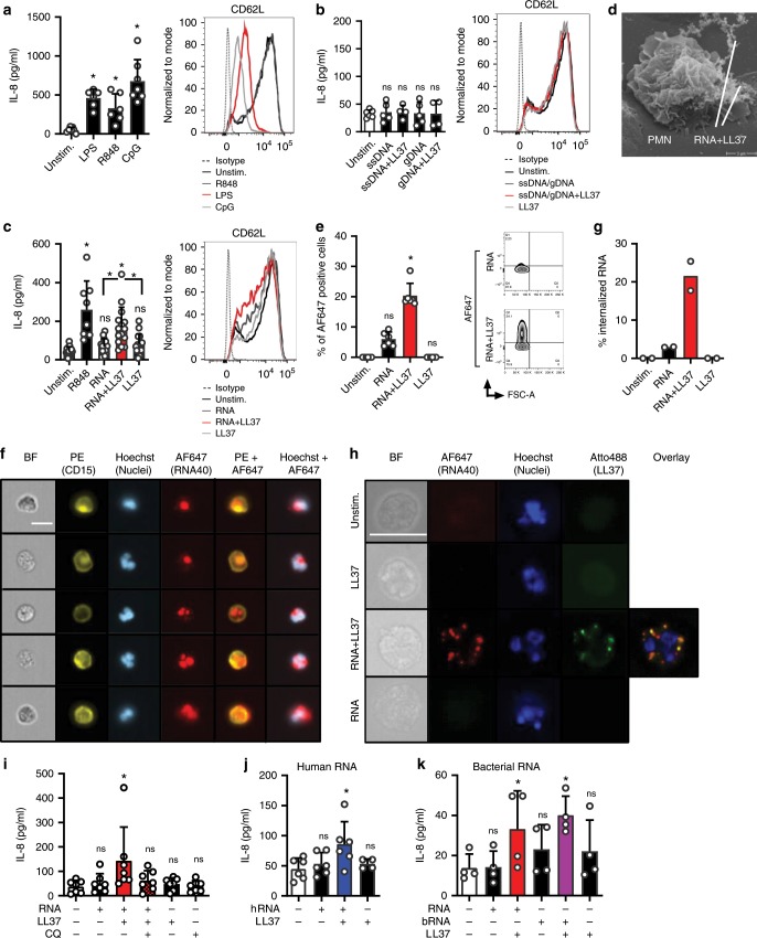
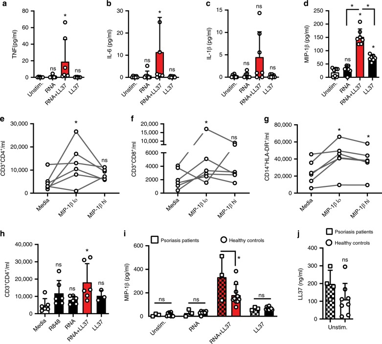
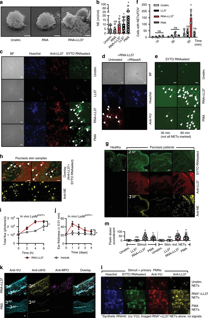
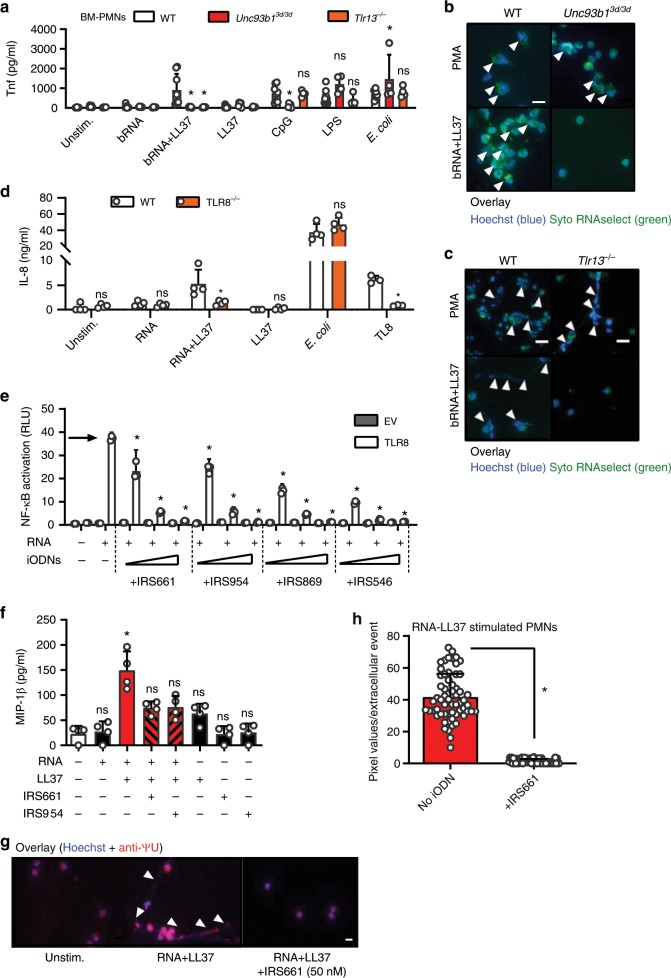
Psoriasis is an inflammatory skin disease with strong neutrophil (PMN) infiltration and high levels of the antimicrobial peptide, LL37. LL37 in complex with DNA and RNA is thought to initiate disease exacerbation via plasmacytoid dendritic cells. However, the source of nucleic acids supposed to start this initial inflammatory event remains unknown. We show here that primary murine and human PMNs mount a fulminant and self-propagating neutrophil extracellular trap (NET) and cytokine response, but independently of the canonical NET component, DNA. Unexpectedly, RNA, which is abundant in NETs and psoriatic but not healthy skin, in complex with LL37 triggered TLR8/TLR13-mediated cytokine and NET release by PMNs in vitro and in vivo. Transfer of NETs to naive human PMNs prompts additional NET release, promoting further inflammation. Our study thus uncovers a self-propagating vicious cycle contributing to chronic inflammation in psoriasis, and NET-associated RNA (naRNA) as a physiologically relevant NET component.


Conflict of interest statement

The authors declare no competing interests.

Figures

Fig. 1
Fig. 1. Human PMNs take up and respond to RNA when complexed to LL37.
ac IL-8 release (ELISA, 4 h stimulation) and CD62L shedding (flow cytometry, 1 or 2 h) by PMNs from healthy donors stimulated with LPS, R848 or CpG ODN (a, n = 6–7), ssDNA and genomic DNA (b, n = 5), or RNA40 (c, n = 8–15), with or without LL37. d Electron microscopy of PMNs incubated with RNA-LL37 for 20 min (n = 2). eh FACS (e, n = 6), ImageStream cytometry images (f) and quantification (g, scale bar = 10 μm) or conventional bright-field microscopy (h, n = 2, scale bar = 10 μm) of PMNs incubated for 60 min with RNA-AF647 complexed with LL37 (n = 6). In (f) five selected cells from one representative donor are shown, in (g) the % of cells with ‘internalized’ features (n = 2, see Methods). In H unmodified LL37 was replaced with Atto488-labeled LL37, one representative donor shown. i as in (c) but including chloroquine (CQ) pre-incubation for 30 min (n = 6–7). j, k as in c but using total human RNA from HEK293T cells (j, n = 4–6) or bacterial RNA isolated from S. aureus (K, n = 4) with and without LL37. ac, e, f, ik represent combined data (mean + SD) from ‘n’ biological replicates (each dot represents one donor). In d, f, and h representatives of ‘n’ biological replicates (donors) is shown (mean + SD). *p < 0.05 according to one-way ANOVA with Dunnett’s correction (a, b, c, i, j) or Friedman test with Dunn’s correction (e, k). Source data is provided as a Source data file.
Fig. 2
Fig. 2. RNA-LL37 promote the release of cytokines and chemokines.
Cytometric bead array (ac) or ELISA (d) for TNF (a), IL-6 (b), IL-1β (c), or MIP-1β (d) secreted from PMNs stimulated for 4 h as indicated (n = 6). Flow cytometric cell counts of migrated CD4+ T cells (e, h), CD8+ T cells (f) and CD14+HLA-DR+ monocytes (g) quantified in transwell assays with total PBMCs in the upper and MIP-1β (30 and 150 pg/ml) (eg, n = 6–7) or R848, RNA with and without LL37 (h, n = 3–7) in the lower chamber. i ELISA of MIP-1β secreted from psoriasis PMNs (n = 3 patients, 10 healthy controls, squares, chequered bars) or PMNs from sex and age-matched healthy donors. Only responses to treatment compared. j as in I but ELISA for LL37 (n = 4 patients, 7 healthy donors). Panels (aj) represent combined data (mean + SD) from ‘n’ biological replicates (each dot represents one donor). *p < 0.05 according to Friedmann (a, b, e, f) or Kruskal–Wallis (c, h) test with Dunn’s correction, or one-way ANOVA with Dunnett’s (d, g, h) or Sidak (i) correction or Student’s t-test (j). Source data is provided as a Source data file.
Fig. 3
Fig. 3. RNA-LL37 trigger the release of NETs containing further RNA, DNA, and LL37.
a EM pictures from PMNs stimulated with RNA-LL37 (n = 2, scale bar = 2 µm). b Neutrophil elastase (NE) release from PMNs stimulated for 3 h (n = 8, each dot represents one donor). Fluorescence microscopy of fixed and Hoechst- anti-LL37- SYTO RNAselect- (c) and/or anti-ΨU-stained (d, with or without RNase A treatment) or live (e) PMNs stimulated as indicated (n = 6, scale bar = 10 µm or n = 4, scale bar = 20 µm). f Quantification of live-cell microscopy shown in Supplementary Movies 1–3. g, h Skin sections from healthy (g) or psoriasis- (g, h) affected skin (n = 12 patients and three healthy controls, scale bar = 20 µm). i, j RNA-LL37-induced PMN influx (i, n = 3 each, measured by GFP in vivo fluorescence) and ear swelling (j, n = 6 each, mm × 0.01) in LysMEGFP/+ mice. k Ear sections from RNA-LL37 and PBS injected mice stained for ΨU, citH3 and MPO (n = 6, scale bar = 20 µm). l PMNs stimulated with transferred NETs (n = 3). m Quantification of (l). AF = autofluorescence. (b), (f), (i), (j), and (m) represent combined data (mean + SD) from ‘n’ biological replicates. In (a), (ch) and (k, l) representative samples of ‘n’ replicates or donors are shown. Arrowheads indicate released RNA/NETs (c–e) or RNA-LL37 co-localization (h). *p < 0.05 according to one-way (b) or two-way ANOVA (f, i, j) with Dunnett’s correction or Kruskal–Wallis test with Dunn’s correction (m). Source data is provided as a Source data file.
Fig. 4
Fig. 4. RNA-LL37 effects on PMNs depend on TLR8 and are blocked by iODNs.
a Tnf release (ELISA, 5 h stimulation as indicated) by BM-PMNs from different mouse strains (n = 4–8 each). b, c Fluorescence microscopy of fixed and Hoechst and SYTO RNAselect-stained stimulated BM-PMNs from different mouse strains (n = 4 Tlr13−/−, n = 5 Unc93b13d/3d, n = 8 WT; scale bar = 10 µm or n = 4, scale bar = 20 µm). d IL-8 and TNF release (ELISA, 18 h stimulation as indicated) by WT and TLR8 CRISPR-edited BLaER1 cells (n = 7). e NF-κB dual luciferase reporter assay in HEK293T cells, transfected with NF-κB firefly luciferase reporter, Renilla control reporter and TLR8 plasmid and subsequently stimulated with RNA without (arrow) or with IRS661, IRS954, IRS869 and IRS546 (0.15–3.5 µM, n = 2 each). MIP-1β (f, n = 4) release from stimulated PMNs with or without IRS661 (1 nM) or IRS954 (50 nM) pre-incubation (30 min) quantified by ELISA. g Fluorescence microscopy of fixed and Hoechst and anti-ΨU-stained RNA-LL37-stimulated PMNs (n = 3, scale bar = 10 µm). h Quantification of (g). Panels (a), (d), (e), (f), and (h) represent combined data (mean + SD) from ‘n’ biological replicates (each dot represents one mouse or donor). In (b), (c), (e), and (g) one representative of ‘n’ replicates is shown (mean + SD of technical triplicates). *p < 0.05 according to two-way ANOVA with Dunnett correction for multiple testing (e, comparison against ‘no inhibitor’ condition, arrow), one-way ANOVA with Sidak correction (a, d) Friedmann test with Dunn correction (f) or Mann–Whitney test (h). Source data is provided as a Source data file.

References

    1. Griffiths CE, Barker JN. Pathogenesis and clinical features of psoriasis. Lancet. 2007;370:263–271. doi: 10.1016/S0140-6736(07)61128-3. - DOI - PubMed
    1. Eberle, F. C., Bruck, J., Holstein, J., Hirahara, K. & Ghoreschi, K. Recent advances in understanding psoriasis. F1000Res5 (2016). - PMC - PubMed
    1. Sen Bilge Bulbul, Rifaioglu Emine Nur, Ekiz Ozlem, Inan Mehmet Ugur, Sen Tugba, Sen Nihat. Neutrophil to lymphocyte ratio as a measure of systemic inflammation in psoriasis. Cutaneous and Ocular Toxicology. 2013;33(3):223–227. doi: 10.3109/15569527.2013.834498. - DOI - PubMed
    1. Schon MP, Broekaert SM, Erpenbeck L. Sexy again: the renaissance of neutrophils in psoriasis. Exp. Dermatol. 2017;26:305–311. doi: 10.1111/exd.13067. - DOI - PubMed
    1. Lande R, et al. Plasmacytoid dendritic cells sense self-DNA coupled with antimicrobial peptide. Nature. 2007;449:564–569. doi: 10.1038/nature06116. - DOI - PubMed

Publication types

MeSH terms