. 2020 Jan 8;12(1):502-517.
doi: 10.18632/aging.102638. Epub 2020 Jan 8.

CXCL9 chemokine promotes the progression of human pancreatic adenocarcinoma through STAT3-dependent cytotoxic T lymphocyte suppression

Affiliations

CXCL9 chemokine promotes the progression of human pancreatic adenocarcinoma through STAT3-dependent cytotoxic T lymphocyte suppression

Hui-Feng Gao et al. Aging (Albany NY). .

Abstract

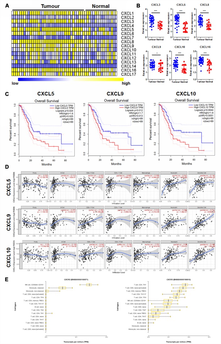
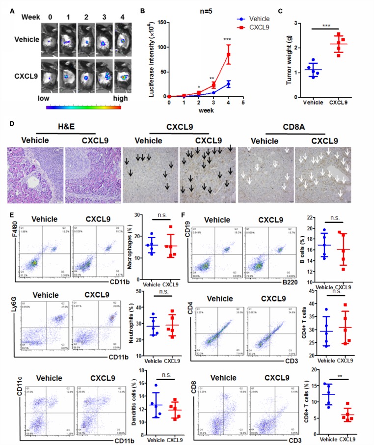
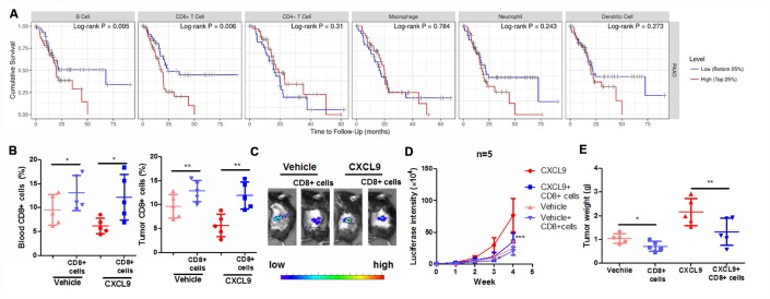
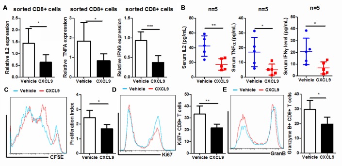
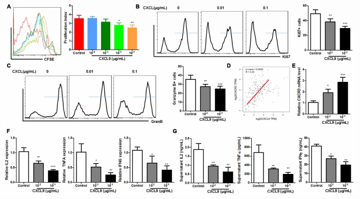
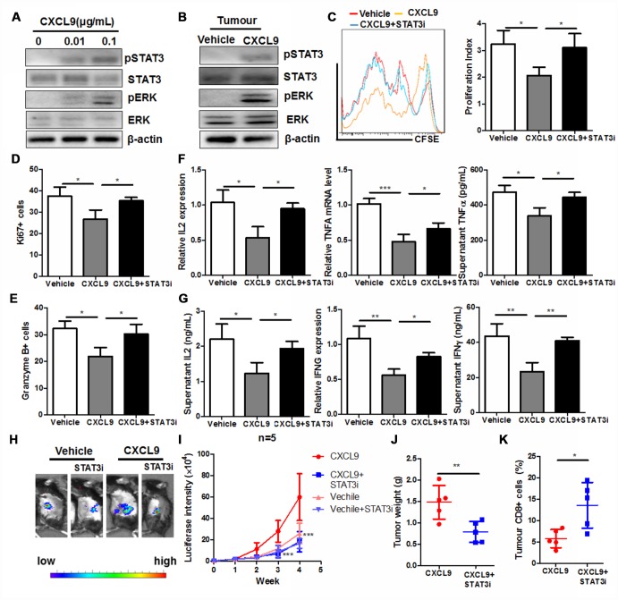
Chemokines play essential roles in the progression of various human cancers; however, the expression and role of CXC chemokines in pancreatic adenocarcinoma (PAAD) have not yet been identified. The aim of this study is to identify the expression patterns, clinical significance and mechanisms of CXC chemokines in regulating tumour microenvironment of PAAD. Three CXC chemokines, including CXCL5, CXCL9, and CXCL10, were significantly overexpressed in PAAD tissues, which were correlated with the poor survival of the patients. CXCL9/10 was associated with change of immune cell pattern in the tumour microenvironment, and supplementation of CXCL9 in the orthotopic murine PAAD model promoted tumour progression. In particular, CXCL9 reduced the CD8+ cytotoxic T lymphocytes in the tumour microenvironment of PAAD, which could be attributed to the reduced CD8+ T cell proliferation, activation, and secretion of anti-tumour cytokines. In vitro treatment of CXCL9 directly led to the suppression of the proliferation, activation, and secretion of anti-tumour cytokines of isolated CD8+ T cells. Inhibition of STAT3 recovered the CXCL9-inhibited proliferation, activation, and secretion of anti-tumour cytokines of CD8+ T cells. Our study indicates CXCL9 as a potential target of immunotherapy in PAAD treatment by regulating the CD8+ T lymphocytes in the tumour microenvironment.

Keywords: CD8+ cytotoxic T cells; CXCL9; STAT3 activation; chemokine; pancreatic adenocarcinoma.


Conflict of interest statement

CONFLICTS OF INTEREST: The authors declare no conflicts of interest.

Figures

Figure 1
Figure 1
CXC chemokines expression was correlated with prognosis and immune cell patterns of PAAD. (A) showed the Heatmap expression patterns of CXC chemokines in PAAD extracted from the GEO database (GDS4102); (B) showed CXC chemokines with significant changes in expression in PAAD; (C) showed CXC chemokines whose expression was correlated with overall survival of PAAD patients; (D) showed the correlation of selected CXC chemokines with the immune cell patterns in the tumour microenvironment; (E) showed the expression pattern of CXC chemokine receptors, CXCR2, and CXCR5, in different types of immune cells. *p<0.05, ***p<0.001 when compared.
Figure 2
Figure 2
CXCL9 promoted tumour progression in murine orthotopic PAAD model. (A) showed CXCL9 accelerated tumour growth of orthotopic murine PAAD in mice; (B) showed the increased luciferin signals detected in mice was speeded up by CXCL9 treatment; (C) showed CXCL9 treatment increased tumour weight of orthotopic murine PAAD; (D) showed that CXCL9 treatment led to more aggressive pattern of tumour cells in orthotopic murine PAAD; immunohistological analysis confirmed that CXCL9 treatment could increase the intratumoral CXCL9 level (black arrow)while reduce the CD8+ cytotoxic T cells (white arrow); (E) showed that CXCL9 treatment had minimal effect on the pattern of innate immune cells in the tumour microenvironment; (F) showed that CXCL9 treatment significantly reduced the CD8+ cytotoxic T cells without affecting other adaptive immune cells in the tumour microenvironment of PAAD. *p<0.05, **p<0.01, ***p<0.001 when compared with control.
Figure 3
Figure 3
Suppression of CD8+ cytotoxic T cells was responsible for the tumour-promoting effect of CXCL9. (A) showed the correlation between survival of PAAD with the pattern expression of different immune cells; (B) showed that the adoptive transfer of CD8+ cytotoxic T cells successfully increased the peripheral and intratumoral CD8+ cytotoxic T cells in orthotopic murine PAAD model; (C) showed that adoptive T cells transfer suppressed the tumour progression induced by CXCL9; (D) showed that the increased luciferin signals by CXCL9 were inhibited by adoptive transfer of CD8+ T cells; (E) showed that adoptive transfer of CD8+ cytotoxic T cells reduces tumour weight of PAAD in CXCL9-treated mice. *p<0.05, **p<0.01, ***p<0.001 when compared with CXCL9 group.
Figure 4
Figure 4
CXCL9 suppressed in vivo proliferation and activation of CD8+ cytotoxic T cells. (A) showed in vivo CXCL9 treatment suppressed mRNA expression of anti-tumour cytokines IL2, TNFα, and IFNγ in CD8+ cytotoxic T cells; (B) showed that in vivo CXCL9 treatment suppressed serum level of anti-tumour cytokines IL2, TNFα, and IFNγ in orthotopic murine PAAD mice; (C) showed that in vivo, CXCL9 treatment significantly retarded the proliferation of CD8+ cytotoxic T cells; (D) showed that in vivo CXCL9 treatment significantly repressed expression of proliferation marker Ki67; (E) showed that in vivo CXCL9 treatment significantly repressed expression of activation marker Granzyme B. *p<0.05, **p<0.01, ***p<0.001 when compared with control.
Figure 5
Figure 5
CXCL9 directly inhibited in vitro activation of CD8+ T cells. (A) showed that in vitro CXCL9 treatment dose-dependently repress division of activated CD8+ T cells; (B) showed that in vitro CXCL9 treatment significantly repressed expression of proliferation marker Ki67; (C) showed that in vitro CXCL9 treatment significantly repressed expression of activation marker Granzyme B; (D) showed a positive correlation between CXCL9 and CXCR3 expression in PAAD tissue; (E) showed that CXCL9 treatment significantly induced CXCR9 expression in CD8+ cytotoxic T cells; (F) showed that in vitro CXCL9 significantly suppressed the mRNA expression of anti-tumour cytokines IL2, TNFα and IFNγ in CD8+ cytotoxic T cells; (G) showed that in vitro CXCL9 treatment suppressed the secretion of anti-tumour cytokines IL2, TNFα and IFNγ in CD8+ cytotoxic T cells. *p<0.05, **p<0.01, ***p<0.001 when compared with control.
Figure 6
Figure 6
STAT3 activation was responsible for the CXCL9-induced suppression of CD8+ T cell activation. (A) showed that CXCL9 dose-dependently induces activation of STAT3 and Ras signalling in CD8+ cytotoxic T cells; (B) showed that CXCL9 treatment could induce STAT3 activity in PAAD tumour; (C) showed that inhibition of STAT3 by selective inhibitor recovered CXCL9-suppressed division of CD8+ cytotoxic T cells; (D) inhibition of STAT3 by selective inhibitor attenuated the suppression of Ki67 expression in CXCL9-treated CD8+ cytotoxic T cells; (E) inhibition of STAT3 by selective inhibitor attenuated the suppression of Granzyme B expression in CXCL9-treated CD8+ cytotoxic T cells; (F) inhibition of STAT3 by selective inhibitor recovered the mRNA expression of anti-tumour cytokines IL2, TNFα and IFNγ in CD8+ cytotoxic T cells; (G) showed that inhibition of STAT3 by selective inhibitor recovered the secretion of anti-tumour cytokines IL2, TNFα and IFNγ in CD8+ cytotoxic T cells. *p<0.05, **p<0.01, ***p<0.001 when compared with CXCL9 treatment only. (H) showed that presence of STAT3 selective inhibitor could signficantly reduce the tumour burden in mice; (I) showed that tumour growth induced by CXCL9 was abolished by STAT3 selective inhibitor; (J) showed that enlarged tumour weight induced by CXCL9 was abolished by STAT3 selective inhibitor; (K) showed that presence of STAT3 selective inhibitor could recover the CD8+ population in PAAD tumour.

References

    1. Hidalgo M. Pancreatic cancer. N Engl J Med. 2010; 362:1605–17. 10.1056/NEJMra0901557 - DOI - PubMed
    1. Lombardi L, Troiano M, Silvestris N, Nanni L, Latiano TP, Di Maggio G, Cinieri S, Di Sebastiano P, Colucci G, Maiello E. Combined modality treatments in pancreatic cancer. Expert Opin Ther Targets. 2012. (Suppl 2); 16:S71–81. 10.1517/14728222.2012.662959 - DOI - PubMed
    1. O’Reilly EM. Refinement of adjuvant therapy for pancreatic cancer. JAMA. 2010; 304:1124–25. 10.1001/jama.2010.1302 - DOI - PubMed
    1. Sun XJ, Jiang TH, Zhang XP, Mao AW. Role of the tumor microenvironment in pancreatic adenocarcinoma. Front Biosci. 2016; 21:31–41. 10.2741/4374 - DOI - PubMed
    1. Ademmer K, Ebert M, Müller-Ostermeyer F, Friess H, Büchler MW, Schubert W, Malfertheiner P. Effector T lymphocyte subsets in human pancreatic cancer: detection of CD8+CD18+ cells and CD8+CD103+ cells by multi-epitope imaging. Clin Exp Immunol. 1998; 112:21–26. 10.1046/j.1365-2249.1998.00546.x - DOI - PMC - PubMed

Publication types

MeSH terms