. 2020 Feb 14;295(7):2113-2124.
doi: 10.1074/jbc.RA119.011752. Epub 2019 Dec 30.

Crystal structures and biochemical analyses of the bacterial arginine dihydrolase ArgZ suggests a "bond rotation" catalytic mechanism

Affiliations

Crystal structures and biochemical analyses of the bacterial arginine dihydrolase ArgZ suggests a "bond rotation" catalytic mechanism

Ningning Zhuang et al. J Biol Chem. .

Abstract

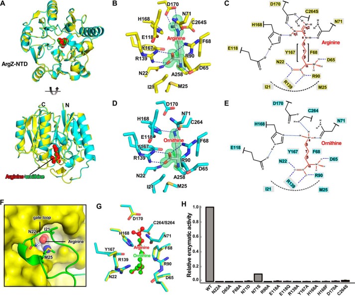
A recently discovered ornithine-ammonia cycle (OAC) serves as a conduit in the nitrogen storage and remobilization machinery in cyanobacteria. The OAC involves an arginine catabolic reaction catalyzed by the arginine dihydrolase ArgZ whose catalytic mechanism is unknown. Here we determined the crystal structures at 1.2-3.0 Å of unliganded ArgZ from the cyanobacterium Synechocystis sp. PCC6803 and of ArgZ complexed with its substrate arginine, a covalently linked reaction intermediate, or the reaction product ornithine. The structures reveal that a key residue, Asn71, in the ArgZ active center functions as the determinant distinguishing ArgZ from other members of the guanidino group-modifying enzyme superfamily. The structures, along with biochemical evidence from enzymatic assays coupled with electrospray ionization MS techniques, further suggest that ArgZ-catalyzed conversion of arginine to ornithine, ammonia, and carbon dioxide consists of two successive cycles of amine hydrolysis. Finally, we show that arginine dihydrolases are broadly distributed among bacteria and metazoans, suggesting that the OAC may be frequently used for redistribution of nitrogen from arginine catabolism or nitrogen fixation.

Keywords: arginine; arginine dihydrolase; arginine metabolism; cyanobacteria; enzyme catalysis; enzyme mechanism; enzyme structure; hydrolase; nitrogen metabolism; ornithine–ammonia cycle.


Conflict of interest statement

The authors declare that they have no conflicts of interest with the contents of this article

Figures

Figure 1.
Figure 1.
Overall structure of full-length ArgZ. A, diagram of enzymatic reactions by ArgZ. B, schematic of ArgZ domains. C, overall structure of ArgZ. The four protomers are shown in different colors; the N-terminal domains (NTD) are shown as in ribbon; and the middle and C-terminal domains (CTD) are shown as in surface. D, the size-exclusion chromatography profile indicates that ArgZ is a tetramer in solution. The calculated mass of the ArgZ tetramer is 313 kDa, and the molecular mass markers used are thyroglobin (669 kDa), apoferritin (443 kDa), β-amylase (200 kDa), alcohol dehydrogenase (150 kDa), albumin (66 kDa), and carbonic anhydrolase (29 kDa). AU, absorption unit. E, the structure of one ArgZ protomer. The dashed line indicates 5-fold pseudosymmetry of the N-terminal AT domain. The colors are as in B.
Figure 2.
Figure 2.
Structure of ArgZ-N. A, side and top views of the superimposition of crystal structures of ArgZ(C264S)-N–arginine (yellow) and ArgZ-N–ornithine (cyan). Arginine (red) and ornithine (green) in the active site are shown as spheres. B, detailed interactions between the substrate arginine and the active center of ArgZ. The carbon atoms of the protein and the substrate arginine are shown in yellow and white, respectively. The N and O atoms are shown in blue and red, respectively. C, schematic of the interactions in B. D, detailed interactions between the product ornithine and the active center of ArgZ. The carbon atoms of the protein are shown in cyan and the rest of colors are as in B. E, schematic of the interactions in D. F, a gate loop (green) covers the active center (yellow surface) and stabilizes the substrate (spheres). G, comparison between substrate and product in the active center of ArgZ Atoms are colored as above. H, enzymatic activities of ArgZ derivatives, determined by GDH-coupled reactions. The experiments were repeated three times, and the results are presented as mean ± S.E. H-bonds and salt bridges are shown as blue dashes and van der Waals interactions are shown in dashed black arcs. Green mesh in B and D represents a simulated Fo-Fc omit polder map contoured at 6.0 σ.
Figure 3.
Figure 3.
Proposed reaction mechanism of ArgZ. A, the active center of substrate-bound ArgZ(C264S). B, the active center of substrate-bound Pseudomonas aeruginosa ADI(C406A) (PDB code 2A9G). C, the active center of Mycoplasma arginini ADI (PDB code 1S9R) with a covalently bound intermediate II. D, the active center of substrate-bound E. coli AstB(C365S) (PDB code 1YNI). The carbon atoms of ArgZ, ADI, and AstB are shown in yellow, green, and light blue, respectively. The N and O atoms are shown in blue and red, respectively. E, the proposed reaction steps of the ArgZ-catalyzed two successive cycles of hydrolysis. The proposed side reaction producing L-citrulline is shown in a pink box. In the first cycle of hydrolysis, deprotonated Cys264 attacks and subsequently covalently links the Cζ of arginine, resulting in a tetrahedral intermediate I. Subsequently, the η1 amine group of the guanidium moiety attracts a proton back from imidazolinium of His168, resulting in collapse of the Nη1-Cζ bond to form intermediate II and release one molecule of ammonia. 180° rotation of the Nϵ-Cζ bond brings the η2 amine group close to His168, forming intermediated II*, and a water molecule deprotonated by the His-Glu pair attacks the Cζ of intermediate II* to form the tetrahedral intermediate III. In the second cycle of hydrolysis, His168 donates a proton to the η2 amine group and releases the second molecule of ammonia. A second water diffuses into the active center, where it is deprotonated by His168. The activated water molecule subsequently attacks Cζ of the covalently linked intermediate IV and forms a tetrahedral intermediate V. In the last step, the final products (CO2 and ornithine) are released, and Cys264 is freed through an unknown mechanism.
Figure 4.
Figure 4.
N71 is the determinant of the dihydrolase activity of ArgZ. A, the deconvoluted ESI-MS spectra of trapped reaction intermediates using WT ArgZ. Top panel, the spectra of ArgZ itself. Bottom panel, the spectra after a 5-min reaction. The calculated mass for unmodified ArgZ-N is 34,399 Da. The expected mass of ArgZ-N with covalently linked intermediate II is 34,557 (red peak with an expected excess mass of 158 Da). The peaks marked with an asterisk are attributable to a smaller fraction of nonenzymatically α-N-gluconoylated protein modified during expression with an expected excess mass of 178. B, the deconvoluted ESI-MS spectra of trapped reaction intermediates using ArgZ-N(N71S). The calculated mass for unmodified ArgZ-N(N71S) is 34,372 Da, and the expected mass of ArgZ-N(N71S) with covalently linked intermediate II is 34,530 (red peak with an expected excess mass of 158 Da). Representative ESI-MS plots are shown and typically have an error of 0.5 Da. C, the HPLC spectra show reaction products of WT or derivatives of ArgZ. The inset shows the m/z values of the products citrulline (Cit) and ornithine (Orn), detected by MS. D, the active center of the crystal structure of ArgZ-N(N71S)–intermediate II. The carbon atoms of protein and the substrate arginine are shown in light blue and white, respectively, and the N and O atoms are shown in blue and red, respectively. Green mesh represents a simulated Fo-Fc omit polder map contoured at 6.0. E, schematic of the active center in D.
Figure 5.
Figure 5.
Broad distribution of arginine dihydrolase in three domains of life. A, the phylogenetic tree of ArgZ homologs in bacteria, eukaryotes, and archaea. Red pentagons label enzymes for subsequent experimental characterization. Blue pentagons label annotated enzymes in the literature. The protein codes are NCBI reference sequence IDs. B, the sequence alignment shows that the top group contains an Asn and the bottom group contains an Asp at the corresponding position of Asn71 of Synechosistis PCC 6803 ArgZ. C, the sequence alignment of proteins from the top group in A. Red stars label catalytic residues. Blue circles label residues making polar interactions with the substrates. Black circles label residues making hydrophobic interactions with the substrate. The gray box indicates the determinant residue Asn71.

Comment in

References

    1. Cunin R., Glansdorff N., Piérard A., and Stalon V. (1986) Biosynthesis and metabolism of arginine in bacteria. Microbiol. Rev. 50, 314–352 - PMC - PubMed
    1. Morris S. M., Jr. (2007) Arginine metabolism: boundaries of our knowledge. J. Nutr. 137, 1602S–1609S 10.1093/jn/137.6.1602S - DOI - PubMed
    1. Mommsen T. P., and Walsh P. J. (1989) Evolution of urea synthesis in vertebrates: the piscine connection. Science 243, 72–75 10.1126/science.2563172 - DOI - PubMed
    1. Winter G., Todd C. D., Trovato M., Forlani G., and Funck D. (2015) Physiological implications of arginine metabolism in plants. Front. Plant Sci. 6, 534 - PMC - PubMed
    1. Simon J. P., Wargnies B., and Stalon V. (1982) Control of enzyme synthesis in the arginine deiminase pathway of Streptococcus faecalis. J. Bacteriol. 150, 1085–1090 10.1128/JB.150.3.1085-1090.1982 - DOI - PMC - PubMed

Publication types

LinkOut - more resources