

A case of Merkel cell carcinoma of the head and neck
- PMID: 31914497
- PMCID: PMC6949494
- DOI: 10.7181/acfs.2019.00542
A case of Merkel cell carcinoma of the head and neck
Abstract
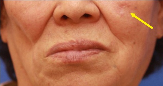
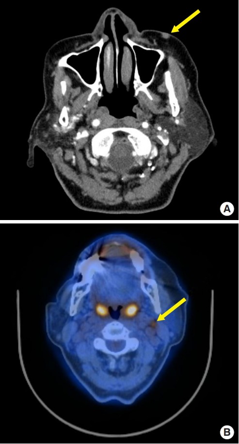
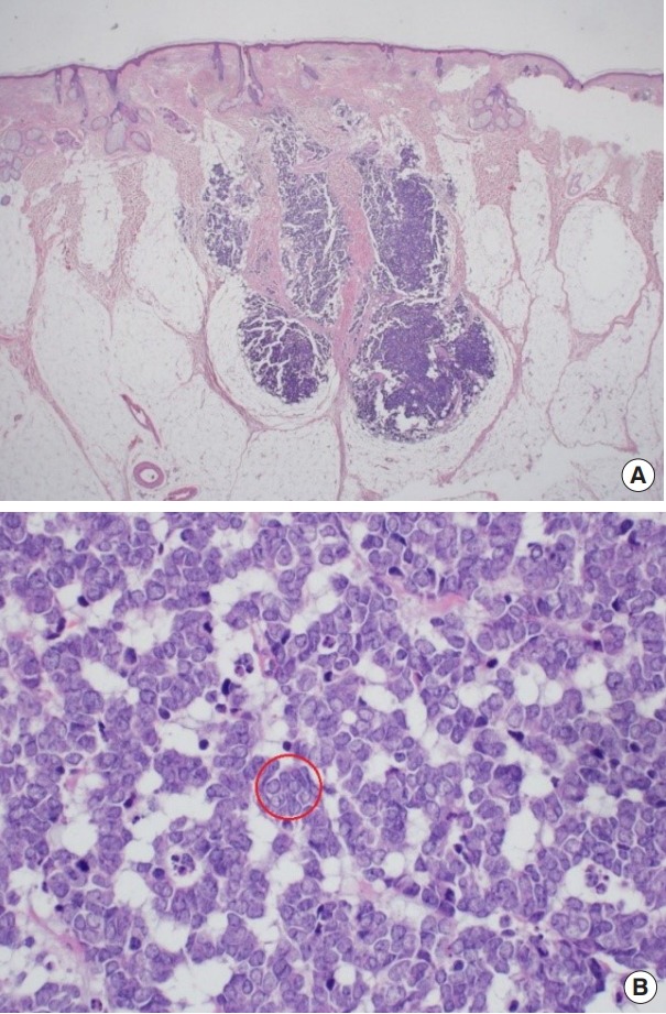
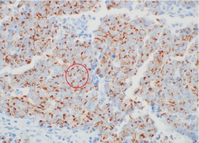
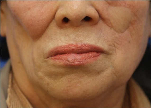
Merkel cell carcinoma (MCC) is a relatively rare and aggressive cutaneous neuroendocrine malignancy. It is characterized by high rates of recurrence and metastasis, both to regional lymph nodes and to distant locations. Its characteristic clinical manifestation is a single, painless, hard, erythematous nodule on a sun-exposed area, particularly in older men. Surgical management of both the primary site and the sentinel lymph node is the standard of care. In this article, we describe the diagnosis and treatment of a case of MCC in the left cheek.
Keywords: Carcinoma; Merkel cells; Skin neoplasm.
Conflict of interest statement
No potential conflict of interest relevant to this article was reported.
Figures
References
-
- Toker C. Trabecular carcinoma of the skin. Arch Dermatol. 1972;105:107–10. - PubMed
-
- Lemos B, Nghiem P. Merkel cell carcinoma: more deaths but still no pathway to blame. J Invest Dermatol. 2007;127:2100–3. - PubMed
-
- Agelli M, Clegg LX, Becker JC, Rollison DE. The etiology and epidemiology of Merkel cell carcinoma. Curr Probl Cancer. 2010;34:14–37. - PubMed
-
- Fitzgerald TL, Dennis S, Kachare SD, Vohra NA, Wong JH, Zervos EE. Dramatic increase in the incidence and mortality from Merkel cell carcinoma in the United States. Am Surg. 2015;81:802–6. - PubMed
-
- Agelli M, Clegg LX. Epidemiology of primary Merkel cell carcinoma in the United States. J Am Acad Dermatol. 2003;49:832–41. - PubMed
Publication types
LinkOut - more resources
Full Text Sources
