. 2020 Jan 8;18(1):12.
doi: 10.1186/s12967-019-02203-z.

Exosomes overexpressing miR-34c inhibit malignant behavior and reverse the radioresistance of nasopharyngeal carcinoma

Affiliations

Exosomes overexpressing miR-34c inhibit malignant behavior and reverse the radioresistance of nasopharyngeal carcinoma

Fang-Zhu Wan et al. J Transl Med. .

Abstract

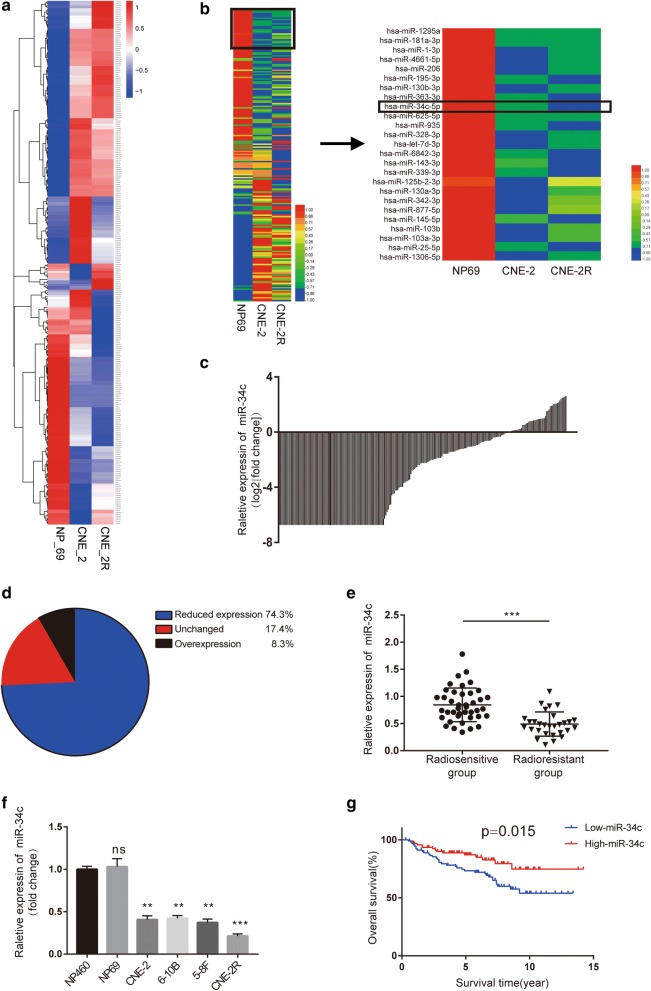
Background: Malignant behavior and radioresistance, which severely limits the efficacy of radiation therapy (RT) in nasopharyngeal carcinoma (NPC), are associated with tumor progression and poor prognosis. Mesenchymal stem cells (MSCs) are used as a therapeutic tool in a variety of tumors. The aim of this study was to reveal the effect of tumor suppressor microRNA-34c-5p (miR-34c) on NPC development and radioresistance, as well as to confirm that exosomes derived from MSCs overexpressing miR-34c restore the sensitivity to radiotherapy in NPCs.

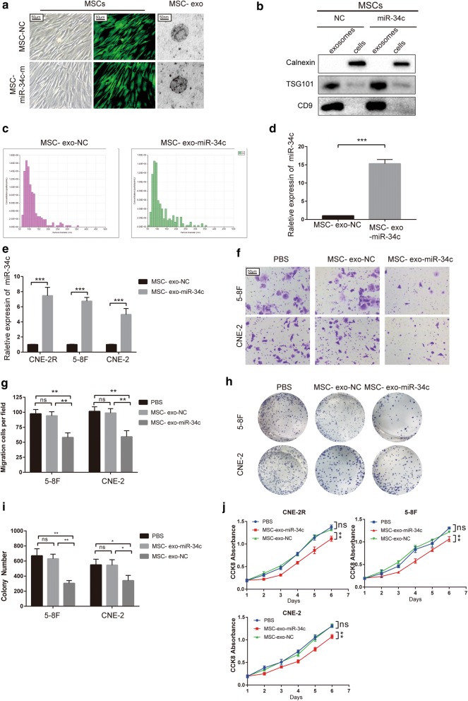
Methods: Potentially active microRNAs were screened by cell sequencing, Gene Expression Omnibus (GEO) database analysis, and analysis of clinical serum samples from 70 patients. The expression of genes and proteins was detected by Western blotting, quantitative reverse transcription PCR (qRT-PCR), and immunohistochemistry (IHC). Proliferation, apoptosis, invasion, migration and radioresistance of NPC were detected. Luciferase reporter assays were used to verify the interactions of microRNAs with their downstream targets. MSCs exosomes were isolated by ultrafiltration and verified by electron microscopy and nanoparticle tracking technology.

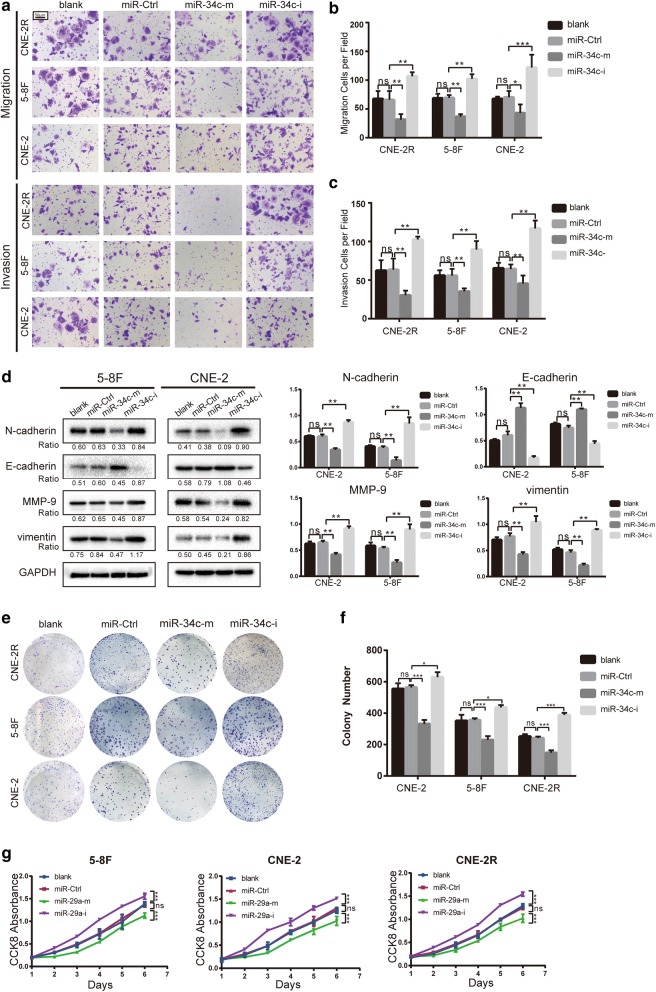
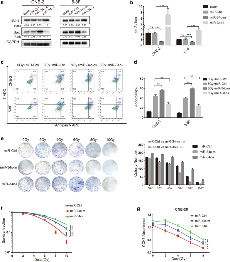
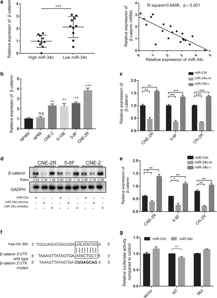
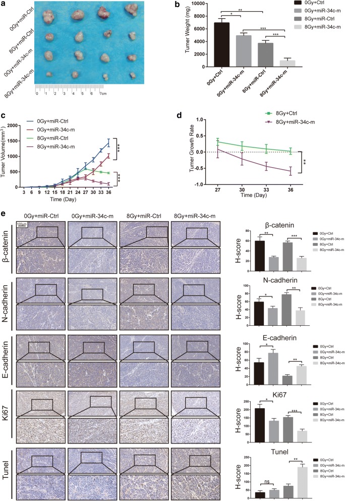
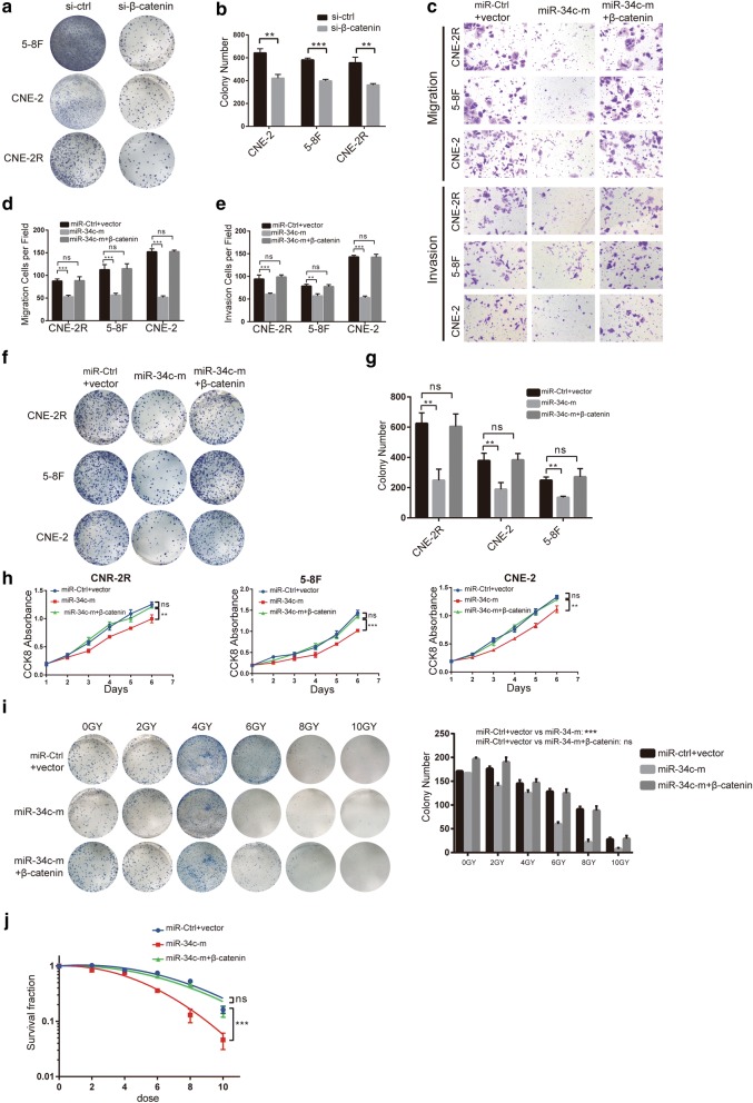
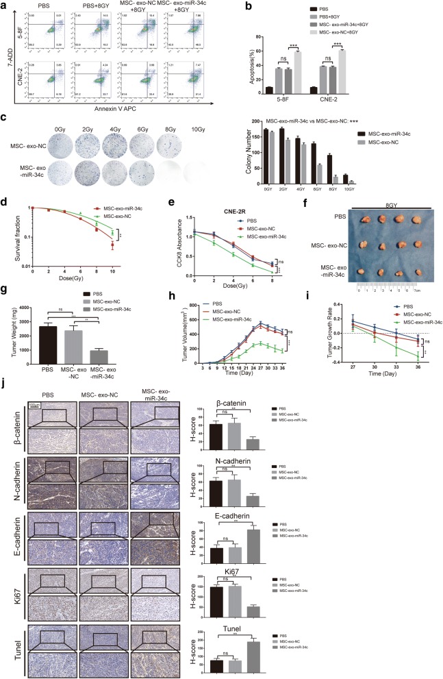
Results: The expression of miR-34c was associated with the occurrence and radiation resistance of NPC. In vitro and in vivo experiments indicated that overexpression of miR-34c inhibit malignant behavior such as invasion, migration, proliferation and epithelial-mesenchymal transition (EMT) in NPCs by targeting β-Catenin. In addition, we found alleviated radioresistance upon miR-34c overexpression or β-catenin knockdown in NPCs. Exosomes derived from miR-34c-transfected MSCs attenuated NPC invasion, migration, proliferation and EMT. Moreover, miR-34c-overexpressing exosomes drastically increased radiation-induced apoptosis in NPC cells.

Conclusion: miR-34c is a tumor suppressor miR in NPC, which inhibits malignant behavior as well as radioresistance of tumor. Therefore, exogenous delivery of miR-34c to NPCs via MSC exosomes inhibits tumor progression and increases the efficiency of RT. Combination IR with miR-34c-overexpressing exosomes may be effective treatment for radioresistant NPCs.

Keywords: EMT; Nasopharyngeal carcinoma; Radioresistance; microRNA-34c; β-Catenin.


Conflict of interest statement

The authors declare that they have no competing interests.

Figures

Fig. 1
Fig. 1
Expression of miR-34c is downregulated in NPC cells and tissues and associated with radioresistance in patients with NPC. a, b Heatmap displaying miRNA expression in NP69, CNE-2, and CNE-2R cells; miR-34c was selected for further study. c Waterfall plot showing the relative expression of miR-34c in NPC tissues (NCBI/GEO/GSE70970). d Pie chart displaying the miR-34c levels in NPC tissues; a fold change > 2 or < 1/2 in relative miR-34c expression was defined as significant. e The miR-34c levels in tissues from radioresistant patients (n = 29) and radiosensitive patients (n = 41) were measured by qRT-PCR. f The miR-34c levels in NP-460, NP69, CNE-2, 6–10B, 5–8F and CNE-2R cells were measured by qRT-PCR. g Kaplan–Meier curve for overall survival in the cohort of 185 NPC patients (NCBI/GEO/GSE70970). Patients were divided into two groups based on miR-34c level in each sample, and the median ratio value was chosen as the cutoff point
Fig. 2
Fig. 2
Overexpression of miR-34c inhibits migration, invasion and proliferation and limits EMT in NPC cells. CNE-2, 5–8F and CNE-2R cells were transfected with miR-34c control (miR-Ctrl), mimics (miR-34c-m) or inhibitors (miR-34c-i). a Cell migration was assessed using Transwell migration and invasion assays (10 times magnification). b, c The number of cells per field in a migration and invasion assay. d Western blot analysis of EMT-related markers in CNE-2 and 5–8F cells transfected with lentivirus from each group. Gray value shows in the right panel. e Photos of colonies of NPC cells in colony formation assays. f The number of colonies in different groups. g Cell proliferation in each group of CNE-2, 5–8F and CNE-2R cells was measured by CCK8 assay (the results were reproduced in three independent experiments.*P < 0.05; **P < 0.01; ***P < 0.001; ns: no statistical significance)
Fig. 3
Fig. 3
Overexpression of miR-34c promotes apoptosis and limits the radioresistance of NPC cells. a Western blot analysis of the apoptosis markers Bcl-2 and bax in CNE-2 and 5–8F cells. b Bar graph showing the ratio of Bcl-2/bax in each group. c The effect of miR-34c on cell apoptosis was analyzed by flow cytometry with APC/7-AAD staining. d The proportion of apoptotic cells. e Colony formation assays assessing the radiosensitivity of each group and the qualified number of colonies in each group. f Curves showing radio resistance were generated and fit with an LQ model. g The effects of miR-34c on the survival of CNE-2R after radiation was determined by CCK-8 assay (the results were reproduced in three independent experiments. *P < 0.05; **P < 0.01; ***P < 0.001; ns: no statistical significance)
Fig. 4
Fig. 4
β-Catenin is a direct target of miR-34c. a Correlation between β-catenin mRNA and miR-34c levels in the serum of 20 NPC patients measured by qRT-PCR; expression was normalized to the mean level in three normal serum samples. b Expression of β-catenin in different NPC cells determined by qRT-PCR. c β-catenin was measured by qRT-PCR in miR-34c-transfected NPC cells. d After 48 h of transfecting miR-34c, β-catenin protein expression was detected by Western blot analysis. e β-Catenin expression level determined by qRT-PCR 48 h after transfection with miR-34c. f Graphical representation indicating miR-34c interaction sites within the 3′-UTR of β-catenin mRNA with boxes. Mutations were generated in the β-catenin 3′-UTR site complementary to the seed sequences of miR-34c. g The β-catenin 3′-UTR with a putative wild-type (WT) miR-34c-binding site or a binding site mutated at the 3′-UTR region (Mut) were cloned downstream of the CMV promoter in the pmiR-REPORT vector. Luciferase activity was measured after cotransfection of the reporter constructs with WT- or Mut-interacting sites or miR-Ctrl in HEK293T cells (data are presented as the mean ± SD of three independent experiments. *P < 0.05; **P < 0.01; ***P < 0.001; ns no statistical significance)
Fig. 5
Fig. 5
Effect of miR-34c overexpression on migration, invasion, proliferation and radioresistance in vivo. a CNE-2R cells stably overexpressing miR-34c or control were injected subcutaneously in nude mice. The mice were treated with RT or not at day 24 (n = 4 in each group). b, c The tumor volumes were monitored every 3 days, and after sacrifice, tumor weights were measured. d Graph of tumor growth rate. e Left panel: Representative images of β-catenin, Ki67, E-cadherin, and vimentin immunohistochemistry (IHC) staining at 20-fold and 40-fold magnification a nd TUNEL assay of tumor samples from each group. Right panel: H-score of each group
Fig. 6
Fig. 6
β-catenin mediates miR-34c-induced inhibition of EMT, proliferation and radioresistance in vitro. a, b Colony assays showing the effect of β-catenin on NPCs proliferation. c Cell movement ability was determined by Transwell assays (10 times magnification). d, e The number of cells per fields in a migration and invasion assay. f Photos of colonies of CNE-2, 5–8F and 6–10B cells in colony formation assays. g Bar graph showing the number of colonies in different groups. h Cell viability in each group of NPC cells was analyzed by CCK8 assay. i Colony formation assays assessing the radiosensitivity the qualified number of colonies of each group. j Curves showing the radiosensitivity of each group were created and fit with an LQ model. The results are presented as the mean ± SD (n = 3) (*P < 0.05; **P < 0.01; ***P < 0.001; ns: no statistical significance)
Fig. 7
Fig. 7
Exosomes derived from MSCs overexpressing miR-34c attenuate malignant behavior in NPC. a Representative images of human MSCs transfected with miR-34c or NC nucleotide sequences and corresponding electron microscopic images of exosomes. b Western blot analysis showing the presence of TSG101 and CD9 and the absence of calnexin in MSC-derived exosomes. c Particle size distribution of exosomes measured by Nanosight. d PCR analysis of miR-34c levels in exosomes. e miR-34c expression in NPCs detected by qRT-PCR after treatment with PBS or miR-34c-overexpressing (MSC-exo-miR-34c) or control (MSC-exo-NC) exosomes. f Migration ability detected by Transwell assay. g The number of cells per field in migration and invasion assays. h Photos of colonies of each group in colony formation assays and i a bar graph showing the number of colonies in different groups. j Cell proliferation in each group was measured by CCK8 assay (the results were reproduced in three independent experiments.*P < 0.05; **P < 0.01; ***P < 0.001; ns: no statistical significance)
Fig. 8
Fig. 8
Exosomes derived from MSCs overexpressing miR-34c attenuate radioresistance of NPC in vivo and in vitro. a Cell apoptosis in each group was analyzed by flow cytometry with APC/7-AAD staining. b Bar graph showing the rate of apoptosis in cells. c Colony formation assays assessing the radiosensitivity and the qualified number of colonies in each group. d Curves showing radioresisitance were generated and fit with an LQ model. e CCK-8 assay assessing the survival of CNE-2R after radiation. f CNE-2R cells were treated with PBS, and exosomes were injected subcutaneously into nude mice (n = 4 in each group). The mice were treated with RT at day 24. h Tumor volumes were monitored every 3 days, and g after sacrifice, tumor weights were measured. i Tumor growth rate was calculated. j Left panel: Representative images showing the IHC staining of tumor samples from different groups for β-catenin, N-cadherin, E-cadherin, Ki67, and TUNEL assays at 20-fold and 40-fold magnification. Right panel: H-score of each group (*P < 0.05; **P < 0.01; ***P < 0.001; ns no statistical significance)

Similar articles

Cited by

References

    1. Shield KD, Ferlay J, Jemal A, Sankaranarayanan R, Chaturvedi AK, Bray F, Soerjomataram I. The global incidence of lip, oral cavity, and pharyngeal cancers by subsite in 2012. CA Cancer J Clin. 2017;67:51–64. doi: 10.3322/caac.21384. - DOI - PubMed
    1. Kwong D, Sham J, Choy D. The effect of loco-regional control on distant metastatic dissemination in carcinoma of the nasopharynx: an analysis of 1301 patients. Int J Radiat Oncol Biol Phys. 1994;30:1029–1036. doi: 10.1016/0360-3016(94)90306-9. - DOI - PubMed
    1. Fang FM, Tsai WL, Chien CY, Chen HC, Hsu HC, Huang TL, Lee TF, Huang HY, Lee CH. Pretreatment quality of life as a predictor of distant metastasis and survival for patients with nasopharyngeal carcinoma. J Clin Oncol. 2010;28:4384–4389. doi: 10.1200/JCO.2010.28.8324. - DOI - PubMed
    1. Floor S, Van Staveren WC, Larsimont D, Dumont JE, Maenhaut C. Cancer cells in epithelial-to-mesenchymal transition and tumor-propagating-cancer stem cells: distinct, overlapping or same populations. Oncogene. 2011;30:4609–4621. doi: 10.1038/onc.2011.184. - DOI - PubMed
    1. Tsai JH, Yang J. Epithelial–mesenchymal plasticity in carcinoma metastasis. Genes Dev. 2013;27:2192–2206. doi: 10.1101/gad.225334.113. - DOI - PMC - PubMed

Publication types

LinkOut - more resources