. 2020 Jan 8;13(1):9.
doi: 10.1186/s13071-020-3883-z.

Leishmania braziliensis prostaglandin F synthase impacts host infection

Affiliations

Leishmania braziliensis prostaglandin F synthase impacts host infection

Eliza Vanessa Carneiro Alves-Ferreira et al. Parasit Vectors. .

Abstract

Background: Prostaglandins (PG) are lipid mediators derived from arachidonic acid metabolism. They are involved in cellular processes such as inflammation and tissue homeostasis. PG production is not restricted to multicellular organisms. Trypanosomatids also synthesize several metabolites of arachidonic acid. Nevertheless, their biological role in these early-branching parasites and their role in host-parasite interaction are not well elucidated. Prostaglandin F synthase (PGF2S) has been observed in the Leishmania braziliensis secreted proteome and in L. donovani extracellular vesicles. Furthermore, we previously reported a positive correlation between L. braziliensis PGF2S (LbrPGF2S) expression and pathogenicity in mice.

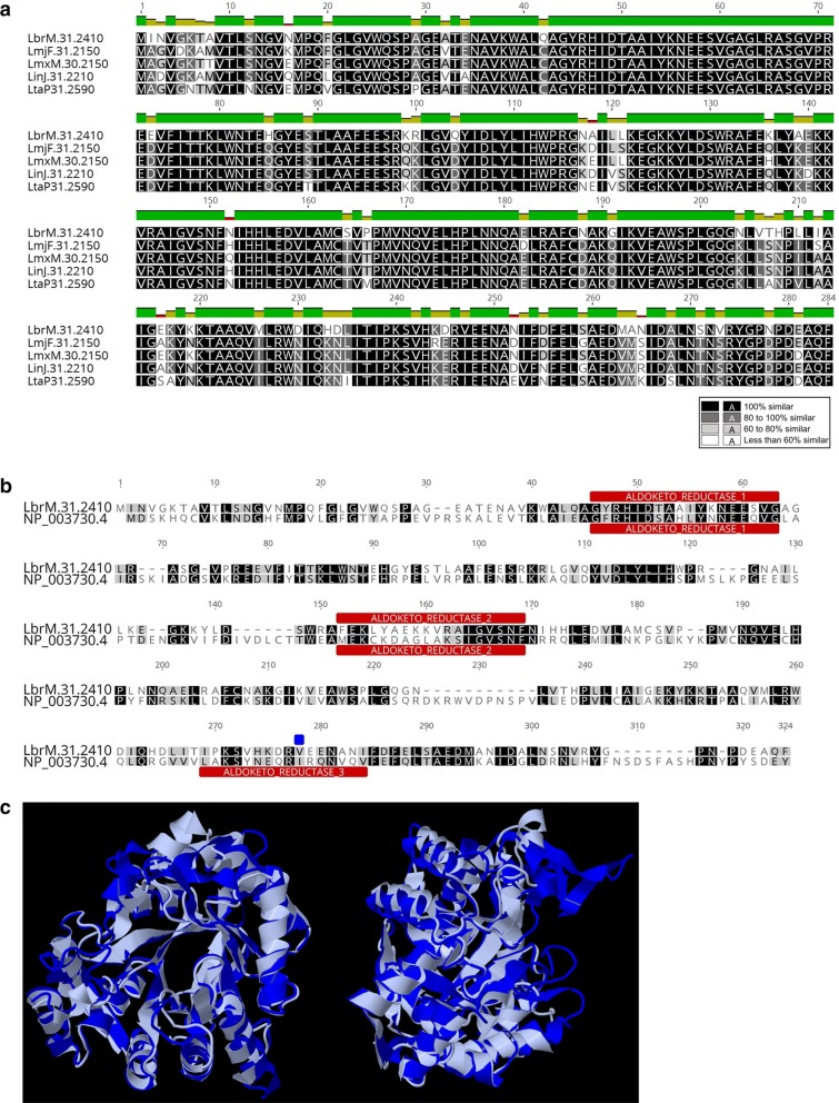
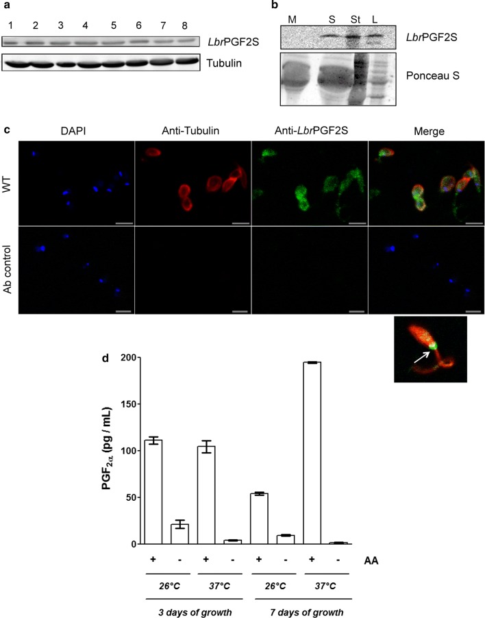
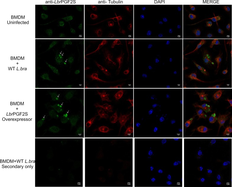
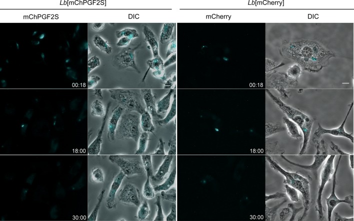
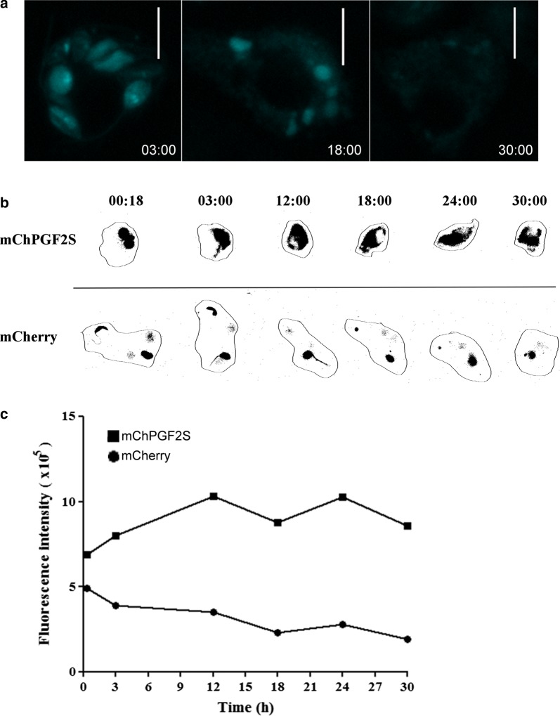
Methods: LbrPGF2S gene expression and PGF2α synthesis in promastigotes were detected and quantified by western blotting and EIA assay kit, respectively. To investigate LbrPGF2S localization in amastigotes during bone marrow-derived macrophage infection, parasites expressing mCherry-LbrPGF2S were generated and followed by time-lapse imaging for 48 h post-infection. PGF2S homolog sequences from Leishmania and humans were analyzed in silico using ClustalW on Geneious v6 and EMBOSS Needle.

Results: Leishmania braziliensis promastigotes synthesize prostaglandin F in the presence of arachidonic acid, with peak production in the stationary growth phase under heat stress. LbrPGF2S is a cytoplasmic protein enriched in the secretory site of the parasite cell body, the flagellar pocket. It is an enzyme constitutively expressed throughout promastigote development, but overexpression of LbrPGF2S leads to an increase of infectivity in vitro. The data suggest that LbrPGF2S may be released from intracellular amastigotes into the cytoplasm of bone marrow-derived macrophages over a 48-hour infection period, using time-lapse microscopy and mCherry-PGF2S (mChPGF2S)-expressing parasites.

Conclusions: LbrPGF2S, a parasite-derived protein, is targeted to the host cell cytoplasm. The putative transfer of this enzyme, involved in pro-inflammatory lipid mediator synthesis, to the host cell suggests a potential role in host-parasite interaction and may partially explain the increased pathogenicity associated with overexpression of LbrPGF2S in L. braziliensis. Our data provide valuable insights to help understand the importance of parasite-derived lipid mediators in pathogenesis.

Keywords: Host-parasite; LbrPGF2S; Leishmania braziliensis; Macrophage; PGF2α.


Conflict of interest statement

The authors declare that they have no competing interests.

Figures

Fig. 1
Fig. 1
Comparative analysis of the LbrPGF2S amino acid sequence. a Multiple sequence alignment of putative PGF2S proteins from L. braziliensis (LbrM.31.2410), L. major (LmjF.31.2150), L. mexicana (LmxM.30.2150), L. infantum (LinJ.31.2210) and L. tarentolae (LtaP.31.2590). b Sequence alignment of L. braziliensis PGF2S and human putative ortholog (AKR1C3, NP_003730.4, gi|24497583) using the ClustalW algorithm. Aldo/keto reductase domains are shown in red and the blue square indicates a I247V mutation. c 3D sequence alignments of protein sequences of LmjPGF2S (PBD ID 4G5D, in grey) and human ortholog AKR1C3 (PDB ID 4YVV in blue), using RCSB PDB’s online comparison tool (rcsb.org/pdb)
Fig. 2
Fig. 2
LbrPGF2S expression and secretion in L. braziliensis promastigotes. a Evaluation of LbrPGF2S protein levels in L. braziliensis promastigotes grown in axenic culture for 8 days using polyclonal anti-LbrPGF2S. Anti-Tubulin antibody was used for protein loading control. b Secretion of LbrPGF2S by promastigotes in the axenic medium. Immunoblots were used to detect LbrPGF2S in logarithmic (L) and stationary (St) phase promastigotes, 3 and 7 days of culture, respectively, or in the supernatant of the stationary phase culture (S) using anti-LbrPGF2S. Parasite-free M199 medium (M) was used as a negative control. Blots were stained with Ponceau S (lower panel) for protein loading control. c Immunofluorescence imaging used to detect LbrPGF2S in wild type promastigotes (at stationary phase). LbrPGF2S (CF488A, green); tubulin (Alexa555, red); DNA was stained with DAPI (blue). An image of a single promastigote is shown bottom right; a strong fluorescence signal appears at the flagellar pocket (white arrow). Ab control: parasites submitted to the same labelling protocol without primary antibodies. d Dosage of prostaglandin F2a synthesized by promastigotes in the presence (+) or absence (−) of 66 µM arachidonic acid (AA), measured by EIA assay. Quantification was performed with promastigotes at day 3 (logarithmic phase) or 7 (stationary phase), at 26 °C or 37 °C, as indicated below the x-axis. Data are means ± standard deviation from three replicates. Scale-bars: c, 5 µm
Fig. 3
Fig. 3
LbrPGF2S detection in L. braziliensis-infected macrophages. Immunofluorescence imaging of uninfected bone marrow-derived macrophages (BMDM, top row), macrophages infected with wild type promastigotes (middle row) and LbrPGF2S-overexpressing transfectants (OE, BA778[pXLbrPGF2S]) (bottom row), for 48 h. DNA was stained with DAPI (blue); LbrPGF2S (CF488A, green); tubulin (Alexa555, red). Scale-bars: 5 μm
Fig. 4
Fig. 4
Parasite-derived mChPGF2S accesses the host cell cytoplasm. Time-lapse images were captured from mouse bone marrow-derived macrophages infected with Lb[mCherry] and Lb[mChPGF2S] promastigotes, as indicated in the figure. Cyan was used as a pseudocolor; numbers at the bottom right of each panel indicate hour:minutes after infection. See videos in Additional files 4, 5, 6, 7, 8, 9. Scale-bar: 5 µm
Fig. 5
Fig. 5
Detection of mChPGF2S in highly-infected macrophages. a Detection of mCherry fluorescence in the host cell cytoplasm. Time-lapse images of macrophages infected with more than three mChPGF2S parasites. Cyan was used as pseudocolor; numbers at the bottom right of each panel indicate hour:minutes after infection. b Close image analysis of mCherry and mChPGF2S localized fluorescence intensity in infected macrophages. Numbers at the top of each image indicate hour:minutes after infection. c Overall intracellular fluorescence in infected macrophages was quantified using FIJI (ImageJ) software up to 30 h pi. Scale-bars: a, 10 µm

Similar articles

Cited by

References

    1. Barral A, Pedral-Sampaio D, Grimaldi Junior G, Momen H, McMahon-Pratt D, Ribeiro de Jesus A, et al. Leishmaniasis in Bahia, Brazil: evidence that Leishmania amazonensis produces a wide spectrum of clinical disease. Am J Trop Med Hyg. 1991;44:536–546. doi: 10.4269/ajtmh.1991.44.536. - DOI - PubMed
    1. Amato VS, Tuon FF, Bacha HA, Neto VA, Nicodemo AC. Mucosal leishmaniasis. Current scenario and prospects for treatment. Acta Trop. 2008;105:1–9. doi: 10.1016/j.actatropica.2007.08.003. - DOI - PubMed
    1. Boaventura VS, Cafe V, Costa J, Oliveira F, Bafica A, Rosato A, et al. Concomitant early mucosal and cutaneous leishmaniasis in Brazil. Am J Trop Med Hyg. 2006;75:267–269. doi: 10.4269/ajtmh.2006.75.267. - DOI - PubMed
    1. Alves-Ferreira EV, Toledo JS, De Oliveira AH, Ferreira TR, Ruy PC, Pinzan CF, et al. Differential gene expression and infection profiles of cutaneous and mucosal Leishmania braziliensis isolates from the same patient. PLoS Negl Trop Dis. 2015;9:e0004018. doi: 10.1371/journal.pntd.0004018. - DOI - PMC - PubMed
    1. Moen SO, Fairman JW, Barnes SR, Sullivan A, Nakazawa-Hewitt S, Van Voorhis WC, et al. Structures of prostaglandin F synthase from the protozoa Leishmania major and Trypanosoma cruzi with NADP. Acta Crystallogr F Struct Biol Commun. 2015;71:609–614. doi: 10.1107/S2053230X15006883. - DOI - PMC - PubMed

MeSH terms

Substances