. 2020 Mar 17;94(7):e01751-19.
doi: 10.1128/JVI.01751-19. Print 2020 Mar 17.

A Genome-Wide CRISPR-Cas9 Screen Identifies the Dolichol-Phosphate Mannose Synthase Complex as a Host Dependency Factor for Dengue Virus Infection

Affiliations

A Genome-Wide CRISPR-Cas9 Screen Identifies the Dolichol-Phosphate Mannose Synthase Complex as a Host Dependency Factor for Dengue Virus Infection

Athena Labeau et al. J Virol. .

Abstract

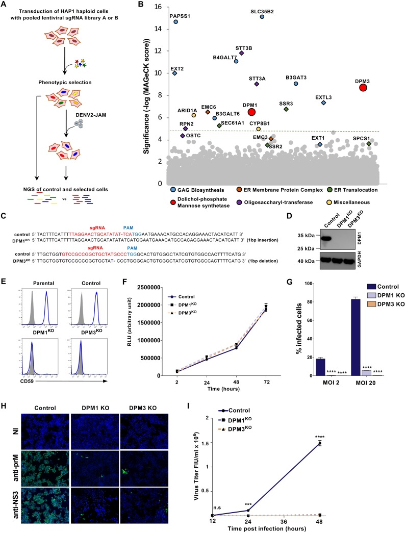
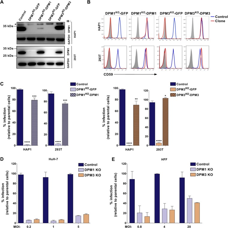
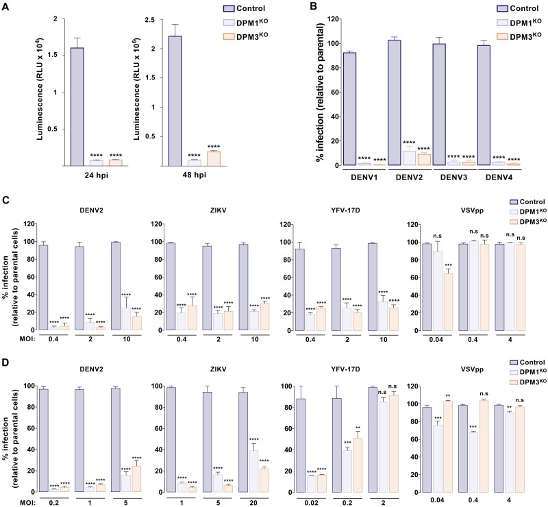
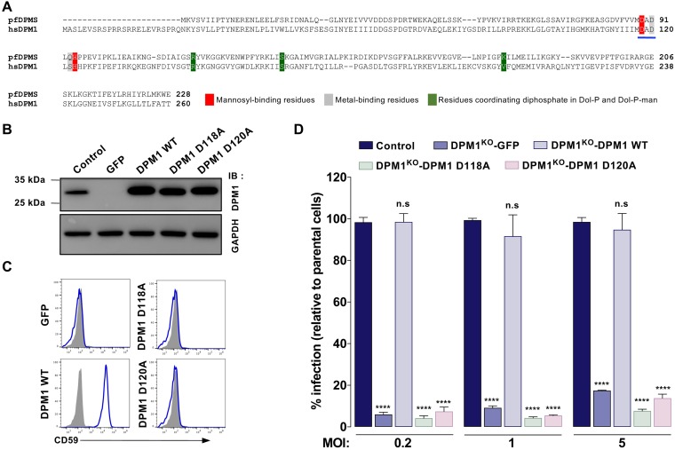
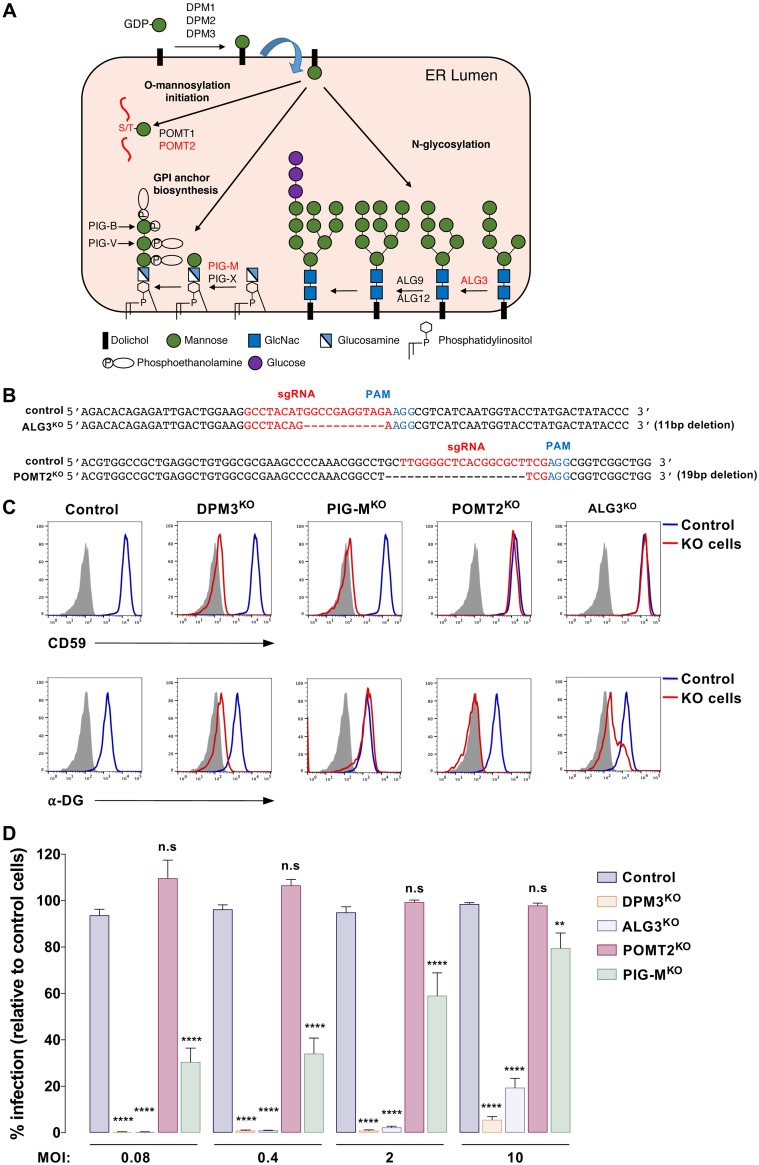
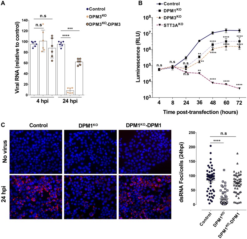
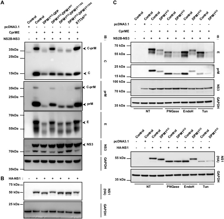
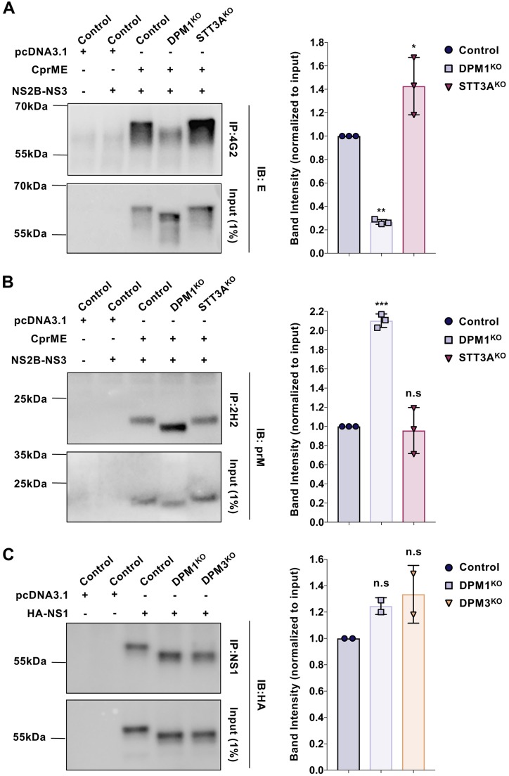
Dengue virus (DENV) is a mosquito-borne flavivirus responsible for dengue disease, a major human health concern for which no specific therapies are available. Like other viruses, DENV relies heavily on the host cellular machinery for productive infection. In this study, we performed a genome-wide CRISPR-Cas9 screen using haploid HAP1 cells to identify host genes important for DENV infection. We identified DPM1 and -3, two subunits of the endoplasmic reticulum (ER) resident dolichol-phosphate mannose synthase (DPMS) complex, as host dependency factors for DENV and other related flaviviruses, such as Zika virus (ZIKV). The DPMS complex catalyzes the synthesis of dolichol-phosphate mannose (DPM), which serves as mannosyl donor in pathways leading to N-glycosylation, glycosylphosphatidylinositol (GPI) anchor biosynthesis, and C- or O-mannosylation of proteins in the ER lumen. Mutation in the DXD motif of DPM1, which is essential for its catalytic activity, abolished DPMS-mediated DENV infection. Similarly, genetic ablation of ALG3, a mannosyltransferase that transfers mannose to lipid-linked oligosaccharide (LLO), rendered cells poorly susceptible to DENV. We also established that in cells deficient for DPMS activity, viral RNA amplification is hampered and truncated oligosaccharides are transferred to the viral prM and E glycoproteins, affecting their proper folding. Overall, our study provides new insights into the host-dependent mechanisms of DENV infection and supports current therapeutic approaches using glycosylation inhibitors to treat DENV infection.IMPORTANCE Dengue disease, which is caused by dengue virus (DENV), has emerged as the most important mosquito-borne viral disease in humans and is a major global health concern. DENV encodes only few proteins and relies on the host cell machinery to accomplish its life cycle. The identification of the host factors important for DENV infection is needed to propose new targets for antiviral intervention. Using a genome-wide CRISPR-Cas9 screen, we identified DPM1 and -3, two subunits of the DPMS complex, as important host factors for the replication of DENV as well as other related viruses such as Zika virus. We established that DPMS complex plays dual roles during viral infection, both regulating viral RNA replication and promoting viral structural glycoprotein folding/stability. These results provide insights into the host molecules exploited by DENV and other flaviviruses to facilitate their life cycle.

Keywords: N-glycosylation; Zika virus; dengue fever; dengue virus; dolichol-phosphate mannose; glycoprotein folding.


Figures

FIG 1
FIG 1
Haploid CRISPR-Cas9 screen identifies DPM1 and -3 as host factors for DENV infection. (A) Schematic representation of the CRISPR-Cas9 screen to identify host factors for DENV2 JAM in HAP1 cells. (B) Results of the DENV2 JAM screen analyzed by MAGeCK. Each circle represents individual gene. Genes of interest were colored according to their biological pathways. The y axis represents the significance of sgRNA enrichment of genes in the selected population compared to the unselected control population. The x axis represents a random distribution of the genes. (C) Sanger sequencing of DPM1 and DPM3 in control and DPM1 and DPM3 KO cells. PAM, protospacer adjacent motif. (D) Immunoblot of DPM1 in control, DPM1KO, and DPM3KO cells. (E) Cell surface CD59 staining on control and DPM1KO or DPM3KO HAP1 cells. (F) Control, DPM1KO, and DPM3KO HAP1 cells were plated and cell viability was assessed over a 72-h period using the CellTiter-Glo assay. (G and H) Control and DPM1 or -3 KO HAP1 cells were challenged with DENV2 JAM (MOIs of 2 and 20 in panel G and MOI of 5 in panel H). Levels of infection were quantified 48 hpi by flow cytometry using MAb 2H2 (G) or by immunofluorescence using MAb 2H2 or antibodies against NS3 (H). (I) Quantification of the viral particles released in the supernatant of inoculated HAP1 cells collected at 48 hpi. Virus titer was determined on Vero E6 cells by flow cytometry. FIU, flow cytometry infectious units. (C, D, E, F, and H) All data are representative of results from at least two independent experiments. (G and I) Data are means ± SD from three independent experiments performed in duplicate. Significance was calculated using a two-way ANOVA with Dunnett’s multiple-comparison test. n.s, nonsignificant. ***, P < 0.001; ****, P < 0.001.
FIG 2
FIG 2
DPM1 and DPM3 trans-complementation restores cell susceptibility to DENV infection. (A) Immunoblot of DPM1 in control, DPM1KO, and DPM3KO cells trans-complemented with the respective cDNA. (B) Cell surface CD59 staining on 293T and HAP1 clones trans-complemented with DPM1 or DPM3 cDNA. (C) 293T and HAP1 trans-complemented clones were challenged with DENV2 JAM (MOI of 5 in 293T cells and MOI of 10 in HAP1 cells). Infection was quantified 48 hpi by flow cytometry using MAb 2H2. Data are means ± SD from three independent experiments performed in duplicate. Significance was calculated using a one-way ANOVA with Dunnett’s multiple-comparison test. *, P < 0.05; **, P < 0.01; ***, P < 0.001; ****, P < 0.001. (D) Control and DPM1 and -3 KO Huh-7 clones were inoculated with increasing MOIs of DENV2 JAM. Levels of infection were quantified 48 hpi by flow cytometry using MAb 2H2. (E) HFF1 cells transfected with control, DPM1, or DPM3 sgRNA were inoculated with increasing MOIs of DENV2 JAM. Levels of infection were quantified 48 hpi by flow cytometry using MAb 2H2 in aCD59-negative population. (A, B, D, and E) Data are representative of results from at least two independent experiments.
FIG 3
FIG 3
DPM1 and -3 are required by the four DENV serotypes and other related flaviruses. (A) Control, DPM1KO, and DPM3KO cells were inoculated with DENV2 16681 Renilla luciferase (Rluc) reporter virus (DVR2A; MOI of 0.5). At 24 and 48 hpi, Rluc activity was measured. RLU, relative light units. (B) Control, DPM1KO, and DPM3KO cells were inoculated with DENV1 KDH (MOI of 2), DENV2 NGC (MOI of 0.2), DENV3 THAI (MOI of 10), and DENV4 1086 (MOI of 0.2). Levels of infection were quantified 48 hpi by flow cytometry using MAb 2H2. Data are means ± SD from three independent experiments performed in duplicate. Significance was calculated using a one-way ANOVA with Tukey’s multiple-comparison test. ****, P < 0.0001. (C and D) Control, DPM1KO, and DPM3KO HAP1 (C) and 293T (D) cells were inoculated with increasing MOIs of DENV2 16681, YFV 17D, ZIKV HD78788, or HIV pseudotyped with VSV-G envelope (VSVpp) expressing red fluorescent protein (RFP). Infection was quantified by flow cytometry 48 hpi by using MAb 4G2 (DENV and ZIKV) or 2D12 (YFV) or by monitoring RFP fluorescence. Data are means ± SD from two independent experiments performed in duplicate. Significance was calculated using a two-way ANOVA with Dunett’s multiple-comparison test. **, P < 0.01; ***, P < 0.001; ****, P < 0.0001.
FIG 4
FIG 4
DENV infection requires DPM1 catalytic activity. (A) Sequence alignment of the catalytic domain of Pyrococcus furiosus DPMS (pfDPMS; UniProt accession number Q8U4M3) and Homo sapiens DPM1 (hsDPM1; UniProt accession number O60762). The DAD motif is underlined in blue. (B and C) Immunoblot analysis of DPM1 expression (B) and staining of cell surface CD59 (C) in DPM1KO 293T cells complemented with the WT or DPM1 mutated in the catalytic site (D118A and D120A). Data are representative of reults from three independent experiments. (D) Control cells, DPM1KO 293T cells, and DPM1KO cells complemented with the WT or catalytically dead mutants of DPM1 were inoculated with increasing MOIs of DENV2 16681. Levels of infection were quantified 48 hpi by flow cytometry using MAb 2H2. Data are means ± SD from three independent experiments performed in duplicate. Significance was calculated using a two-way ANOVA with Dunett’s multiple-comparison test. ****, P < 0.0001.
FIG 5
FIG 5
DENV infection requires transfer of mannose from DPM to lipid-linked oligosaccharide by ALG3 mannosyltransferase. (A) Schematic representation of the ER DPM-dependent pathways and their related mannosyltransferases. Mannosyltransferases invalidated in this study are depicted in red. (B) Sanger sequencing of ALG3 and POMT2 in control and ALG3KO or POMT2KO HAP1 clones. (C) Cell surface staining of CD59 and glycosylated α-dystoglycan on HAP1 cells deficient for DPM3, PIG-M, and POMT2. The blue line represents staining with the respective Ab and gray shading staining with the matching isotypes in control cells. Data are representative of results from three independent experiments. (D) Control and HAP1 cells with KO of the indicated genes were inoculated with increasing MOIs of DENV2 11681, and levels of infection were quantified 48 hpi by flow cytometry using MAb 2H2. Data are means ± SD from three independent experiments performed in duplicate. Significance was calculated using a two-way ANOVA with Dunett’s multiple-comparison test. *, P < 0.05; **, P < 0.01; ***, P < 0.001; ****, P < 0.0001.
FIG 6
FIG 6
The DPMS complex is required for optimal viral RNA replication. (A) Control, DPM3KO, and DPM3KO 293T cells complemented with DPM3 were inoculated with DENV 11681 (MOI of 10). At 4 and 24 hpi, cells were treated with trypsin to remove cell surface-bound virus and viral RNA was quantified by RT-qPCR. Data are means ± SD from two independent experiments performed in triplicate. Significance was calculated using a one-way ANOVA with Tukey’s multiple-comparison test. ***, P < 0.001; ****, P < 0.0001). (B) Control, DPM1KO, DPM3KO, and STT3AKO 293T cells were transfected with in vitro-transcribed subgenomic DENV2 RNA expressing the Renilla luciferase. Rluc activity was monitored at the indicated time points. Data are means ± SD from two independent experiments performed in quadruplicate. Significance was calculated using a two-way ANOVA with Dunett’s multiple-comparison test. **, P < 0.01; ****, P < 0.0001. (C) Control, DPM1KO, and DPM1KO 293T cells complemented with DPM1 were inoculated with DENV2 16681 (MOI of 100). Left, representative images of the cells stained with anti-dsRNA MAb J2 at 24 hpi. Right, quantification of the number of foci per cell using Icy software. Data are means ± SD from a representative experiment with n = 50 independent cells. Significance was calculated using a one-way ANOVA with Dunnett’s multiple-comparison test. ****, P < 0.0001.
FIG 7
FIG 7
Effect of DPM1 depletion on DENV viral protein glycosylation. Control, DPM1KO, and STT3AKO 293T cells and DPM1KO cells complemented with the WT or catalytically dead mutants of DPM1 were cotransfected with DENV C-prM-E and NS2B-NS3 plasmids (1:1 ratio) (A) or transfected with DENV hemagglutinin (HA)-tagged NS1 plasmid (B). Cell lysates were subjected to immunoblot analysis with anti-capsid, anti-prM, anti-E, and anti-HA antibodies. Data are representative of results from three independent experiments. (C) Control and DPM1KO 293T cells were cotransfected with DENV C-prM-E and NS2B-NS3 plasmids or HA-NS1 plasmid. Twenty micrograms of total proteins was subjected to deglycosylation with either PNGase F or endo H (25 kU ml−1) for 1 h at 37°C and analyzed by immunoblotting with anti-prM, anti-E, and anti-HA antibodies. As a positive control for deglycosylation, cells were also transfected in the continuous presence of tunicamycin (Tun; 5 μg ml−1). For all panels, blots are representative of those from three independent experiments. NT, nontreated.
FIG 8
FIG 8
DPM1 deficiency affects E and prM glycoprotein epitope accessibility. Control, DPM1KO, and STT3AKO 293T cells were cotransfected with DENV C-prM-E and NS2B-NS3 plasmids (1:1 ratio). (A) Relatively equivalent amounts of viral glycoproteins were immunoprecipitated with MAb 4G2, followed by immunoblot analysis with anti-E antibodies. (B) Relatively equivalent amounts of viral glycoproteins were immunoprecipitated with MAb 2H2, followed by immunoblot analysis with anti-prM antibodies. (C) Control, DPM1KO, and DPM3KO 293T cells were transfected with DENV HA-NS1 plasmid. Relatively equivalent amounts of viral glycoproteins were immunoprecipitated with commercially available (Abcam; ab41623) anti-NS1, followed by immunoblot analysis with anti-HA antibody. Blots are representative of those from three independent experiments. Bar graphs represent quantification of the chemiluminescent band intensities relative to E, prM, and NS1 expression in control cells. Each point plotted corresponds to the quantification from one transfection experiment. Data are means ± SD from from three (A and B) or two (C) independent transfection experiments. Significance was calculated using a one-way ANOVA with Dunnett’s multiple-comparison test. *, P < 0.05; **, P < 0.01; ***, P < 0.001.

Similar articles

Cited by

References

    1. Holbrook MR, Holbrook M. 2017. Historical perspectives on flavivirus research. Viruses 9:97. doi:10.3390/v9050097. - DOI - PMC - PubMed
    1. Halstead SB. 2007. Dengue. Lancet 370:1644–1652. doi:10.1016/S0140-6736(07)61687-0. - DOI - PubMed
    1. Brady OJ, Gething PW, Bhatt S, Messina JP, Brownstein JS, Hoen AG, Moyes CL, Farlow AW, Scott TW, Hay SI. 2012. Refining the global spatial limits of dengue virus transmission by evidence-based consensus. PLoS Negl Trop Dis 6:e1760. doi:10.1371/journal.pntd.0001760. - DOI - PMC - PubMed
    1. Bhatt S, Gething PW, Brady OJ, Messina JP, Farlow AW, Moyes CL, Drake JM, Brownstein JS, Hoen AG, Sankoh O, Myers MF, George DB, Jaenisch T, Wint GR, Simmons CP, Scott TW, Farrar JJ, Hay SI. 2013. The global distribution and burden of dengue. Nature 496:504–507. doi:10.1038/nature12060. - DOI - PMC - PubMed
    1. Hadinegoro SR, Arredondo-Garcia JL, Capeding MR, Deseda C, Chotpitayasunondh T, Dietze R, Muhammad Ismail HI, Reynales H, Limkittikul K, Rivera-Medina DM, Tran HN, Bouckenooghe A, Chansinghakul D, Cortes M, Fanouillere K, Forrat R, Frago C, Gailhardou S, Jackson N, Noriega F, Plennevaux E, Wartel TA, Zambrano B, Saville M, CYD-TDV Dengue Vaccine Working Group. 2015. Efficacy and long-term safety of a dengue vaccine in regions of endemic disease. N Engl J Med 373:1195–1206. doi:10.1056/NEJMoa1506223. - DOI - PubMed

Publication types