Review
. 2019 Dec 28;7(4):352-361.
doi: 10.14218/JCTH.2019.00025. Epub 2019 Nov 13.

The Direct Contribution of Astrocytes and Microglia to the Pathogenesis of Hepatic Encephalopathy

Affiliations
Review

The Direct Contribution of Astrocytes and Microglia to the Pathogenesis of Hepatic Encephalopathy

Victoria Jaeger et al. J Clin Transl Hepatol. .

Abstract

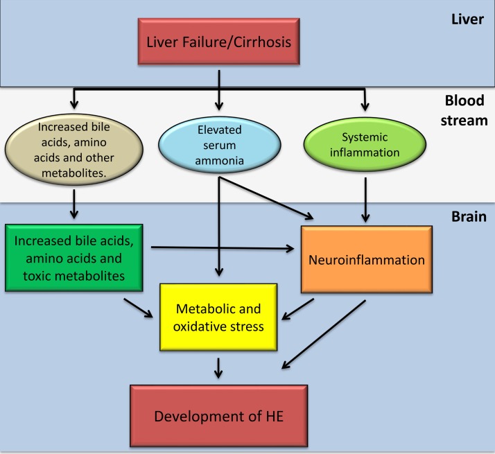
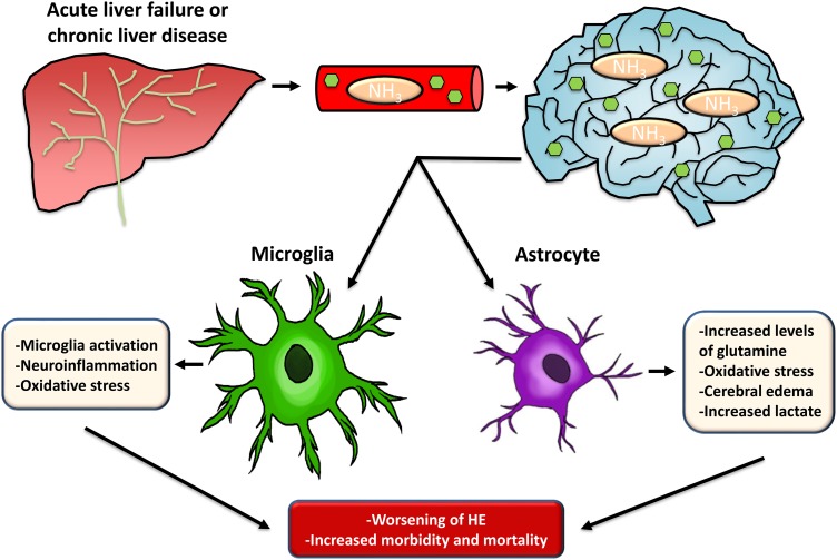
Hepatic encephalopathy is a neurological complication resulting from loss of hepatic function and is associated with poor clinical outcomes. During acute liver failure over 20% of mortality can be associated with the development of hepatic encephalopathy. In patients with liver cirrhosis, 1-year survival for those that develop overt hepatic encephalopathy is under 50%. The pathogenesis of hepatic encephalopathy is complicated due to the multiple disruptions in homeostasis that occur following a reduction in liver function. Of these, elevations of ammonia and neuroinflammation have been shown to play a significant contributing role to the development of hepatic encephalopathy. Disruption of the urea cycle following liver dysfunction leads to elevations of circulating ammonia, which enter the brain and disrupt the functioning of astrocytes. This results in dysregulation of metabolic pathways in astrocytes, oxidative stress and cerebral edema. Besides ammonia, circulating chemokines and cytokines are increased following liver injury, leading to activation of microglia and a subsequent neuroinflammatory response. The combination of astrocyte dysfunction and microglia activation are significant contributing factors to the pathogenesis of hepatic encephalopathy.

Keywords: Acute liver failure; Ammonia; Astroglia; Microglia; Neuroinflammation.


Conflict of interest statement

The authors have no conflict of interests related to this publication.

Figures

Fig. 1.
Fig. 1.. Pathophysiology of HE.
Following the development of liver failure or decompensated liver cirrhosis, there is an increase of circulating bile acids, amino acids, serum ammonia, and toxic metabolites as well as an increase of systemic inflammation. The systemic elevation of these factors leads to an increase of their concentrations in the brain, leading to metabolic and oxidative stress as well as increased neuroinflammation. Elevation of circulating cytokines and chemokines is associated with increased activation of microglia in the brain and subsequent neuroinflammation. The increase of neuroinflammation as well as the metabolic and oxidative stress present under these conditions promote the development of HE.
Fig. 2.
Fig. 2.. Summary of the involvement of microglia and astrocytes in HE pathology.
The liver can lose its ability to function during acute liver failure or chronic liver disease. When this occurs, toxins, such as ammonia, enter the circulation and can enter the brain. After accumulating in neural tissue, they cause a disruption of astrocyte and microglia cellular function. Astrocytes metabolize ammonia, leading to an increase of glutamine, cell swelling, cerebral edema, oxidative stress and hyperlactatemia. Microglia become activated leading to increased neuroinflammation due to pro-inflammatory cytokine release and oxidative stress. Together, these changes in neural cellular function lead to increased HE pathology, resulting in increased morbidity and mortality. Abbreviations: HE, hepatic encephalopathy; NH3, ammonia.

References

    1. Martino ME, Fernández-Lorente J, Romero-Vives M, Bárcena R, Gaztelu JM. Brain oscillatory activity during sleep shows unknown dysfunctions in early encephalopathy. J Physiol Biochem. 2014;70:821–835. doi: 10.1007/s13105-014-0351-2. - DOI - PubMed
    1. Nardelli S, Allampati S, Riggio O, Mullen KD, Prakash R, Gioia S, et al. Hepatic encephalopathy is associated with persistent learning impairments despite adequate medical treatment: A multicenter, international study. Dig Dis Sci. 2017;62:794–800. doi: 10.1007/s10620-016-4425-6. - DOI - PubMed
    1. Hirode G, Vittinghoff E, Wong RJ. Increasing burden of hepatic encephalopathy among hospitalized adults: An analysis of the 2010-2014 national inpatient sample. Dig Dis Sci. 2019;64:1448–1457. doi: 10.1007/s10620-019-05576-9. - DOI - PubMed
    1. Suraweera D, Sundaram V, Saab S. Evaluation and management of hepatic encephalopathy: Current status and future directions. Gut Liver. 2016;10:509–519. doi: 10.5009/gnl15419. - DOI - PMC - PubMed
    1. Patidar KR, Bajaj JS. Covert and overt hepatic encephalopathy: Diagnosis and management. Clin Gastroenterol Hepatol. 2015;13:2048–2061. doi: 10.1016/j.cgh.2015.06.039. - DOI - PMC - PubMed