

Three-dimensional comparison of 2 digital models obtained from cone-beam computed tomographic scans of polyvinyl siloxane impressions and plaster models
- PMID: 31915610
- PMCID: PMC6941835
- DOI: 10.5624/isd.2019.49.4.257
Three-dimensional comparison of 2 digital models obtained from cone-beam computed tomographic scans of polyvinyl siloxane impressions and plaster models
Abstract
Purpose: This study was performed to evaluate the dimensional accuracy of digital dental models constructed from cone-beam computed tomographic (CBCT) scans of polyvinyl siloxane (PVS) impressions and cast scan models.
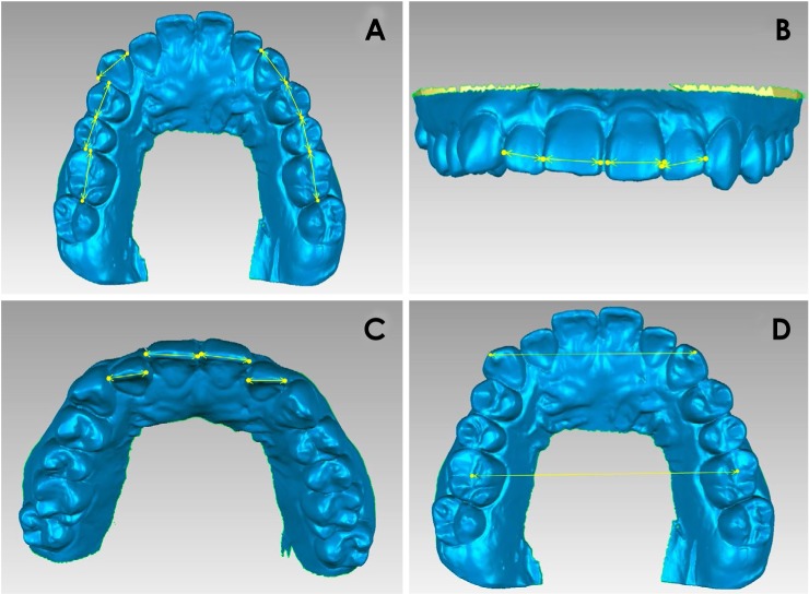
Materials and methods: A pair of PVS impressions was obtained from 20 subjects and scanned using CBCT (resolution, 0.1 mm). A cast scan model was constructed by scanning the gypsum model using a model scanner. After reconstruction of the digital models, the mesio-distal width of each tooth, inter-canine width, and inter-molar width were measured, and the Bolton ratios were calculated and compared. The 2 models were superimposed and the difference between the models was measured using 3-dimensional analysis.
Results: The range of mean error between the cast scan model and the CBCT scan model was -0.15 mm to 0.13 mm in the mesio-distal width of the teeth and 0.03 mm to 0.42 mm in the width analysis. The differences in the Bolton ratios between the cast scan models and CBCT scan models were 0.87 (anterior ratio) and 0.72 (overall ratio), with no significant difference (P>0.05). The mean maxillary and mandibular difference when the cast scan model and the CBCT scan model were superimposed was 53 µm.
Conclusion: There was no statistically significant difference in most of the measurements. The maximum tooth size difference was 0.15 mm, and the average difference in model overlap was 53 µm. Digital models produced by scanning impressions at a high resolution using CBCT can be used in clinical practice.
Keywords: Cone-Beam Computed Tomography; Dental Models; Orthodontics.
Copyright © 2019 by Korean Academy of Oral and Maxillofacial Radiology.
Conflict of interest statement
Conflicts of Interest: None
Figures


Similar articles
-
[Degree of reliability of the assessment of the Bolton analysis in three-dimensional virtual models versus plaster models. a review].Rev Cient Odontol (Lima). 2023 Jun 29;11(2):e155. doi: 10.21142/2523-2754-1102-2023-155. eCollection 2023 Apr-Jun. Rev Cient Odontol (Lima). 2023. PMID: 38288455 Free PMC article. Review. Spanish.
-
Accuracy of Bolton analysis measured in laser scanned digital models compared with plaster models (gold standard) and cone-beam computer tomography images.Korean J Orthod. 2016 Jan;46(1):13-9. doi: 10.4041/kjod.2016.46.1.13. Epub 2016 Jan 25. Korean J Orthod. 2016. PMID: 26877978 Free PMC article.
-
Assessment of the accuracy of laser-scanned models and 3-dimensional rendered cone-beam computed tomographic images compared to digital caliper measurements on plaster casts.Imaging Sci Dent. 2021 Dec;51(4):429-438. doi: 10.5624/isd.20210142. Epub 2021 Oct 15. Imaging Sci Dent. 2021. PMID: 34988004 Free PMC article.
-
Validity, reliability, and reproducibility of linear measurements on digital models obtained from intraoral and cone-beam computed tomography scans of alginate impressions.Am J Orthod Dentofacial Orthop. 2013 Jan;143(1):140-7. doi: 10.1016/j.ajodo.2012.06.018. Am J Orthod Dentofacial Orthop. 2013. PMID: 23273370
-
[Exploring a new method for superimposition of pre-treatment and post-treatment mandibular digital dental casts in adults].Beijing Da Xue Xue Bao Yi Xue Ban. 2018 Apr 18;50(2):271-278. Beijing Da Xue Xue Bao Yi Xue Ban. 2018. PMID: 29643526 Chinese.
Cited by
-
[Degree of reliability of the assessment of the Bolton analysis in three-dimensional virtual models versus plaster models. a review].Rev Cient Odontol (Lima). 2023 Jun 29;11(2):e155. doi: 10.21142/2523-2754-1102-2023-155. eCollection 2023 Apr-Jun. Rev Cient Odontol (Lima). 2023. PMID: 38288455 Free PMC article. Review. Spanish.
-
Mesiodistal Measurements for Dental Implant Planning: A Prospective Clinical Study of Linear Measurements on Cone-Beam Computed Tomography Images in Comparison with Caliper-Based Measurements on Plaster Casts.Dent J (Basel). 2022 Sep 7;10(9):169. doi: 10.3390/dj10090169. Dent J (Basel). 2022. PMID: 36135164 Free PMC article.
-
Comparative Evaluation of Digitization of Diagnostic Dental Cast (Plaster) Models Using Different Scanning Technologies.Dent J (Basel). 2020 Aug 2;8(3):79. doi: 10.3390/dj8030079. Dent J (Basel). 2020. PMID: 32748890 Free PMC article.
-
Scanning Accuracy of Bracket Features and Slot Base Angle in Different Bracket Materials by Four Intraoral Scanners: An In Vitro Study.Materials (Basel). 2021 Jan 13;14(2):365. doi: 10.3390/ma14020365. Materials (Basel). 2021. PMID: 33451075 Free PMC article.
-
The precision of two alternative indirect workflows for digital model production: an illusion or a possibility?Clin Oral Investig. 2023 Jul;27(7):3787-3797. doi: 10.1007/s00784-023-04996-2. Epub 2023 Apr 13. Clin Oral Investig. 2023. PMID: 37046002 Free PMC article.
References
-
- Fleming PS, Marinho V, Johal A. Orthodontic measurements on digital study models compared with plaster models: a systematic review. Orthod Craniofac Res. 2011;14:1–16. - PubMed
-
- Leifert MF, Leifert MM, Efstratiadis SS, Cangialosi TJ. Comparison of space analysis evaluations with digital models and plaster dental casts. Am J Orthod Dentofacial Orthop. 2009;136:16.e1–16.e4. - PubMed
-
- Mullen SR, Martin CA, Ngan P, Gladwin M. Accuracy of space analysis with emodels and plaster models. Am J Orthod Dentofacial Orthop. 2007;132:346–352. - PubMed
-
- Reuschl RP, Heuer W, Stiesch M, Wenzel D, Dittmer MP. Reliability and validity of measurements on digital study models and plaster models. Eur J Orthod. 2016;38:22–26. - PubMed
LinkOut - more resources
Full Text Sources
Research Materials
Miscellaneous
