Case Reports
. 2019 Oct-Dec;63(4):397-402.

Multimodal imaging in a child with severe posterior scleritis

Affiliations
Case Reports

Multimodal imaging in a child with severe posterior scleritis

Sibel Inan et al. Rom J Ophthalmol. 2019 Oct-Dec.

Abstract

Objective: Posterior scleritis in a child is a rare condition. High-resolution imaging techniques in the course of posterior scleritis have not been published extensively in literature. The authors reported a case of posterior scleritis in a 12-year-old child to demonstrate multimodal imaging techniques in the course of development and improvement of the disease. Methods: Case report that included fundus photography, spectral domain optical coherence tomography with enhanced depth imaging, blue-peak autofluorescence, multicolor imaging, fluorescein angiography, indocyanine green angiography, and ultrasonography. Results: A twelve-year-old healthy boy presented with ocular pain and mild vision loss. His visual acuity was 20/ 32. There was no sign of inflammation on the ocular surface. There were no cells in the anterior chamber or vitreous. Ultrasonography revealed the diagnosis of posterior scleritis. When he was seen the next day for multimodal imaging techniques, he presented with exudative retinal detachment with visual acuity of 20/ 100. One week after the beginning of the therapy, ocular symptoms, and findings resolved and visual acuity improved to 20/ 20. Conclusion: Multimodal imaging techniques, which are important for the diagnosis of posterior scleritis, before and after the treatment, are presented in this case report.

Keywords: exudative retinal detachment; multimodal imaging; posterior scleritis.


Figures

Fig. 1
Fig. 1
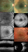
Color photograph of right eye at the presentation. Note radial retinochoroidal striae, tiny retinal deposits, and slight retinal elevation just inferotemporal to the optic disc (A). Some subretinal and intraretinal fluids are seen near the optic disc in OCT (Spectralis OCT; Heidelberg Engineering, Heidelberg, Germany) (B). A granular irregularity of autofluorescence (AF) temporal to the fovea is noted in blue-peak AF (C). Fellow eye shows a normal appearance except for retinal tiny-yellowish deposits in color photograph and a slight increase in choroidal thickness (D and E). Color photograph shows marked elevation of the retina inferior to the disc and macula 7 days after the first presentation (F). OCT shows development of submacular fluid, multiple intraretinal and subretinal hyperreflective dot spots, and convex elevation of RPE-choriocapillaris interface (G)
Fig. 2
Fig. 2
Color photograph the next day shows further elevation in the retina (A). Blue-peak AF shows hypo-AF area suggesting the site of inflammation (B). Early phase of FFA and ICG-A (Spectralis HRA; Heidelberg Engineering, Heidelberg, Germany) also shows a hypo-fluorescence dark area at the presumed site of inflammation (C). Initial-mid phase of FFA and ICG-A show multiple tiny fluorescein leakage spots and enlargement of hypo-fluorescence, respectively (D). Middle-mid phase FFA and ICG-A show enlargement of sites of leakage and appearance of leakage spots, respectively (E). Late-mid phase of FFA and ICG-A show leakage areas confined to the half of the retinal detachment area and enlargement of sites, respectively (F). Late phase of FFA and ICG-A show filling of fluorescence dye and more leakage, respectively (G). Late phase wide-field FFA and ICG-A show complete staining of retinal detachment area in the presumed inflammation site but a hypo-fluorescence in retinal detachment area inferior to the disc (H). Wide field OCT shows multiple detachment areas on different sections (J). EDI-OCT shows enlargement of choroidal layers with elevation of RPE plane possibly due to compression by scleral swelling (K). Middle phase of FFA and ICG-A in the fellow eye show multiple tiny hypo-fluorescent spots (L). Multicolor imaging shows a tiny hypo-pigmentary spot possibly reflecting involvement and leakage of RPE sites (M). Blue-peak AF shows area of inflammatory involvement with hypo-fluorescence and multiple dark spots (N). Ocular ultrasonography (USG, Aviso ultrasound unit, Quantel Medical Systems Inc. Cedex, France) shows marked scleral thickening with T sign and normal ultrasonography in the fellow eye in the smaller picture (O).
Fig. 3
Fig. 3
Fig. 3 Color photograph shows resolution subretinal fluid with residual retinal striae 1 week after the initiation of the therapy (A). Infrared reflectance imaging shows multiple dot spots (B). Blue-peak AF shows some hyper-fluorescence granular AF at the site of presumed inflammation (C). OCT shows a decrease in subretinal fluid with residual hyper-reflective material at the site of presumed inflammation and decrease in choroidal thickness (D). OCT shows complete resolution of subretinal fluid with residual hyperreflective subretinal material and residual multiple subretinal hyperreflective dots 2 weeks after the initiation of the therapy (E). Color photograph shows normal retinal appearance but some hypo-pigmentary spots and darker yellow appearance in the temporal to the fovea (F). Early phase of control FFA shows hypo-fluorescent dark spots and a few areas of choroidal filling defects 1 month after the therapy initiation (G). Multicolor imaging shows hypo-pigmentary changes in the original site of the presumed inflammation possibly reflecting RPE involvement at this area (H). OCT shows normal retinal and choroidal configuration at month 1 (J). Control USG shows normalized appearance of scleral wall at month 1.

References

    1. McCluskey PJ, Watson PG, Lightman S, et al. Posterior scleritis: clinical features, systemic associations, and outcome in a large series of patients. Ophthalmology. 1999;106:2380–2386. - PubMed
    1. Gonzalez-Gonzalez LA, Molina-Prat N, Doctor P, Tauber J, Sainz de la Maza M, Foster CS. Clinical features and presentation of posterior scleritis: a report of 31 cases. Ocul Immunol Inflamm. 2014;22:203–207. - PubMed
    1. Lavric A, Gonzalez-Lopez JJ, Majumder PD, et al. Posterior Scleritis: Analysis of Epidemiology, Clinical Factors, and Risk of Recurrence in a Cohort of 114 Patients. Ocul Immunol Inflamm. 2015;2:1–10. - PubMed
    1. Cheung CM, Chee SP. Posterior scleritis in children: clinical features and treatment. Ophthalmology. 2012;119:59–65. - PubMed
    1. Dave V, Mathai A. Posterior scleritis in a 9-year-old boy: a case report. Retin Cases Brief Rep. 2012;6:30–32. - PubMed

Publication types

LinkOut - more resources