. 2020 Apr;27(4):1002-1007.
doi: 10.1007/s43032-019-00099-0. Epub 2020 Jan 8.

Androgens Modulate Rat Granulosa Cell Steroidogenesis

Affiliations

Androgens Modulate Rat Granulosa Cell Steroidogenesis

Manuel Doblado et al. Reprod Sci. 2020 Apr.

Abstract

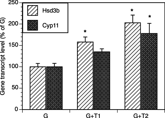
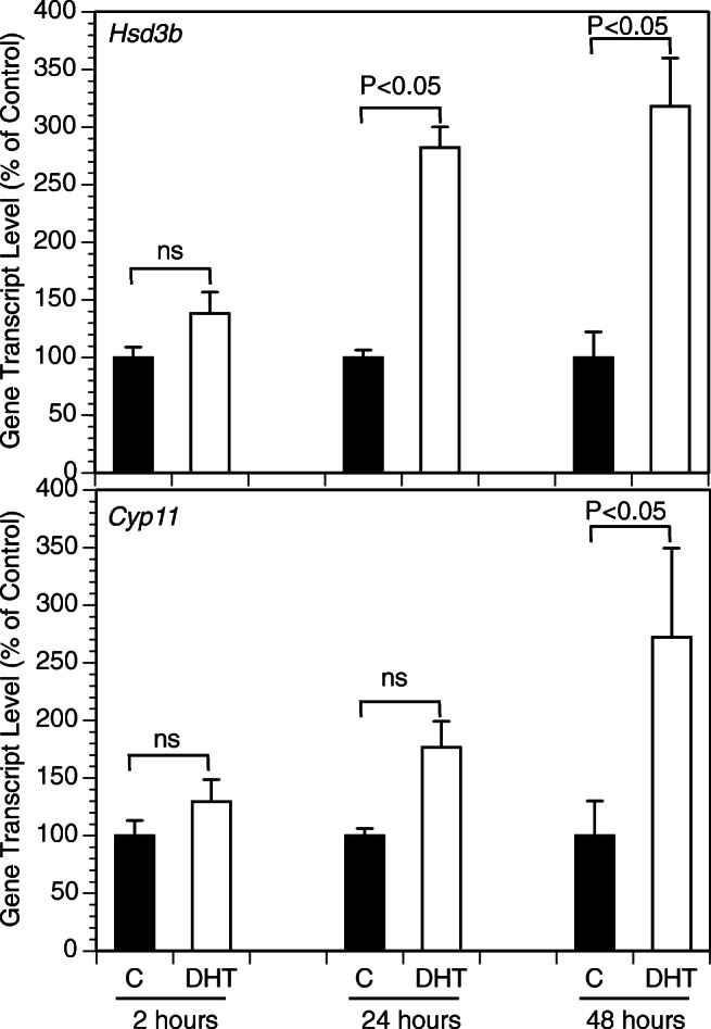
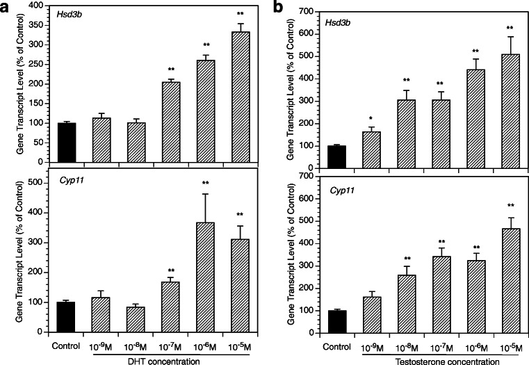
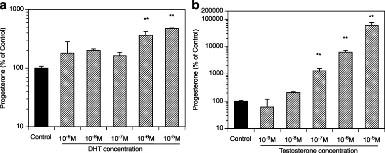
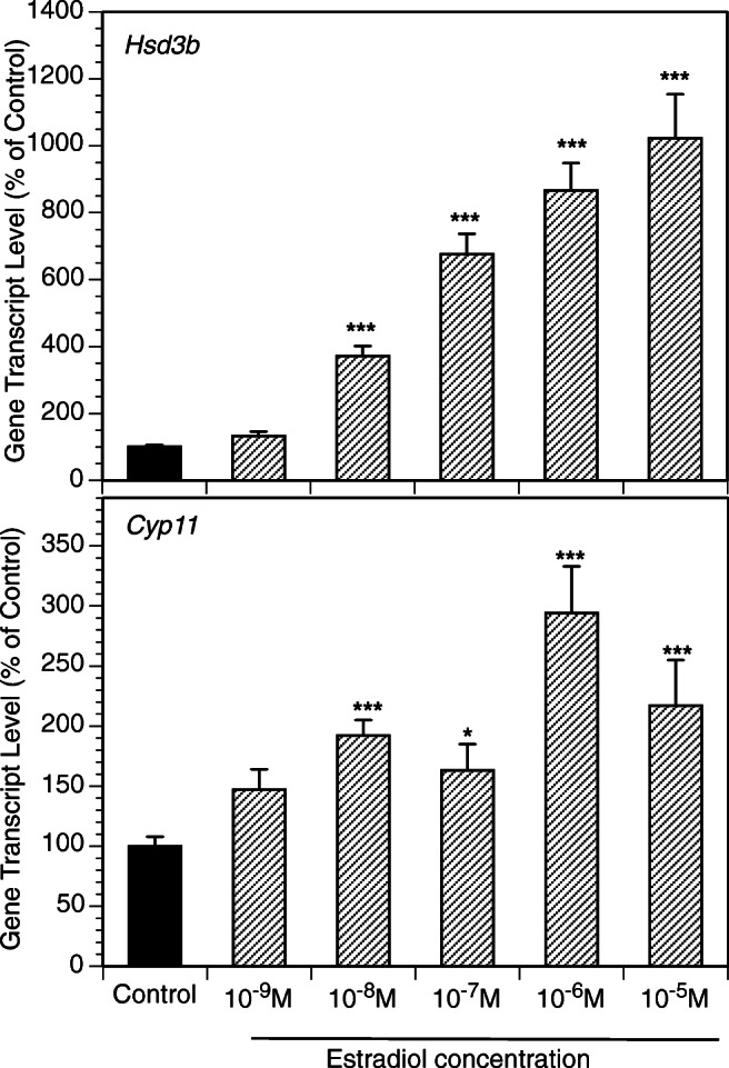
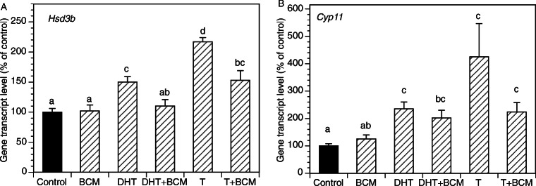
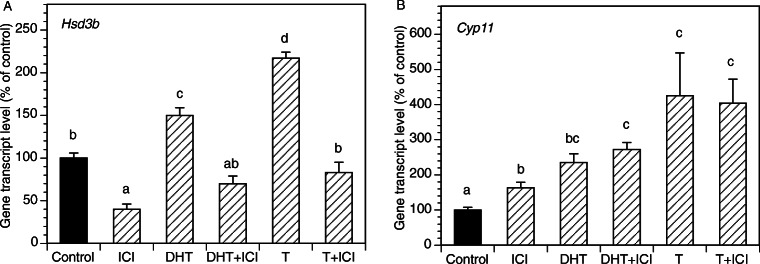
Paracrine interactions between ovarian theca-interstitial cells (TICs) and granulosa cells (GCs) play an important role in the regulation of follicular steroidogenesis. Androgens serve as substrates for aromatization as well as affect GC function. This study evaluated the effects of co-culture of GC with TICs and the role of testosterone (T) and 5-alpha-dihydrotestosterone (DHT), and estradiol (E2) in modulation of GC expression of genes involved in the production of progesterone: 3β-hydroxysteroid dehydrogenase/Δ5-4 isomerase (Hsd3b) and cholesterol side-chain cleavage (Cyp11). GCs obtained from immature Sprague-Dawley rats and were cultured in chemically defined media without or with TICs, DHT, or T. Hsd3b and Cyp11 transcripts were analyzed by qt-PCR. Co-culture of GCs with TICs stimulated Hsd3b and CYP11 expression in GCs. DHT and T induced a concentration-dependent upregulation of Hsd3b and CYP11 expression, as well as increased progesterone concentrations in spent media. E2 also increased expression of Hsd3b, and Cyp11. Effects of androgens were abrogated in the presence of an anti-androgen bicalutamide and the antiestrogen ICI 182780 (ICI). In conclusion, present findings demonstrate that androgens upregulate production of progesterone in GCs; these effects are likely due to a combination of direct action on androgen receptors and effects mediated by estrogen receptors.

Keywords: Androgens; Gene expression; Granulosa cells; Rat.


Figures

Fig. 1
Fig. 1
Effect of co-culture of TICs with GCs on mRNA expression of Hsd3b and Cyp11 in GCs. GCs (200,000 cells/well) were cultured in the absence (G) or in the presence of TICs at a concentration of 100,000 cells per well (G + T1) or 200,000 cells/well (G + T2). TICs were placed in the fibronectin-coated 24-well plates, while Gs were placed in the matrigel-coated inserts. All cultures were carried out for 48 h in chemically defined media. Each bar represents mean ± SEM; * denotes means significantly different (P < 0.05) from control cultures (G)
Fig. 2
Fig. 2
Time course of effects of DHT on mRNA expression of select genes by GCs cultured for 2, 24, and 48 h in the absence (C) or in the presence of DHT (10 μM). GCs were plated in fibronectin-coated 24-well plates and incubated in chemically defined media. Each bar represents mean ± SEM; significant differences are denoted by P-values above brackets
Fig. 3
Fig. 3
Effects of various concentrations of DHT (A, left panel) and T (B, right panel) on mRNA expression of Hsd3b and Cyp11 by GCs. All cultures were carried out for 48 h in chemically defined media. Each bar represents mean ± SEM; means significantly different from cultures in the absence of androgens (Control) are denoted by * (P < 0.05) or ** (P < 0.01)
Fig. 4
Fig. 4
Effects of various concentrations of DHT (A, left panel) and T (B, right panel) on progesterone level in culture media. All cultures were carried out for 48 h in chemically defined media. Each bar represents mean ± SEM; means significantly different from cultures in the absence of androgens (Control) are denoted by ** (P < 0.01)
Fig. 5
Fig. 5
Effects of an anti-androgen BCM on basal and androgen-induced mRNA expression of Hsd3b and Cyp11 by GCs. All cultures were carried out for 48 h in chemically defined media. Each bar represents mean ± SEM; means with no superscripts in common are significantly different (P < 0.05)
Fig. 6
Fig. 6
Effects of various concentrations of E2 on mRNA expression of Hsd3b and Cyp11 by GCs. All cultures were carried out for 48 h in chemically defined media. Each bar represents mean ± SEM; means significantly different from cultures in the absence of androgens (control) are denoted by * (P < 0.05), ** (P < 0.01), or *** (P < 0.001)
Fig. 7
Fig. 7
Effects of an anti-androgen ICI on basal and androgen-induced mRNA expression of Hsd3b and Cyp11 by GCs. All cultures were carried out for 48 h in chemically defined media. Each bar represents mean ± SEM; means with no superscripts in common are significantly different (P < 0.05)

References

    1. Hillier SG, Whitelaw PF, Smyth CD. Follicular oestrogen synthesis: the 'two-cell, two-gonadotrophin' model revisited. Mol Cell Endocrinol. 1994;100:51–54. doi: 10.1016/0303-7207(94)90278-X. - DOI - PubMed
    1. Wang H, Andoh K, Hagiwara H, Xiaowei L, Kikuchi N, Abe Y, Yamada K, Fatima R, Mizunuma H. Effect of adrenal and ovarian androgens on type 4 follicles unresponsive to FSH in immature mice. Endocrinology. 2001;142:4930–4936. doi: 10.1210/endo.142.11.8482. - DOI - PubMed
    1. Vendola KA, Zhou J, Adesanya OO, Weil SJ, Bondy CA. Androgens stimulate early stages of follicular growth in the primate ovary. J Clin Invest. 1998;101:2622–2629. doi: 10.1172/JCI2081. - DOI - PMC - PubMed
    1. Sen A, Hammes SR. Granulosa cell-specific androgen receptors are critical regulators of ovarian development and function. Mol Endocrinol. 2010;24:1393–1403. doi: 10.1210/me.2010-0006. - DOI - PMC - PubMed
    1. Murray AA, Gosden RG, Allison V, Spears N. Effect of androgens on the development of mouse follicles growing in vitro. J Reprod Fertil. 1998;113:27–33. doi: 10.1530/jrf.0.1130027. - DOI - PubMed

Publication types