. 2020 Mar;27(3):940-953.
doi: 10.1007/s43032-019-00098-1. Epub 2020 Jan 8.

Declining BRCA-Mediated DNA Repair in Sperm Aging and its Prevention by Sphingosine-1-Phosphate

Affiliations

Declining BRCA-Mediated DNA Repair in Sperm Aging and its Prevention by Sphingosine-1-Phosphate

Robert Stobezki et al. Reprod Sci. 2020 Mar.

Abstract

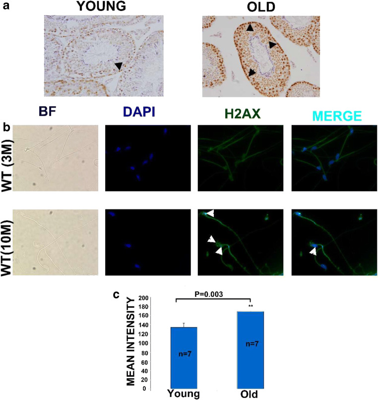
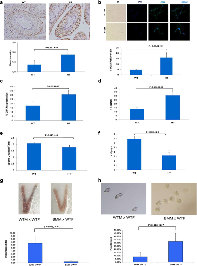
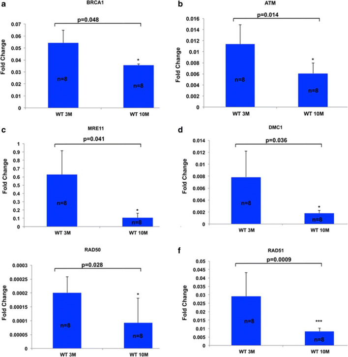
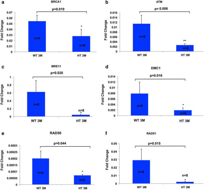
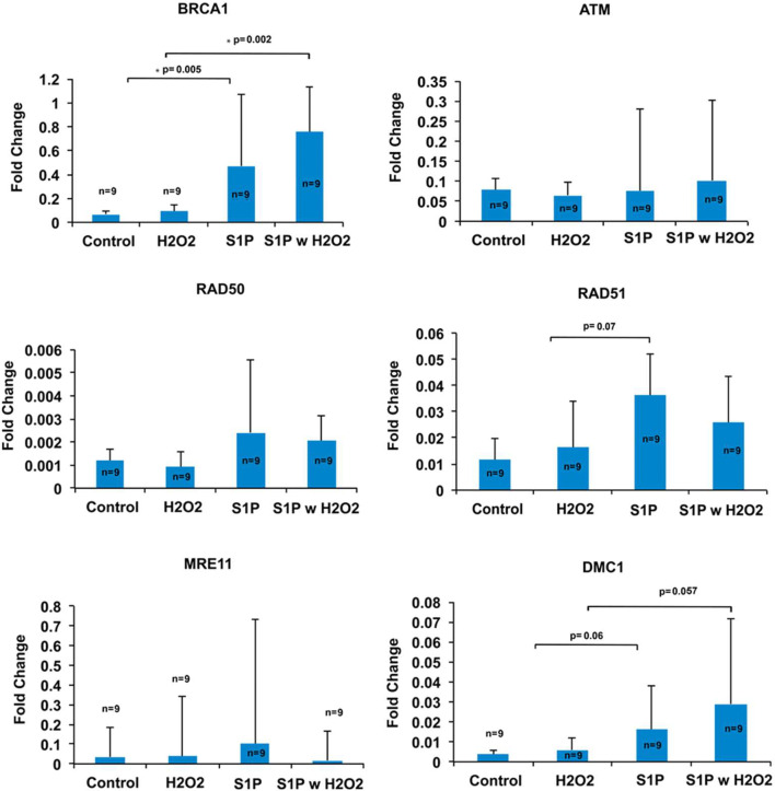
Recent data suggest that paternal age can have major impact on reproductive outcomes, and with increased age, there is increased likelihood of chromosomal abnormalities in the sperm. Here, we studied DNA damage and repair as a function of male aging and assessed whether sphingosine-1-phosphate (S1P), a ceramide-induced death inhibitor, can prevent sperm aging by enhancing DNA double-strand breaks (DSB) repair. We observed a significant increase in DNA damage with age and this increase was associated with a decline in the expression of key DNA DSB repair genes in mouse sperm. The haploinsufficiency of BRCA1 male mice sperm showed significantly increased DNA damage and apoptosis, along with decreased chromatin integrity when compared to similar age wild type (WT) mice. Furthermore, haploinsufficiency of BRCA1 male mice had lower sperm count and smaller litter size when crossed with WT females. The resulting embryos had a higher probability of growth arrest and reduced implantation. S1P treatment decreased genotoxic-stress-induced DNA damage in sperm and enhanced the expressions of key DNA repair genes such as BRCA1. Co-treatment with an ATM inhibitor reversed the effects of S1P, implying that the impact of S1P on DNA repair is via the ATM-mediated pathway. Our findings indicate a key role for DNA damage repair mechanism in the maintenance of sperm integrity and suggest that S1P can improve DNA repair in sperm. Further translational studies are warranted to determine the clinical significance of these findings and whether S1P can delay male reproductive aging. There is mounting evidence that sperm quality declines with age, similar to that of the oocyte. However, the reasons behind this decline are poorly understood and there is no medical intervention to improve sperm quality. Our study suggests a strong role for DNA damage repair in maintenance of sperm quality, and for the first time, a potential pharmaceutical approach to prevent sperm aging.

Keywords: Aging; DNA fragmentation; Gene expression; Sperm.


Conflict of interest statement

The authors declare that they have no competing interests.

Figures

Fig. 1
Fig. 1
Age-related DNA damage in testicular tissue and mature sperm cells. a Representative photomicrographs show γH2AX immunostaining of sperm cells of young and old mice at × 40 magnification. b Photomicrographs represent γH2AX foci (green) and counterstaining of DAPI (blue) of sperm cells from young and old WT mice at × 40 magnification. Arrowheads show increased numbers of γH2AX foci in old WT mice as compared to young WT mice. c Bar graph shows a higher expression of γH2AX as represented by the intensity of the γH2AX immunofluorescence in the spermatogonium and spermatocytes in testicular tissue of old (169.2 ± 3.0 vs. 134.6 ± 8.9, n = 7; P = 0.003; Student’s t test) compared to young. All bar graphs show means ± SEM
Fig. 2
Fig. 2
Age-related decline in sperm quality. Aged mice showed an a increase in DNA damage as reported by γH2AX expression through utilizing laser scanning cytometry with at least 1000 cells counted per sample (4.6 ± 0.6 vs. 15.4 ± 3.5, n = 12; P = 0.006, Student’s t test), b increased susceptibility of DNA of sperm cells to denaturation by the acridine orange assay (17.4 ± 5.3 vs. 32.6 ± 3.6, n = 10; P = 0.027, Student’s t test), c increased percentage of apoptotic cells detected by the FLICA analysis (13.9 ± 1.5 vs. 35.5 ± 3.7, n = 10; P = 7.9 × 10−5, Student’s t test), and d lower sperm concentration (2.1 × 106 ± 0.07 × 106 vs. 1.7 × 106 ± 0.12 × 106, n = 8; P = 0.05, Student’s t test) as compared to young. e Old male mice resulted in smaller litter size when mated with WT females compared to when WT males were mated with WT females (young vs. old: 5.2 ± 0.72 vs. 6.9 ± 0.42, n = 9; P = 0.05, Student’s t test). All bar graphs show the means ± SEM
Fig. 3
Fig. 3
Increased sperm DNA damage in in haploinsufficient BRCA1 male mouse. a Representative photomicrographs show γH2AX staining of haploinsufficient BRCA1 mice testicular tissue at × 40 magnification. Bar graph shows a higher expression of γH2AX as represented by the intensity of the γH2AX immunofluorescence in the spermatogonium and spermatocytes in testicular tissue of haploinsufficient BRCA1 mice testis by laser scanning cytometry (155.2 ± 3.4 vs. 134.6 ± 8.9, n = 7; P = 0.05, Student’s t test). b Photomicrographs represent γH2AX foci (green) and counterstaining of DAPI (blue) of sperm cells from young and old WT mice at × 40 magnification. Bar graph shows an increase in DNA damage as reported by γH2AX expression (15.9 ± 4.3 vs. 4.6 ± 0.6, n = 12; P = 0.03, Student’s t test), c increased susceptibility of DNA of sperm cells to denaturation by the acridine orange assay (30.6 ± 3.4 vs. 17.4 ± 5.3, n = 10; P = 0.05, Student’s t test), d increased percentage of apoptosis via FLICA analysis (30.7 ± 5.4 vs. 13.9 ± 1.5, n = 10; P < 0.014, Student’s t test), and e lower sperm concentration compared to WT (1.8 × 106 ± 0.1 × 106 vs. 2.1 × 106 ± 0.07 × 106, n = 8; P = 0.045, Student’s t test). f Haploinsufficiency of BRCA1 male mice resulted in smaller litter size when mated with WT females compared to when WT males were mated with WT females (3.2 ± 0.77 vs. 6.9 ± 0.42, n = 9; P = 0.0008, Student’s t test). g Representative photomicrographs show implantation sites produced by crossing BRCA1 WT mice with WT females and haploinsufficient BRCA1 male mice with WT females. Bar graph shows implantation sites were significantly decreased in WT females crossed with haploinsufficient BRCA1 male mice (0.43 ± 0.30 vs. 6.60 ± 2.34, n = 7; P = 0.05, Student’s t test) when compared to crossing with WT males. h Representative photomicrographs show increased percentage of arrested embryos produced by the crosses mentioned from the implantation sites at × 20 magnification. Bar graph shows a higher percentage of arrested embryos produced by crossing haploinsufficient BRCA1 male mice with WT females (32.14% ± 0.13% vs. 8.33% ± 0.07%, n = 7; P = 0.026, Student’s t test) as compared to WT mice. All results are mean ± SEM
Fig. 4
Fig. 4
Age-related decline in the expression of DNA repair genes in mouse sperm. Significant decreases in the expression of DNA repair genes in old WT mice as compared to young shown by real-time PCR. All results are mean ± SEM (n = 8 per group). Bar graphs represent the gene expressions, which are significantly lower for key DNA repair genes with age, including a BRCA1 (0.05 ± 0.01 vs. 0.04 ± 0.001, P = 0.048, Student’s t test), b ATM (0.011 ± 0.003 vs. 0.006 ± 0.002, P = 0.014, Student’s t test), c MRE11 (0.6 ± 0.3 vs. 0.1 ± 0.05, P = 0.041, Student’s t test), d DMC1 (0.008 ± 0.004 vs. 0.002 ± 0.004, P = 0.036, Student’s t test), e RAD50 (2.2 × 10−4 ± 5 × 10−5 vs. 9.2 × 10−5 ± 8 × 10−5, P = 0.028, Student’s t test), and f RAD51 (0.3 ± 0.4 vs. 0.01 ± 0.4, P = 0.0009, Student’s t test)
Fig. 5
Fig. 5
Compromised sperm DNA repair gene function in haploinsufficient BRCA1 male mouse. All results are mean ± SEM (n = 8 per group). Bar graphs represent the expressions of key DNA repair genes by qRT-PCR, which are significantly lower for a BRCA1 (0.028 ± 0.012 vs. 0.05 ± 0.01, P = 0.019, Student’s t test), b ATM (0.002 ± 0.0005 vs. 0.011 ± 0.003, P = 0.006, Student’s t test), c MRE11 (0.045 ± 0.018 vs. 0.6 ± 0.3, P = 0.02, Student’s t test), and d DMC1 (0.002 ± 9 × 10−4 vs. 0.008 ± 0.004, P = 0.02, Student’s t test), e RAD50 (9.7 × 10−5 ± 8.9 × 10−6 vs. 2.2 × 10−4 ± 5 × 10−5, P = 0.044, Student’s t test), and f RAD51 (0.002 ± 8 × 10−4 vs. 0.03 ± 0.4, P = 0.015, Student’s t test) compared to WT.
Fig. 6
Fig. 6
S1P treatment enhances DNA DSB repair in sperm. S1P treatment induced the expression of BRCA1 compared to baseline (0.471 ± 0.602 vs. 0.068 ± 0.031, n = 9; P = 0.005, Student’s t test) and H2O2-treated sperm (0.755 ± 0.377 vs. 0.090 ± 0.053, n = 9; P = 0.002 Student’s t test). Furthermore, there was a trend for increase in the expression of Rad51 (0.036 ± 0.015 vs. 0.012 ± 0.008, n = 9; P = 0.07, Student’s t test) and DMC1 (0.016 ± 0.02 vs. 0.003 ± 0.002, n = 9; P = 0.06, Student’s t test) in the S1P treated samples when compared to the controls. Other genes tested showed no significant difference in the S1P treated sperm when compared to the control. All results are mean ± SEM

Similar articles

Cited by

References

    1. Herbert M, Kalleas D, Cooney D, Lamb M, Lister L. Meiosis and maternal aging: insights from aneuploid oocytes and trisomy births. Cold Spring Harb Perspect Biol. 2015;7(4):a017970. doi: 10.1101/cshperspect.a017970. - DOI - PMC - PubMed
    1. Titus S, Li F, Stobezki R, et al. Impairment of BRCA1-related DNA double-strand break repair leads to ovarian aging in mice and humans. Sci Transl Med. 2013;5(172):172ra121. doi: 10.1126/scitranslmed.3004925. - DOI - PMC - PubMed
    1. Kuhnert B, Nieschlag E. Reproductive functions of the ageing male. Hum Reprod Update. 2004;10(4):327–339. doi: 10.1093/humupd/dmh030. - DOI - PubMed
    1. Johnson SL, Dunleavy J, Gemmell NJ, Nakagawa S. Consistent age-dependent declines in human semen quality: a systematic review and meta-analysis. Ageing Res Rev. 2015;19:22–33. doi: 10.1016/j.arr.2014.10.007. - DOI - PubMed
    1. Belloc S, Benkhalifa M, Cohen-Bacrie M, Dalleac A, Amar E, Zini A. Sperm deoxyribonucleic acid damage in normozoospermic men is related to age and sperm progressive motility. Fertil Steril. 2014;101(6):1588–1593. doi: 10.1016/j.fertnstert.2014.02.006. - DOI - PubMed

Publication types