. 2020 Mar 24;141(12):968-983.
doi: 10.1161/CIRCULATIONAHA.119.042640. Epub 2020 Jan 10.

Small Extracellular Microvesicles Mediated Pathological Communications Between Dysfunctional Adipocytes and Cardiomyocytes as a Novel Mechanism Exacerbating Ischemia/Reperfusion Injury in Diabetic Mice

Affiliations

Small Extracellular Microvesicles Mediated Pathological Communications Between Dysfunctional Adipocytes and Cardiomyocytes as a Novel Mechanism Exacerbating Ischemia/Reperfusion Injury in Diabetic Mice

Lu Gan et al. Circulation. .

Abstract

Background: Diabetes mellitus exacerbates myocardial ischemia/reperfusion (MI/R) injury by incompletely understood mechanisms. Adipocyte dysfunction contributes to remote organ injury. However, the molecular mechanisms linking dysfunctional adipocytes to increased MI/R injury remain unidentified. The current study attempted to clarify whether and how small extracellular vesicles (sEV) may mediate pathological communication between diabetic adipocytes and cardiomyocytes, exacerbating MI/R injury.

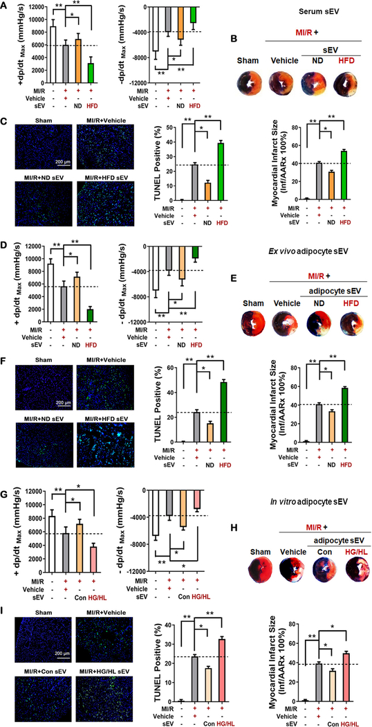
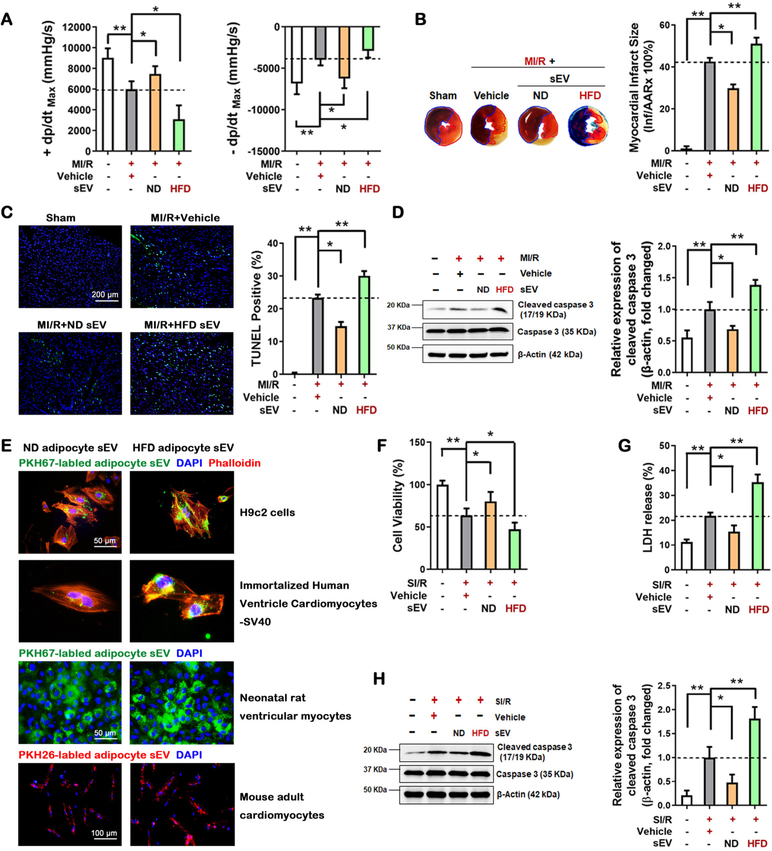
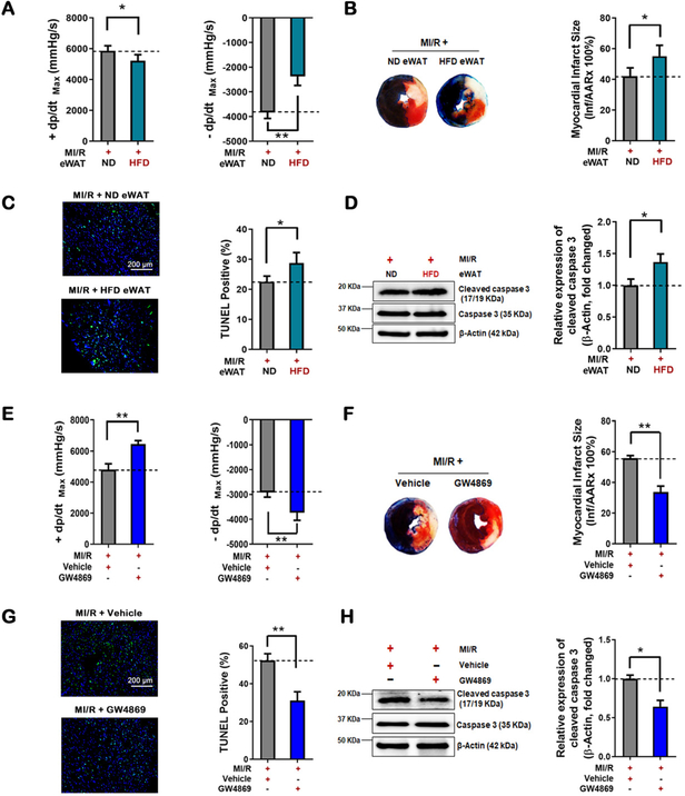
Methods: Adult male mice were fed a normal or a high-fat diet for 12 weeks. sEV (from diabetic serum, diabetic adipocytes, or high glucose/high lipid-challenged nondiabetic adipocytes) were injected intramyocardially distal of coronary ligation. Animals were subjected to MI/R 48 hours after injection.

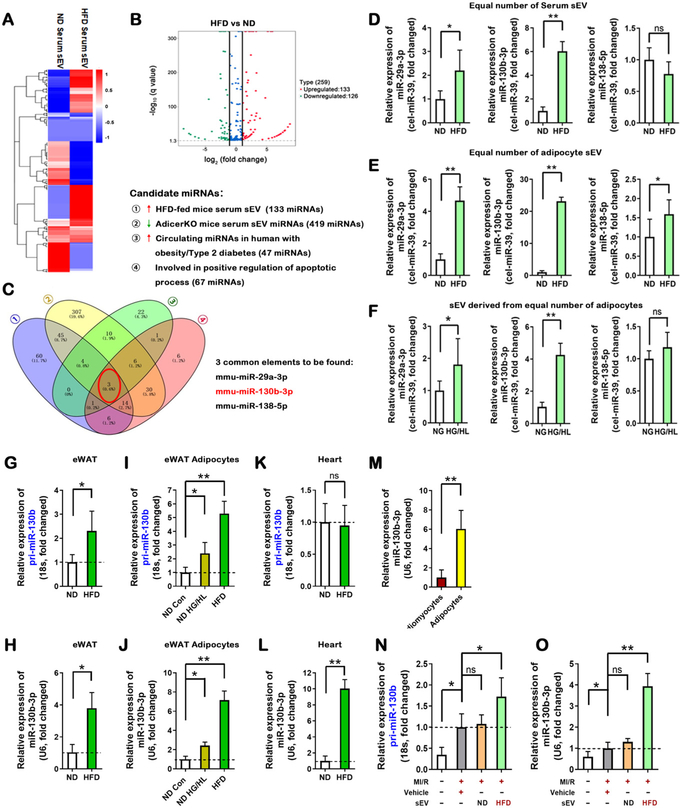
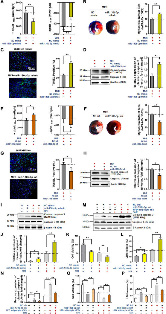
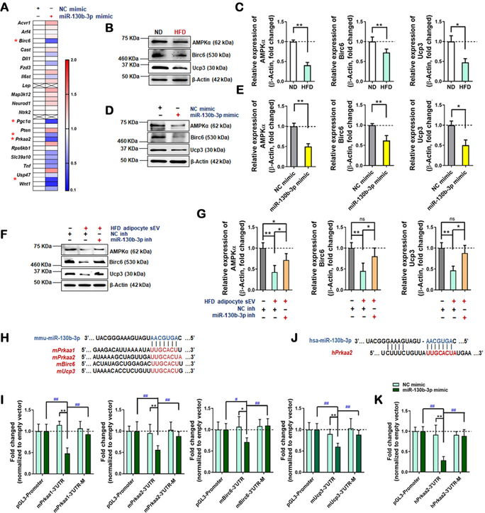
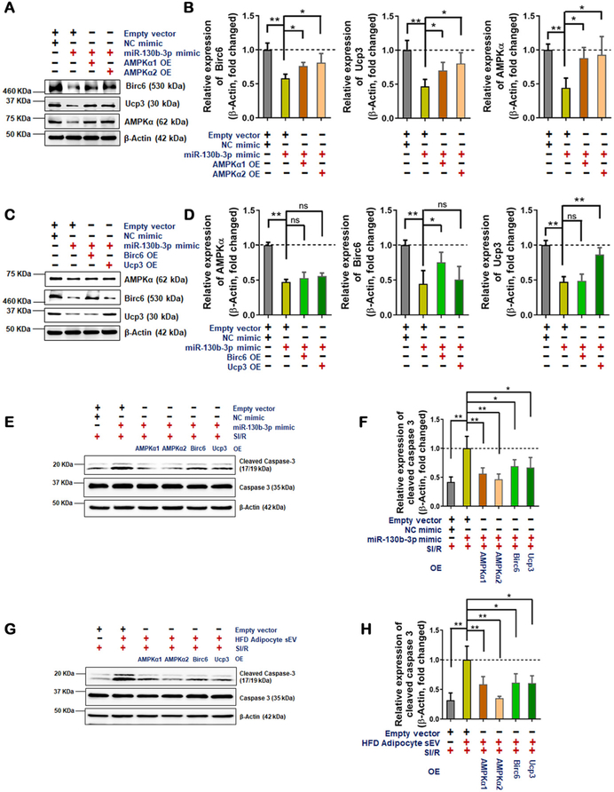
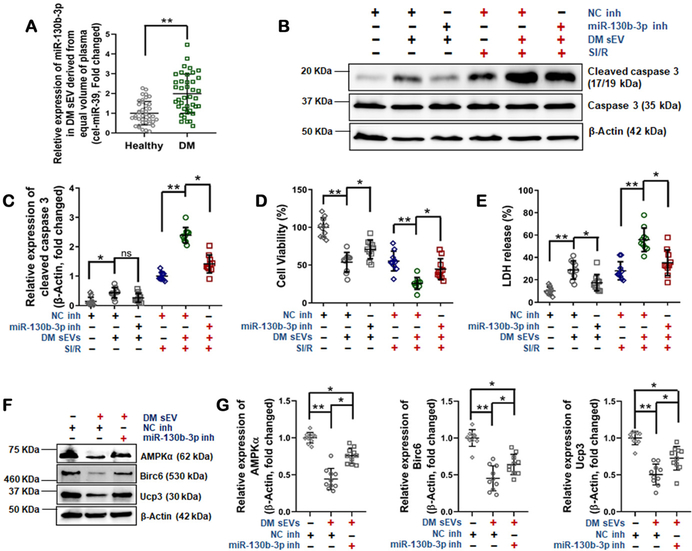
Results: Intramyocardial injection of diabetic serum sEV in the nondiabetic heart significantly exacerbated MI/R injury, as evidenced by poorer cardiac function recovery, larger infarct size, and greater cardiomyocyte apoptosis. Similarly, intramyocardial or systemic administration of diabetic adipocyte sEV or high glucose/high lipid-challenged nondiabetic adipocyte sEV significantly exacerbated MI/R injury. Diabetic epididymal fat transplantation significantly increased MI/R injury in nondiabetic mice, whereas administration of a sEV biogenesis inhibitor significantly mitigated MI/R injury in diabetic mice. A mechanistic investigation identified that miR-130b-3p is a common molecule significantly increased in diabetic serum sEV, diabetic adipocyte sEV, and high glucose/high lipid-challenged nondiabetic adipocyte sEV. Mature (but not primary) miR-130b-3p was significantly increased in the diabetic and nondiabetic heart subjected to diabetic sEV injection. Whereas intramyocardial injection of a miR-130b-3p mimic significantly exacerbated MI/R injury in nondiabetic mice, miR-130b-3p inhibitors significantly attenuated MI/R injury in diabetic mice. Molecular studies identified AMPKα1/α2, Birc6, and Ucp3 as direct downstream targets of miR-130b-3p. Overexpression of these molecules (particularly AMPKα2) reversed miR-130b-3p induced proapoptotic/cardiac harmful effect. Finally, miR-130b-3p levels were significantly increased in plasma sEV from patients with type 2 diabetes mellitus. Incubation of cardiomyocytes with diabetic patient sEV significantly exacerbated ischemic injury, an effect blocked by miR-130b-3p inhibitor.

Conclusions: We demonstrate for the first time that miR-130b-3p enrichment in dysfunctional adipocyte-derived sEV, and its suppression of multiple antiapoptotic/cardioprotective molecules in cardiomyocytes, is a novel mechanism exacerbating MI/R injury in the diabetic heart. Targeting miR-130b-3p mediated pathological communication between dysfunctional adipocytes and cardiomyocytes may be a novel strategy attenuating diabetic exacerbation of MI/R injury.

Keywords: apoptosis; diabetes mellitus; extracellular vesicles; ischemia-reperfusion injury; microRNA.


Figures

Figure 1.
Figure 1.. Deleterious effects of adipocyte sEV derived from obese/diabetic mice in MI/R-injury.
48 hours after sEV intramyocardial injection, mice were subjected to MI/R. (A, D, G) 24 hours after reperfusion, cardiac function was evaluated by hemodynamic testing (n≥15). (B, E, H) Cardiac injury was identified by myocardial Evans blue/TTC double stain (n≥8). (C, F, I) 3 hours after reperfusion, cardiomyocyte apoptosis was determined by TUNEL assay (n=8). (All experiment groups were compared to MI/R+Vehicle group via One-way ANOVA, *p<0.05, **p<0.01) Abbreviations: MI/R, myocardial ischemia/reperfusion; sEV, small extracellular vesicles; ND, normal diet; HFD, high fat diet; HG/HL, high glucose and high lipid administration.
Figure 2.
Figure 2.. The content shift of HFD adipocyte sEV was responsible for MI/R injury exacerbation.
48 hours after tantamount sEV intramyocardial injection, mice were subjected to MI/R. 24 hours after reperfusion. (A) cardiac function was evaluated by hemodynamic testing (n≥15). (B) Cardiac injury was identified by myocardial Evans blue/TTC double stain (n=8). (C, D) 3 hour after reperfusion, cardiomyocyte apoptosis was determined by (C) TUNEL assay (n=8) and (D) cleaved caspase-3 assay by Western blot (n=5). (E) In vitro adipocyte sEV uptaken analysis. PKH67-labled or PKH26-labled adipocyte sEV were harvested from primary adipocyte culture medium, and subsequently incubated with H9c2 cells, human cardiomyocytes, and adult mouse cardiomyocytes for 6 hours. (F) Neonatal rat ventricular myocyte (NRVM) cell viability was evaluated by MTT assay in vitro with adipocyte sEV plus simulated ischemia/reoxygenation (SI/R) administration (simulated ischemia 6 hours and reoxgenation 3 hours, n=5). (G) Cell death was determined by LDH release (n=5). (H) Cell apoptosis was determined by cleaved caspase-3 assay by Western blot (n=5). (All experiment groups were compared to MI/R+Vehicle group via One-way ANOVA, *p<0.05, **p<0.01).
Figure 3.
Figure 3.. Deleterious effects of dysfunctional adipose tissue and systemic sEV from HFD-fed mice in MI/R-injury.
(A-D) The epididymal adipose tissues (eWAT) were isolated from HFD mice or ND donor mice, and transplanted into 8-week-old male non-diabetic recipient mice removed epidydimal fat depots. 7 days after eWAT transplantation, MI/R was performed. (E-H) The HFD-fed mice were intraperitoneal injected GW4869 (2 mg/kg) or vehicle 3 times a week for 12 weeks. Then, MI/R was performed. (A, E) Cardiac function was evaluated by hemodynamic testing (n≥15). (B, F) Cardiac injury was identified by myocardial Evans blue/TTC double stain (n≥8). Cardiomyocyte apoptosis was determined by (C, G) TUNEL assay (n=8) and (D, H) cleaved caspase-3 assay by Western blot (n=5). (All experiment groups were compared to MI/R+ ND eWAT group or MI/R+Vehicle group via upaired t-test, *p<0.05, **p<0.01). Abbreviation: eWAT, epididymal white adipose tissue.
Figure 4.
Figure 4.. Increased miR-130b-3p present in HFD adipocyte sEV.
(A, B) Differentially expressed miRNAs were selected from sequencing data of serum sEV miRNAs, shown in (A) and (B). (C) Venn analysis selected three candidate miRNAs satisfying four conditions. (D, E) The relative expression of three mature miRNAs in equal numbers of serum sEV and eWAT adipocyte sEV. (F) The relative expression of three mature miRNAs, in adipocyte sEV derived from the same number of adipocytes (n=6, unpaired t-test, *p<0.05, **p<0.01). (G, H) The relative expression of pri-miR-130b and mature miR-130b-3p in eWAT from ND-fed and HFD-fed mice (12 weeks), analyzed by real-time PCR. (I, J) The relative expression of pri-miR-130b and mature miR-130b-3p in eWAT adipocytes from ND-fed and HFD-fed mice, or ND adipocytes treated with HG/HL, analyzed by real-time PCR. (K, L) The relative expression of pri-miR-130b and mature miR-130b-3p in heart tissue from ND-fed and HFD-fed mice, analyzed by real-time PCR. (M) The relative expression of miR-130b-3p in cardiomyocytes and adipocytes. (N, O) The relative expression of pri-miR-130b and mature miR-130b-3p in cardiac tissue +/− adipocyte sEV intramyocardial injection, analyzed by real-time PCR. (All n≥5, unpaired t-test and One-way ANOVA, *p<0.05, **p<0.01,). Abbreviations: ns, not statistically significant.
Figure 5.
Figure 5.. Deleterious effects of miR-130b-3p upon MI/R-injury in obese/diabetic mice and in cultured cells.
(A-D) miR-130b-3p increased MI/R-injury in non-diabetic mice. 48 hours after miR-130b-3p mimic and NC mimic transfection of the myocardium of non-diabetic mice, MI/R commenced. 24 hours after reperfusion, cardiac function was evaluated by hemodynamic testing (A, n=15, unpaired t-test, *p<0.05, **p<0.01). Cardiac injury was identified by myocardial Evans blue/TTC double stain (B, n=5); cardiomyocyte apoptosis was determined by TUNEL assay (C, n=8) and cleaved caspase-3 assay by Western blot (D, n=5). (E-H) miR-130b-3p-antagonism reduced MI/R-injury in obese/diabetic mice. 48 hours after miR-130b-3p inhibitor (miR-130b-3p inh) and NC inhibitor (NC inh) transfection of the myocardium of HFD-fed mice, MI/R commenced. 24 hours after reperfusion, cardiac function were evaluated by hemodynamic testing (E, n=15, unpaired t-test, *p<0.05, **p<0.01). Cardiac injury was identified by myocardial Evans blue/TTC double stain (F, n=5); cardiomyocyte apoptosis was determined by TUNEL assay (G, n=8) and cleaved caspase-3 assay by Western blot (H, n=5). (Unpaired t-test, *p<0.05, **p<0.01). miR-130b-3p activated cardiomyocyte apoptosis in vitro. Expression of cleaved caspase-3 (I and J), cell viability (K) and LDH release (L) was detected in NRVM cells transfected by mimics of miR-130b-3p or NC for 24 hours and received SI/R administration. Expression of cleaved caspase-3 (M and N), cell viability (O) and LDH release (P) in NRVM cells overexpressing miR-130b-3p inhibitor before HFD adipocyte sEV + SI/R administration. The cells were transfected with miR-130b-3p inh or NC inh 24 hours before HFD adipocyte sEV treatment, and then subjected to SI/R performance 24 hours after HFD adipocyte sEV treatment. (n≥5, One-way ANOVA, *p<0.05, **p<0.01)
Figure 6.
Figure 6.. miR-130b-3p decreased expression of AMPKα and downstream molecules.
(A) Real-time PCR analysis detected expressions of the predicted targets of miR-130b-3p involved in negative regulation of apoptosis in heart tissue with or without miR-130b-3p administration (n=3, multiple unpaired t-test, *p<0.05, **p<0.01) (B and C) Proteins levels were detected by Western blot in cardiac tissue from ND-fed and HFD-fed mice (n=5, unpaired t-test, *p<0.05, **p<0.01). (D and E) Proteins levels were detected in NRVM cells +/− miR-130b-3p mimic or NC mimic (n=5, unpaired t-test, *p<0.05, **p<0.01). (F and G) Protein levels were detected in NRVM cells overexpressing miRNA inhibitors before HFD adipocyte sEV administration. The cells were transfected with miR-130b-3p inh or NC inh 24 hours before HFD adipocyte sEV treatment, and 24 hours after HFD adipocyte sEV treatment, the protein levels were detected. (n=9, One-way ANOVA, *p<0.05, **p<0.01) (H and I) The direct effects of miR-130b-3p upon mouse AMPKα1/2, Birc6, and Ucp3 were identified by reporter gene analysis. (J and K) The direct effects of miR-130b-3p upon human AMPKα2 were identified by reporter gene analysis. The reported plasmids containing the gene mRNA 3’UTR regions (including binding sites) are shown in E and G (mutated binding sites were reverse in sequence). NRVM cells or HEK cells were cotransfected by the miRNA mimics and reporter plasmids for 48 hours. The regulatory effects were evaluated by firefly/renilla luciferase activity. Fold-change was calculated by dividing the value of firefly/renilla luciferase activity in each group transfected with miR-130b-3p mimic by the value obtained from the group transfected with the same reporter constructs and NC mimic. The empty vector (pGL3-Promoter) transfected group served as control, and its value was set as 1.0 (n≥6, two-way ANOVA, comparisons to NC mimic group, *p<0.05, **p<0.01; for mutated reporter plasmid and miR-130b-3p mimic values compared to groups with normal reporter plasmid transfection + miR-130b-3p mimic, #p<0.05, ##p<0.01).
Figure 7.
Figure 7.. miR-130b-3p-mediated AMPKα downregulation contributed to the cardiomyocyte apoptosis induced by HFD adipocyte sEV.
(A and B) AMPKα1 and AMPKα2 overexpression restored the Birc6 and Ucp3 protein levels in NRVM cells with miR-130b-3p mimic administration. (C and D) Birc6 and Ucp3 overexpression did not restored the AMPKα protein levels in NRVM cells with miR-130b-3p mimic administration. (n=5, One-way ANOVA, comparisons to empty vector + miR-130b-3p mimic group *p<0.05, **p<0.01) (E-H) AMPKα1/2, Birc6 and Ucp3 overexpression downregulated miR-130b-3p mimic + SI/R administration-induced (E and F) or HFD adipocyte sEV + SI/R administration-induced (G and H) cleaved caspase3 expression. Western blot detected protein levels in NRVM cells with plasmid-mediated exogenous genes infusion after miR-130b-3p mimic transfection or HFD adipocyte sEV administration. 24 hours after miRNA mimic transfection or sEV administration, the plasmids were transfected. 24 hours later, SI/R was performed. Plasmids overexpressed AMPKα1/2, Birc6 and Ucp3. Empty vector + miR-130b-3p mimic transfected group with SI/R treatment served as control, and its value was set at 1.0. (n=5, One-way ANOVA, *p<0.05, **p<0.01)
Figure 8.
Figure 8.
Plasma sEV from diabetic patients promoted SI/R-induced NRVM apoptosis. (A) miR-130b-3p expression in plasma sEV derived from patients with type 2 diabetes (DM sEV) and healthy controls (n=40, unpaired t-test, *p<0.05, **p<0.01). (B-E) DM sEV exacerbated SI/R-induced NRVM death, an effect attenuated by overexpressing miR-130b-3p inhibitor (B/C: cleaved caspase-3; D: cell viability; E: LDH release). (n=10/group, One-way ANOVA, *p<0.05, **p<0.01). (F and G) DM sEV inhibited AMPKα1/2, Birc6, and Ucp3 expression, an effect reversed by overexpressing miR-130b-3p inhibitor. (n=10, One-way ANOVA, *p<0.05, **p<0.01).

Comment in

References

    1. Oikonomou EK and Antoniades C. The role of adipose tissue in cardiovascular health and disease. Nat Rev Cardiol. 2019;16:83–99. - PubMed
    1. Mazzone T Intensive Glucose Lowering and Cardiovascular Disease Prevention in Diabetes: Reconciling the Recent Clinical Trial Data. Circulation. 2010;122:2201–2211. - PMC - PubMed
    1. Duckworth W, Abraira C, Moritz T, Reda D, Emanuele N, Reaven PD, Zieve FJ, Marks J, Davis SN, Hayward R, Warren SR, Goldman S, McCarren M, Vitek ME, Henderson WG and Huang GD. Glucose control and vascular complications in veterans with type 2 diabetes. N Engl J Med. 2009;360:129–139. - PubMed
    1. Katayama T, Nakashima H, Takagi C, Honda Y, Suzuki S, Iwasaki Y and Yano K. Clinical outcomes and left ventricular function in diabetic patients with acute myocardial infarction treated by primary coronary angioplasty. Int Heart J. 2005;46:607–618. - PubMed
    1. Ansley DM and Wang B. Oxidative stress and myocardial injury in the diabetic heart. J Pathol. 2013;229:232–241. - PMC - PubMed

Publication types