. 2020 Jan 9;18(1):10.
doi: 10.1186/s12951-019-0563-2.

Engineered exosomes for targeted co-delivery of miR-21 inhibitor and chemotherapeutics to reverse drug resistance in colon cancer

Affiliations

Engineered exosomes for targeted co-delivery of miR-21 inhibitor and chemotherapeutics to reverse drug resistance in colon cancer

Gaofeng Liang et al. J Nanobiotechnology. .

Abstract

Background: 5-Fluorouracil (5-FU) has been commonly prescribed for patients with colorectal cancer (CRC), but resistance to 5-FU is one of the main reasons for failure in CRC. Recently, microRNAs (miRNAs) have been established as a means of reversing the dilemma by regulating signaling pathways involved in initiation and progression of CRC. However, how to safely and effectively deliver miRNA to target cells becomes a main challenge.

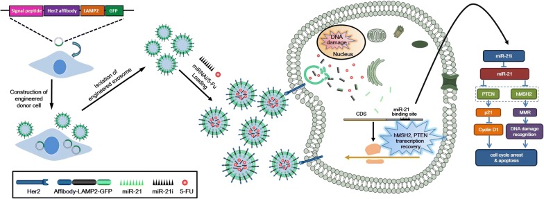
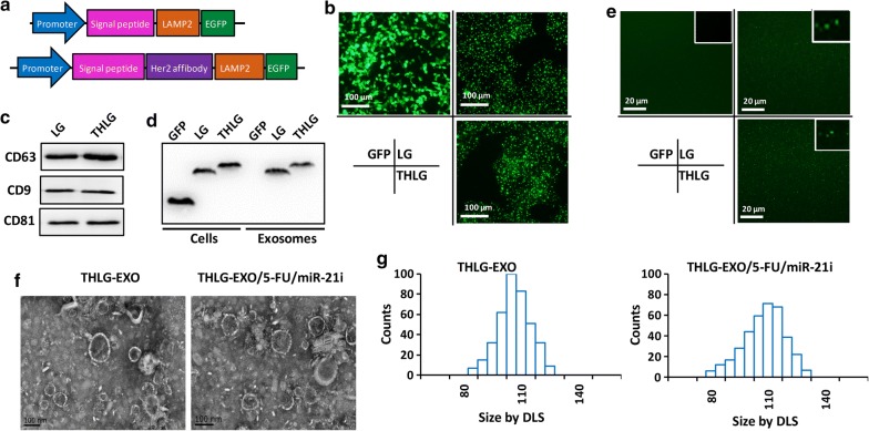
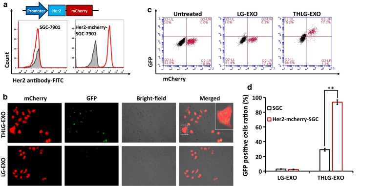
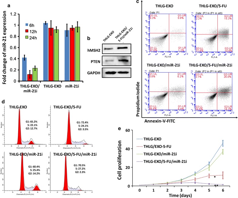
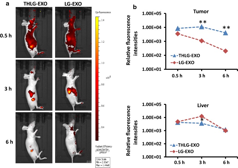
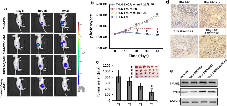
Results: In this study, Engineered exosomes were exploited to simultaneously deliver an anticancer drug 5-FU and miR-21 inhibitor oligonucleotide (miR-21i) to Her2 expressing cancer cells. Purified engineered exosomes from the donor cells loaded with 5-FU and miR-21i via electroporation to introduce into 5-FU-resistant colorectal cancer cell line HCT-1165FR. Furthermore, systematic administration of 5-FU and miR-21i loaded exosomes in tumor bearing mice indicated a significantly anti-tumor effect. The results showed that the engineered exosome-based 5-FU and miR-21i co-delivery system could efficiently facilitate cellular uptake and significantly down-regulate miR-21 expression in 5-FU resistant HCT-1165FR cell lines. Consequently, the down-regulation of miR-21 induced cell cycle arrest, reduced tumor proliferation, increased apoptosis and rescued PTEN and hMSH2 expressions, regulatory targets of miR-21. Of particular importance was the significant reduction in tumor growth in a mouse model of colon cancer with systematic administration of the targeting miR-21i. More excitedly, the combinational delivery of miR-21i and 5-FU with the engineered exosomes effectively reverse drug resistance and significantly enhanced the cytotoxicity in 5-FU-resistant colon cancer cells, compared with the single treatment with either miR-21i or 5-FU.

Conclusion: The strategy for co-delivering the functional small RNA and anticancer drug by exosomes foreshadows a potential approach to reverse the drug resistance in CRC and thus to enhance the efficacy of the cancer treatment.

Keywords: 5-FU; Delivery system; Drug resistance; Exosomes; miR-21 inhibitor.


Conflict of interest statement

The authors declare that they have no competing interests.

Figures

Scheme 1
Scheme 1
Engineered exosomes based nanocarrier for 5-FU and miR-21i simultaneously deliver to HCT-1165FR human colon cancer cells for enhancing chemotherapy efficacy
Fig. 1
Fig. 1
Isolation and molecular characterization of engineered exosomes. a Schematics of DNA constructs used for the production of fusion proteins LG (upper) and THLG (lower). b Fluorescence images of the GFP, LG and THLG in 293T cells ( ×400). c Biomarkers of exosomes were determined by western blot analysis of exosomes. d Western blot analysis of GFP in exosomes obtained from supernatants of GFP-293T cells, LG-293T cells and THLG-293T cells. e Confocal microscopy of exosomes isolated from 0.22 filtered conditioned medium of GFP-293T cells, LG-293T cells and THLG-293T cells ( ×1000). f Transmission electron micrograph of THLG-EXO and THLG-EXO/5-FU/miR-21i. g Size distribution of THLG-EXO and THLG-EXO/5-FU/miR-21i measured by DLS
Fig. 2
Fig. 2
The cellular tropism of THLG-EXO in vitro. a Schematics of DNA constructs used for the production of fusion proteins Her2-mCherry (upper panel); flow cytometric analyses of Her2 from SGC-7901 WT cells and Her2-mcherry-SGC-7901 cells with a FITC labeled anti-Her2 monoclonal antibody (lower panel). b Confocal microscopy images of cellular uptake of THLG-EXO or LG-EXO after 3 h incubation with SGC-7901 WT cells and Her2-mcherry-SGC-7901 cells co-culture model. red shows Her2-mcherry-SGC-7901 cells. Green represents THLG-EXO or LG-EXO. Right panel shows enlarged graphs with frame indicating internalized exosomes in cells (×400). c Flow cytometry analysis of co-culture cells after incubation with THLG-EXO or LG-EXO (upper panel); d quantification of exosomes internalization based on flow cytometry analysis (lower panel). Data are expressed as mean ± SD. n = 5; **p < 0.01
Fig. 3
Fig. 3
The results of HCT-1165FR cells treated with THLG-EXO/5-FU/miR-21i in vitro. a Real-time fluorescent quantitative PCR analysis of relative expression change of miR-21 level in HCT-1165FR cells treated with THLG-EXO/5-FU/miR-21i, THLG-EXO or miR-21i. Data were expressed as mean ± SD. n = 3. b Western blot analysis of hMSH2 and PTEN protein levels of HCT-1165FR cells treated with THLG-EXO/5-FU/miR-21i. c Flow cytometric analysis of AnnexinV-FITC/PI stained HCT-1165FR cells treated with THLG-EXO, THLG-EXO/miR-21i, THLG-EXO/5-FU or THLG/5-FU/miR-21i. d Cell cycle profiles of HCT-1165FR cells after treatment as described in c; e the proliferation of HCT-1165FR cells after treatment as described in c. Data were expressed as means ± SD. n = 5;*: significantly different from other groups (p < 0.05), **: extremely significantly different from other groups (p < 0.01)
Fig. 4
Fig. 4
Tumor targeting ability of THLG-EXO in a nude mouse xenograft model using HCT-1165FR cells. a In vivo fluorescent images of HCT-1165FR-bearing nude mice after intravenous injection of DiR-labeled THLG-EXO (left panel) or LG-EXO (right panel). b Quantitative analysis showed the relative fluorescence intensities in the tumor region (upper panel) and the liver (lower panel) following administration of THLG-EXO or LG-EXO. Data were expressed as means ± SD. n = 5; *P < 0.05, **P < 0.01 versus the LG-EXO group at the same time point
Fig. 5
Fig. 5
Antitumor activity of THLG-EXO/5-FU/miR-21i in nude mice xenograft using HCT-1165FR cells. a Representative bioluminescent images of tumor growth in nude mice treated with THLG-EXO, THLG-EXO/miR-21i, THLG-EXO/5-FU or THLG-EXO/5-FU/miR-21i at different time point. Signals were adjusted to the same color scale for the entire time course. b The mean bioluminescence intensity (BLI) for each mouse/group, line chart represented quantification of the relative bioluminescent intensity in tumor site. c Tumor weights of different treated groups. T1, T2, T3 and T4 represented the treatment of THLG-EXO, THLG-EXO/miR-21i, THLG-EXO/5-FU, THLG-EXO/5-FU/miR-21i, respectively. d TUNEL assessments of tumor tissues treated with THLG-EXO, THLG-EXO/miR-21i, THLG-EXO/5-FU, THLG-EXO/5-FU/miR-21i, respectively. e Western blot of hMSH2 and PTEN in the tissue of tumor region were performed for 24 h after administration. Data were expressed as means ± SD. n = 6; *P < 0.05, **P < 0.01 versus the THLG-EXO groups or THLG-EXO/miR-21i group, and #P < 0.05 versus the THLG-EXO/5-FU groups
Fig. 6
Fig. 6
The systematic toxicity assessment of THLG-EXO. Histopathological analysis of heart, lung, liver, kidney and spleen sections stained with hematoxylin and eosin of BALB/c mice post-intravenous injection of 20 mg/kg THLG-EXO or PBS for 7 days (one dose every other day). Images were obtained under Nikon Ti microscope using a  × 40 objective

References

    1. Siegel RL, Miller KD, Jemal A. Cancer statistics, 2018. CA Cancer J Clin. 2018;68:7–30. doi: 10.3322/caac.21442. - DOI - PubMed
    1. Krishna R, Mayer LD. Multidrug resistance (MDR) in cancer. Mechanisms, reversal using modulators of MDR and the role of MDR modulators in influencing the pharmacokinetics of anticancer drugs. Eur J Pharm Sci. 2000;11:265–283. doi: 10.1016/S0928-0987(00)00114-7. - DOI - PubMed
    1. Baker EK, El-Osta A. The rise of DNA methylation and the importance of chromatin on multidrug resistance in cancer. Exp Cell Res. 2003;290:177–194. doi: 10.1016/S0014-4827(03)00342-2. - DOI - PubMed
    1. To KK, Polgar O, Huff LM, Morisaki K, Bates SE. Histone modifications at the ABCG2 promoter following treatment with histone deacetylase inhibitor mirror those in multidrug-resistant cells. Mol Cancer Res. 2008;6:151–164. doi: 10.1158/1541-7786.MCR-07-0175. - DOI - PMC - PubMed
    1. Wang S, Su X, Bai H, Zhao J, Duan J, An T, Zhuo M, Wang Z, Wu M, Li Z, et al. Identification of plasma microRNA profiles for primary resistance to EGFR-TKIs in advanced non-small cell lung cancer (NSCLC) patients with EGFR activating mutation. J Hematol Oncol. 2015;8:127. doi: 10.1186/s13045-015-0210-9. - DOI - PMC - PubMed

MeSH terms