

Persistent Proarrhythmic Neural Remodeling Despite Recovery From Premature Ventricular Contraction-Induced Cardiomyopathy
- PMID: 31918815
- PMCID: PMC7006705
- DOI: 10.1016/j.jacc.2019.10.046
Persistent Proarrhythmic Neural Remodeling Despite Recovery From Premature Ventricular Contraction-Induced Cardiomyopathy
Abstract
Background: The presence and significance of neural remodeling in premature ventricular contraction-induced cardiomyopathy (PVC-CM) remain unknown.
Objectives: This study aimed to characterize cardiac sympathovagal balance and proarrhythmia in a canine model of PVC-CM.
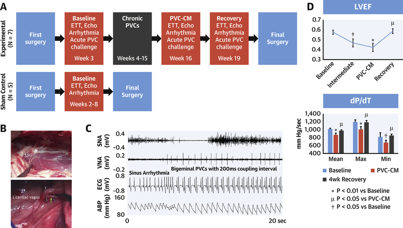
Methods: In 12 canines, the investigators implanted epicardial pacemakers and radiotelemetry units to record cardiac rhythm and nerve activity (NA) from the left stellate ganglion (SNA), left cardiac vagus (VNA), and arterial blood pressure. Bigeminal PVCs (200 ms coupling) were applied for 12 weeks to induce PVC-CM in 7 animals then disabled for 4 weeks to allow complete recovery of left ventricular ejection fraction (LVEF), versus 5 sham controls.
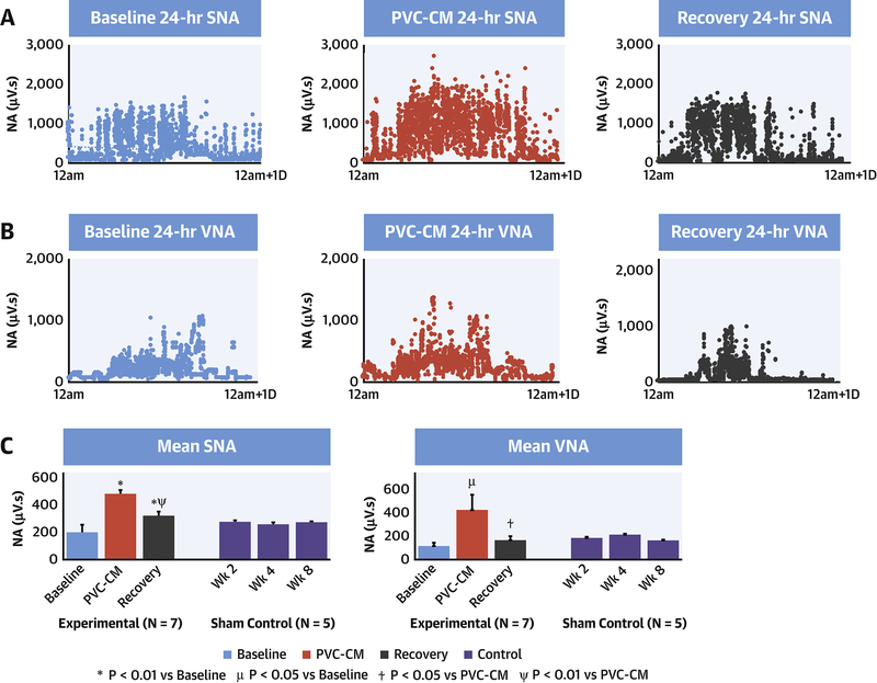
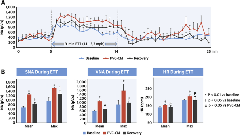
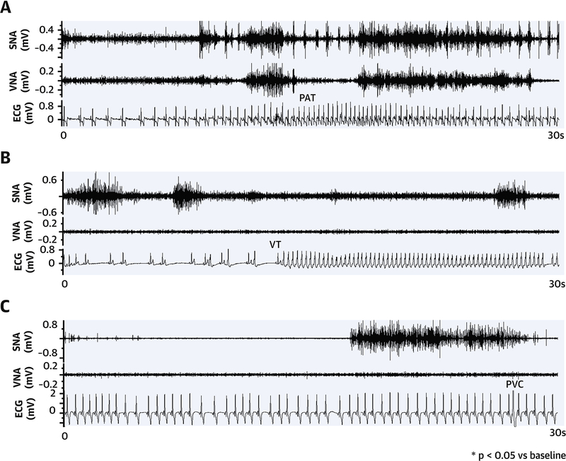
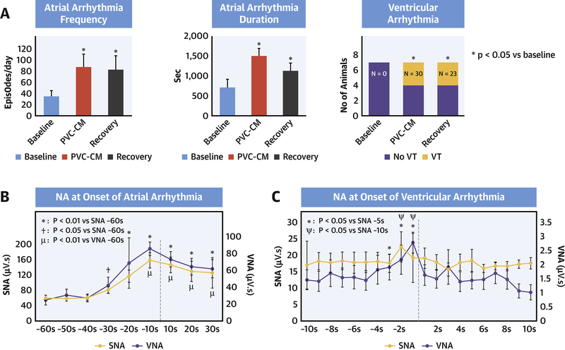
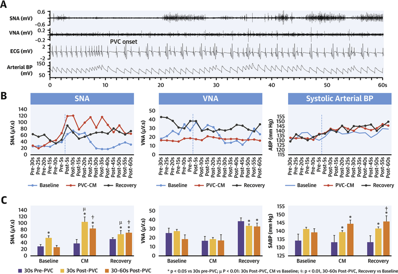
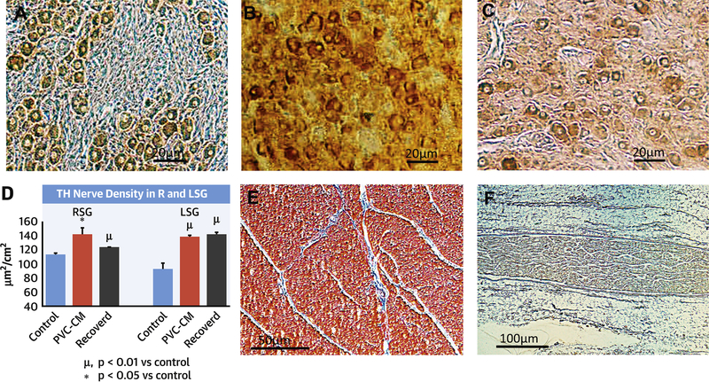
Results: After 12 weeks of PVCs, LVEF (p = 0.006) and dP/dT (p = 0.007) decreased. Resting SNA (p = 0.002) and VNA (p = 0.04), exercise SNA (p = 0.01), SNA response to evoked PVCs (p = 0.005), heart rate (HR) at rest (p = 0.003), and exercise (p < 0.04) increased, whereas HR variability (HRV) decreased (p = 0.009). There was increased spontaneous atrial (p = 0.02) and ventricular arrhythmias (p = 0.03) in PVC-CM. Increased SNA preceded both atrial (p = 0.0003) and ventricular (p = 0.009) arrhythmia onset. Clonidine suppressed SNA and abolished all arrhythmias. After disabling PVC for 4 weeks, LVEF (p = 0.01), dP/dT (p = 0.047), and resting VNA (p = 0.03) recovered to baseline levels. However, SNA, resting HR, HRV, and atrial (p = 0.03) and ventricular (p = 0.03) proarrhythmia persisted. There was sympathetic hyperinnervation in stellate ganglia (p = 0.02) but not ventricles (p = 0.2) of PVC-CM and recovered animals versus sham controls.
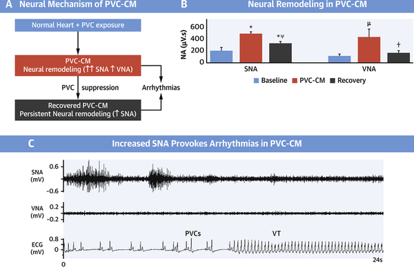
Conclusions: Neural remodeling in PVC-CM is characterized by extracardiac sympathetic hyperinnervation and sympathetic neural hyperactivity that persists despite normalization of LVEF. The altered cardiac sympathovagal balance is an important trigger and substrate for atrial and ventricular proarrhythmia.
Keywords: autonomic nervous system; cardiomyopathy; idiopathic ventricular arrhythmia; nonsustained ventricular tachycardia.
Published by Elsevier Inc.
Figures
Comment in
-
Is the Hitman in Cardiac Death Hidden in the Sympathetic Nervous System Remodeling?J Am Coll Cardiol. 2020 Jan 7;75(1):14-16. doi: 10.1016/j.jacc.2019.11.018. J Am Coll Cardiol. 2020. PMID: 31918829 No abstract available.
References
-
- Yarlagadda RK, Iwai S, Stein KM, et al. Reversal of cardiomyopathy in patients with repetitive monomorphic ventricular ectopy originating from the right ventricular outflow tract. Circulation 2005;112:1092–1097. - PubMed
-
- Penela D, Van Huls Van Taxis C, Van Huls Vans Taxis C, et al. Neurohormonal, structural, and functional recovery pattern after premature ventricular complex ablation is independent of structural heart disease status in patients with depressed left ventricular ejection fraction: a prospective multicenter study. J. Am. Coll. Cardiol 2013;62:1195–1202. - PubMed
-
- Noda T, Shimizu W, Taguchi A, et al. Malignant entity of idiopathic ventricular fibrillation and polymorphic ventricular tachycardia initiated by premature extrasystoles originating from the right ventricular outflow tract. J. Am. Coll. Cardiol 2005;46:1288–1294. - PubMed
Publication types
MeSH terms
Grants and funding
LinkOut - more resources
Full Text Sources
Medical
Research Materials
