. 2020 Feb 21;295(8):2212-2226.
doi: 10.1074/jbc.RA119.011319. Epub 2020 Jan 9.

A fluorescence-activatable reporter of flavivirus NS2B-NS3 protease activity enables live imaging of infection in single cells and viral plaques

Affiliations

A fluorescence-activatable reporter of flavivirus NS2B-NS3 protease activity enables live imaging of infection in single cells and viral plaques

Jorge L Arias-Arias et al. J Biol Chem. .

Abstract

The genus Flavivirus in the family Flaviviridae comprises many medically important viruses, such as dengue virus (DENV), Zika virus (ZIKV), and yellow fever virus. The quest for therapeutic targets to combat flavivirus infections requires a better understanding of the kinetics of virus-host interactions during infections with native viral strains. However, this is precluded by limitations of current cell-based systems for monitoring flavivirus infection in living cells. In the present study, we report the construction of fluorescence-activatable sensors to detect the activities of flavivirus NS2B-NS3 serine proteases in living cells. The system consists of GFP-based reporters that become fluorescent upon cleavage by recombinant DENV-2/ZIKV proteases in vitro A version of this sensor containing the flavivirus internal NS3 cleavage site linker reported the highest fluorescence activation in stably transduced mammalian cells upon DENV-2/ZIKV infection. Moreover, the onset of fluorescence correlated with viral protease activity. A far-red version of this flavivirus sensor had the best signal-to-noise ratio in a fluorescent Dulbecco's plaque assay, leading to the construction of a multireporter platform combining the flavivirus sensor with reporter dyes for detection of chromatin condensation and cell death, enabling studies of viral plaque formation with single-cell resolution. Finally, the application of this platform enabled the study of cell-population kinetics of infection and cell death by DENV-2, ZIKV, and yellow fever virus. We anticipate that future studies of viral infection kinetics with this reporter system will enable basic investigations of virus-host interactions and facilitate future applications in antiviral drug research to manage flavivirus infections.

Keywords: NS2B–NS3; Zika virus (ZIKV); cell death; cell-based reporter; dengue virus (DENV); flavivirus; fluorescence; internal cleavage; plaque assay; protease.


Conflict of interest statement

The authors declare that they have no conflicts of interest with the contents of this article

Figures
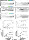
Figure 1.
Figure 1.
Fluorescence-activatable GFP-based reporters for flavivirus NS2B–NS3 protease activity become fluorescent upon cleavage by DENV-2/ZIKV recombinant proteases in vitro. The flavivirus-activatable GFP reporters contain a quenching peptide (QP) at the C terminus of GFP joined by a linker consisting of a cleavage site for the flavivirus NS2B–NS3 proteases. When the viral proteases cleave the linker, the quenching peptide is removed, and the GFP adopts a conformation promoting chromophore maturation. Three variants of this reporter were developed by changing the linker sequence: ZIKVA-GFP (ZIKV polyprotein NS2B/NS3 cleavage site linker), DENV2A-GFP (DENV-2 polyprotein NS2B/NS3 cleavage site linker), and FlaviA-GFP with the internal NS3 cleavage site linker, which is present in many members of the Flavivirus genus. A, in vitro cleavage kinetics of the flavivirus-activatable GFP reporter. Purified reporter proteins were mixed with purified DENV-2 NS2B–NS3 protease (left panels) or ZIKV NS2B–NS3 protease (right panels) at a molar ratio of 1:1 and incubated for given times. The reactions were quenched by thermal treatment in SDS loading buffer, and samples were analyzed by SDS-PAGE and staining of the gels with Coomassie Blue. tRep/control is an engineered cleaved variant of the FlaviA-GFP protein and was used as size marker of cleaved reporters. Representative cropped images from three independent experiments are shown. B, cleavage efficiency kinetics of the purified flavivirus-activatable GFP reporter proteins treated with purified DENV-2 NS2B–NS3 protease (left panel) and ZIKV NS2B–NS3 protease (right panel). C, time-resolved fluorescence signal-to-noise ratio of the purified flavivirus-activatable GFP reporter proteins treated with purified DENV-2 NS2B–NS3 protease (left panel) and ZIKV NS2B–NS3 protease (right panel). The data are expressed as means ± S.D. of three independent experiments. **, p < 0.001 compared with the other two reporter variants at 20 h post-treatment.
Figure 2.
Figure 2.
The FlaviA-GFP sensor reports the highest fluorescence increase in stably transduced mammalian cells upon DENV-2/ZIKV infection. We generated three BHK-21 stable cell lines expressing the flavivirus-activatable GFP reporters, each with one of the previously tested linker sequences. After cell sorting of subpopulations with homogeneous expression of each reporter, the cells were grown and infected with either infectious or UV-inactivated DENV-2 13538/ZIKV CIET-01 at a low MOI of 0.25, for the specified time periods. A, an automated image analysis protocol was constructed in CellProfiler 2.0 for the quantification of live (white outline), dead (red outline), and activated FlaviA-GFP fluorescent cells (green). A representative experiment is shown for the FlaviA-GFP stable cell line infected with DENV-2 (n = three independent experiments, magnification of 200×; scale bar, 100 μm). B, the flavivirus-activatable GFP reporter activation is represented by scatter plots showing the time-resolved fluorescence of the population of single live (blue) and dead (red) reporter cells after the exposure to infectious or UV-inactivated DENV-2 (left panels) or ZIKV (right panels). The population mean values for each condition are represented by the green continuous lines. Representative scatter plots are shown (n = three independent experiments). C, the cell population kinetics of the flavivirus-activatable GFP sensors fluorescence across multiple experiments confirmed that the flavivirus internal NS3 cleavage site linker (AAQRRGRIG) confers the highest signal-to-noise ratio to report the infection with both DENV-2 (left panel) and ZIKV (right panel) in stably transduced BHK-21 cells. The data are expressed as means ± S.D. of three independent experiments. *, p < 0.05 compared with both ZIKVA-GFP and DENV2A-GFP at 96 h postinfection.
Figure 3.
Figure 3.
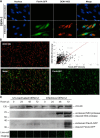
The FlaviA-GFP reporter becomes cleaved and fluorescent in stably transduced BHK-21 cells upon DENV-2 infection, which correlates with the viral NS3 protease synthesis and autoproteolysis. Stable BHK-21 cells expressing the FlaviA-GFP reporter were infected with either infectious or UV-inactivated DENV-2 13538 at a low MOI of 0.25, for the specified time periods postinoculation. A, comparison of DENV-2 infection detection by the FlaviA-GFP reporter (green) and immunostaining with an anti-DENV NS3 protease antibody (orange) in stably transduced BHK-21 cells at 72 h postinfection with DENV-2. Images from a representative experiment are shown (n = three independent experiments, magnification of 600×; scale bar, 30 μm). B, correlation of the total cell fluorescence intensity given by the FlaviA-GFP reporter (green) and an immunofluorescence assay (IFA) with an anti-DENV NS3 protease antibody (orange) in stably transduced BHK-21 cells at 72 h postinfection with DENV-2. A representative experiment is shown (n = three independent experiments, magnification of 200×; scale bar, 300 μm). C, cleavage and fluorescence kinetics of the FlaviA-GFP reporter in stable BHK-21 cells upon DENV-2 infection and subsequent viral NS3 protease production and activity. A representative Western blotting kinetics with its corresponding live-cell images set is shown (n = three independent experiments, magnification of 200×; scale bar, 100 μm).
Figure 4.
Figure 4.
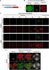
The FlaviA-mNeptune is a far-red version of the flavivirus reporter that enables earlier detection of the cell infection kinetics in a DENV-2/ZIKV plaque assay. We generated two BHK-21 stable cell lines expressing either a green or a far-red version of the flavivirus reporter. Both versions were compared by plaque assay upon infection with DENV-2 13538 and ZIKV CIET-01 at the specified time periods. A, the FlaviA-mNeptune reporter is a quenched version of the fluorescent protein mNeptune which contains a quenching peptide (QP) at the N terminus joint by a linker consisting of the internal NS3 cleavage site (AAQRRGRIG), which is conserved among many members of the Flavivirus genus. When the flavivirus protease cleaves the linker, the quenching peptide is removed, and the mNeptune adopts its fluorescent structural conformation. B, BHK-21 cells stably transduced with the FlaviA-GFP reporter and infected with DENV-2 required a contrast enhancement procedure to reveal viral plaques at 120 h postinfection but with a poor correlation when compared with the same plaques labeled with an anti-DENV NS3 protease antibody. C, the plaque assay kinetics showed that BHK-21 cells stably transduced with the FlaviA-mNeptune reporter accumulate enough intensity to reveal fluorescent plaques by 48 and 72 h postinfection with ZIKV and DENV-2, respectively, much earlier than their counterparts transduced with the FlaviA-GFP reporter. D, the performance of both GFP and mNeptune reporters was further evaluated by comparing the size of their resulting fluorescent plaques to the signal reported by an anti-DENV NS3 protease antibody and crystal violet staining, confirming that the FlaviA-mNeptune reporter has a better correlation to both the infection front (DENV NS3 immunostaining) and the cytopathic effect (crystal violet staining), compared with the FlaviA-GFP reporter. A representative experiment for each condition is shown (n = three independent experiments, magnification of 40×; scale bar, 1000 μm).
Figure 5.
Figure 5.
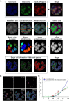
The FlaviA-mNeptune reporter enables analysis of the kinetics of ZIKV infection and induced cell death in single plaques and single cells. We constructed an image analysis protocol using the software CellProfiler 2.0 to characterize the kinetics of infection and cell death by the quantification of fluorescence features of cells imaged to assess DNA/chromatin condensation (Hoechst 33342), cell death (SYTOX green), and viral infection (FlaviA-mNeptune reporter). A, high resolution images were stitched into a single image of the well for all three fluorescent channels, which intensities were added into a single image to have a redundant single-cell identification. A neighbor count and threshold were performed to filter the cells belonging to plaques and achieve single plaque identification. The single cells previously identified were related to their respective parent plaques to quantify single-cell parameters within each plaque, including chromatin condensation (blue), cell death (green), and the intensity of the FlaviA-mNeptune reporter (red). B, the image analysis protocol was applied to analyze a representative kinetics of ZIKV plaque formation during 120 h postinfection (hpi), achieving the identification of individual plaques and the categorization of single cells within each plaque as live-infected (red), live-infected with chromatin condensation (blue), and dead (green). The 120-h postinfection image was reused from point A, because its generation was used in Fig. 5A to exemplify the image analysis pipeline applied to analyze the viral plaques at every postinfection time depicted in B. C, time-resolved kinetics of ZIKV infection described by parameters of percentage of live-infected cells (red), live-infected cells with chromatin condensation (blue), and dead cells (green) within the viral plaques. Images and data from a representative analyzed experiment are shown (n = three independent experiments, magnification of 40×; scale bar, 1000 μm). The data are expressed as means ± S.D. for the number of plaques identified at each time point depicted.
Figure 6.
Figure 6.
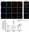
The FlaviA-mNeptune reporter enables the comparative characterization of the infection produced by flaviviruses in terms of viral replication and induced cell death at a single-cell level. We applied our image analysis protocol to characterize the DENV, ZIKV, and YFV infection and induced cell death by the quantification of fluorescence features of cells imaged to assess DNA/chromatin condensation (Hoechst 33342), cell death (SYTOX green), and viral infection (FlaviA-mNeptune reporter). A, the image analysis protocol was applied to achieve the identification of viral plaques produced by DENV, ZIKV, and YFV at 120 h postinfection and the categorization of single cells within each plaque as live-infected (red), live-infected with chromatin condensation (blue), and dead (green). B, DENV, ZIKV, and YFV infection described by parameters of percentage of live-infected cells (red), live-infected cells with chromatin condensation (blue), and dead cells (green) within the viral plaques at 120-h postinfection. The images and data from a representative analyzed experiment are shown (n = three independent experiments, magnification of 40×; scale bar, 1000 μm). The data are expressed as means ± S.D. for the number of plaques generated by each virus. *, p < 0.05; **, p < 0.001 compared with the other two cell populations conforming the plaques of the same viral species.

Similar articles

Cited by

References

    1. Gould E. A., and Solomon T. (2008) Pathogenic flaviviruses. Lancet 371, 500–509 10.1016/S0140-6736(08)60238-X - DOI - PubMed
    1. Lindenbach B., Murray C. L., Thiel H.-J., and Rice C. M. (2013) Flaviviridae. In Fields Virology, pp. 712–746, Lippincott Williams & Wilkins, Philadelphia, Pennsylvania
    1. Teo K. F., and Wright P. J. (1997) Internal proteolysis of the NS3 protein specified by dengue virus 2. J. Gen. Virol. 78, 337–341 10.1099/0022-1317-78-2-337 - DOI - PubMed
    1. Lindenbach B. D., and Rice C. M. (2003) Molecular biology of flaviviruses. Adv. Virus Res. 59, 23–61 10.1016/S0065-3527(03)59002-9 - DOI - PubMed
    1. Bera A. K., Kuhn R. J., and Smith J. L. (2007) Functional characterization of cis and trans activity of the flavivirus NS2B–NS3 protease. J. Biol. Chem. 282, 12883–12892 10.1074/jbc.M611318200 - DOI - PubMed

Publication types

Substances

LinkOut - more resources