. 2020 Jan 21;117(3):1722-1730.
doi: 10.1073/pnas.1919133117. Epub 2020 Jan 9.

A promiscuous inflammasome sparks replication of a common tumor virus

Affiliations

A promiscuous inflammasome sparks replication of a common tumor virus

Eric M Burton et al. Proc Natl Acad Sci U S A. .

Abstract

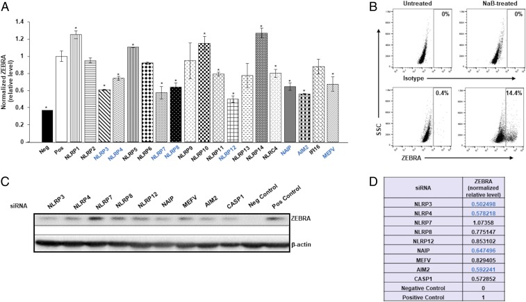
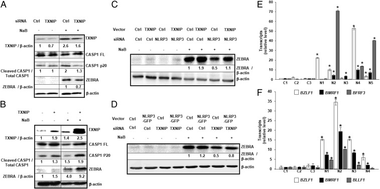
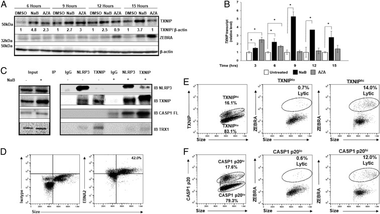
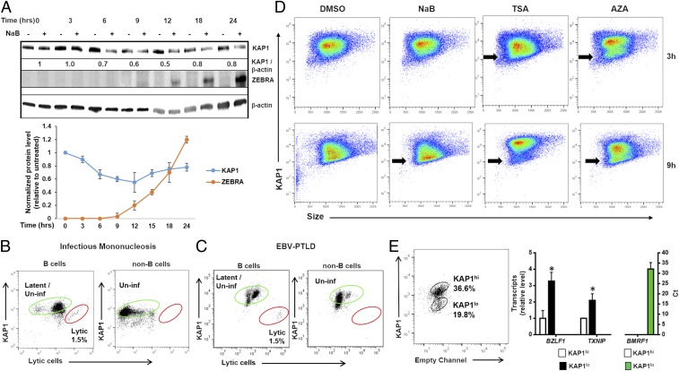
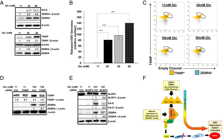
Viruses activate inflammasomes but then subvert resulting inflammatory responses to avoid elimination. We asked whether viruses could instead use such activated or primed inflammasomes to directly aid their propagation and spread. Since herpesviruses are experts at coopting cellular functions, we investigated whether Epstein-Barr virus (EBV), an oncoherpesvirus, exploits inflammasomes to activate its replicative or lytic phase. Indeed, our experiments reveal that EBV exploits several inflammasome sensors to actually activate its replicative phase from quiescence/latency. In particular, TXNIP, a key inflammasome intermediary, causes assembly of the NLRP3 inflammasome, resulting in caspase-1-mediated depletion of the heterochromatin-inducing epigenetic repressor KAP1/TRIM28 in a subpopulation of cells. As a result, only TXNIPhiKAP1lo cells, that is, in a primed/prolytic state, turn expression of the replication/lytic/reactivation switch protein on to enter the replicative phase. Our findings 1) demonstrate that EBV dovetails its escape strategy to a key cellular danger-sensing mechanism, 2) indicate that transcription may be regulated by KAP1 abundance aside from canonical regulation through its posttranslational modification, 3) mechanistically link diabetes, which frequently activates the NLRP3 inflammasome, to deregulation of a tumor virus, and 4) demonstrate that B lymphocytes from NOMID (neonatal onset multisystem inflammatory disease) patients who have NLRP3 mutations and suffer from hyperactive innate responses are defective in controlling a herpesvirus.

Keywords: Epstein−Barr virus; KAP1; TXNIP; diabetes; inflammasome.


Conflict of interest statement

The authors declare no competing interest.

Figures

Fig. 1.
Fig. 1.
Inflammasome sensors regulate the EBV replication switch. (A) HH514-16 BL cells were exposed to control siRNA or inflammasome sensor-directed siRNAs, treated with the replicative/lytic cycle-inducing agent NaB 24 h later, and harvested after another 24 h to assay intracellular levels of the EBV replication switch protein ZEBRA and GAPDH by in-cell Western. (B) HH514-16 BL cells were treated with NaB (versus untreated control), harvested 24 h later, stained with anti-ZEBRA antibody, and subjected to flow cytometry. ZEBRA+ gate was set based on staining with isotype control antibody; numbers represent percent ZEBRA+ cells. (C) Cells treated and harvested as in A were immunoblotted for ZEBRA and β-actin. Negative (Neg) control, untreated cells exposed to control siRNA; positive (Pos) control, NaB-treated cells exposed to control siRNA in A and C. Data in A represent means of three independent experiments; error bars, SEM; *P ≤ 0.05. Sensors whose knockdown resulted in at least 25% reduction in ZEBRA levels compared to control (Pos) in A were tested in C. (D) ZEBRA levels (normalized to β-actin) in siRNA-knocked down cells relative to control siRNA but NaB-treated cells (Pos; shown in C). Inflammasome components whose knockdown resulted in at least 25% loss of ZEBRA in A and D (via in-cell Western and immunoblot) are indicated in blue.
Fig. 2.
Fig. 2.
NLRP3 activates the EBV replication switch via TXNIP. (AD) HH514-16 cells were transfected with (A) TXNIP-directed (or control) siRNA, (B) TXNIP (or control) plasmid, and (C and D) combinations of plasmids and siRNAs, exposed to NaB 18 h later, and harvested after another (A and B) 12 h or (C and D) 24 h for immunoblotting with indicated antibodies. Numbers represent ratios between indicated proteins. (E) PBMC from healthy subjects (controls [C]) and NOMID patients (N) were infected with EBV in the presence of FK506 and analyzed 24 h later for transcripts from EBV lytic cycle genes BZLF1, BMRF1, and BFRF3 by qRT-PCR. Transcript levels in each sample were compared to those in control sample 1. (F) Three-week-old EBV-transformed LCL derived from healthy subjects (C) and NOMID patients (N) were harvested for analysis of EBV lytic transcripts BZLF1, BMRF1, and BLLF1 by qRT-PCR. Data represent averages of three independent experiments; error bars, SEM; *P ≤ 0.05.
Fig. 3.
Fig. 3.
Early increase in TXNIP and activation of the NLRP3 inflammasome precedes expression of the replication switch. (A and B) HH514-16 cells were exposed to lytic triggers NaB and 5-AZA-2′-deoxycytidine (versus dimethyl sulfoxide [DMSO] control) and harvested at indicated time points for immunoblotting with (A) indicated antibodies or (B) qRT-PCR. Numbers in A represent ratios between indicated proteins. Experiments were performed at least three times, and data in B represent averages of three independent experiments; error bars, SEM; *P ≤ 0.05. (C) HH514-16 cells were treated with NaB (versus untreated control) for 6 h, cell lysates were immunoprecipitated using indicated antibodies (IP), and bound proteins were detected by immunoblotting with indicated antibodies (IB). Input lysates are shown on the left. (DF) CD19+ B cells from the blood of a patient with PTLD were analyzed by flow cytometry for EBNA2 and size to demarcate EBNA2+ “blast-like” large cells for further analysis; number indicates percent EBNA2+ cells (D). EBV-infected B blast-like cells were analyzed by flow cytometry for TXNIP (E) or cleaved caspase-1 (CASP1 p20; F) expression (Left dot plots), and percent TXNIPhi versus percent TXNIPlo cells (E) or percent CASP1 p20hi versus percent CASP1 p20lo cells (F) expressing the lytic switch protein ZEBRA are shown (Right dot plots). Data from a representative of 3 patients is shown.
Fig. 4.
Fig. 4.
KAP1 loss precedes activation of the EBV replication switch in lytic cells. (A) (Upper) HH514-16 cells were treated with NaB and harvested at different time points for immunoblotting with indicated antibodies. Numbers represent ratios between indicated proteins. (Lower) Mean KAP1 and ZEBRA levels from three independent time course experiments are graphically displayed; error bars, SEM. (B and C) PBMC from the blood of patients with (B) infectious mononucleosis and (C) PTLD were analyzed by flow cytometry for expression of KAP1 and ZEBRA (lytic cells). Percent latent/uninfected and lytic cells are depicted in green and red gates, respectively. (D) HH514-16 cells were exposed to lytic triggers NaB, TSA, or AZA (versus DMSO control) and examined 3 or 9 h later for KAP1 expression by flow cytometry; black arrows, KAP1lo subpopulations. (E) HH514-16 cells were exposed to TSA, FACS-sorted 12 h later into KAP1hi and KAP1lo subpopulations, and followed by qRT-PCR for relative levels of TXNIP transcript and EBV lytic transcript BZLF1, as well as Ct values for lytic transcript BMRF1 in each subpopulation. Data represent averages of three independent experiments; error bars, SEM; *P ≤ 0.05.
Fig. 5.
Fig. 5.
High glucose triggers the EBV replication switch via TXNIP. (A and B) Mutu I BL cells were seeded in medium containing standard glucose (11 mM) or in the presence of increasing concentrations of glucose. (A) Cells were harvested after 6 h for immunoblotting for TXNIP and ZEBRA levels (Lower), and 24 h for immunoblotting for ZEBRA and EA-D (early lytic gene product) (Upper), while culture media were harvested after 72 h, treated with DNase, and (B) examined for released virus particles by qPCR of the EBV BALF5 gene. Data represent averages of three independent experiments; error bars, SEM; ***P ≤ 0.0005. (C) HH514-16 cells were seeded in media containing indicated concentrations of glucose and harvested 6 h later for staining for TXNIP and ZEBRA followed by flow cytometry. (D) HH514-16 cells were transfected with control or TXNIP-directed siRNA, placed in standard or high-glucose medium for 24 h, and subjected to immunoblotting. Numbers represent ratios between indicated proteins. (E) HH514-16 cells were transfected with control or NLRP3-directed siRNA, placed in standard or high-glucose medium for 24 h, and subjected to immunoblotting. Numbers represent ratios between indicated proteins. (F) Model depicting NLRP3 inflammasome-mediated activation of the EBV replication switch during the “prolytic state.”

References

    1. Broz P., Dixit V. M., Inflammasomes: Mechanism of assembly, regulation and signalling. Nat. Rev. Immunol. 16, 407–420 (2016). - PubMed
    1. Johnson K. E., et al. , IFI16 restricts HSV-1 replication by accumulating on the hsv-1 genome, repressing HSV-1 gene expression, and directly or indirectly modulating histone modifications. PLoS Pathog. 10, e1004503 (2014). - PMC - PubMed
    1. Xiao X., Qi J., Lei X., Wang J., Interactions between enteroviruses and the inflammasome: New insights into viral pathogenesis. Front. Microbiol. 10, 321 (2019). - PMC - PubMed
    1. Brodsky I. E., et al. , A Yersinia effector protein promotes virulence by preventing inflammasome recognition of the type III secretion system. Cell Host Microbe 7, 376–387 (2010). - PMC - PubMed
    1. Scott R. S., Epstein-Barr virus: A master epigenetic manipulator. Curr. Opin. Virol. 26, 74–80 (2017). - PMC - PubMed

Publication types

MeSH terms