. 2020 Jan 9;10(1):92.
doi: 10.1038/s41598-019-56542-4.

Melanoma cell-derived exosomes in plasma of melanoma patients suppress functions of immune effector cells

Affiliations

Melanoma cell-derived exosomes in plasma of melanoma patients suppress functions of immune effector cells

Priyanka Sharma et al. Sci Rep. .

Abstract

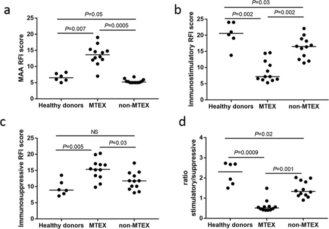
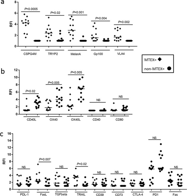
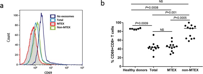
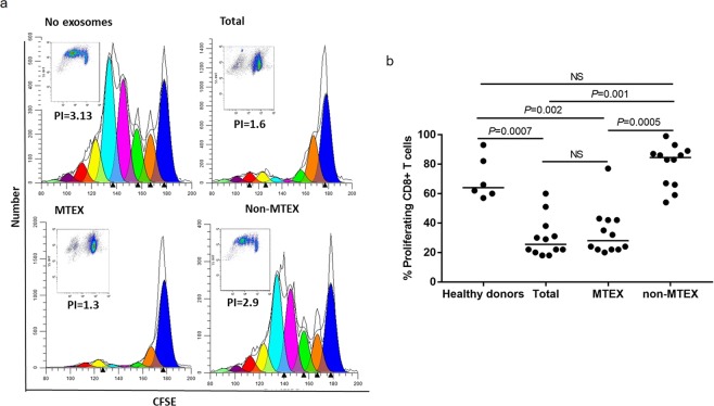
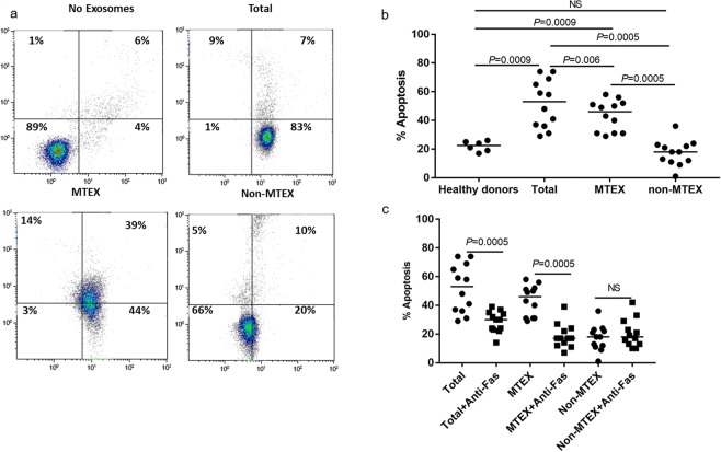
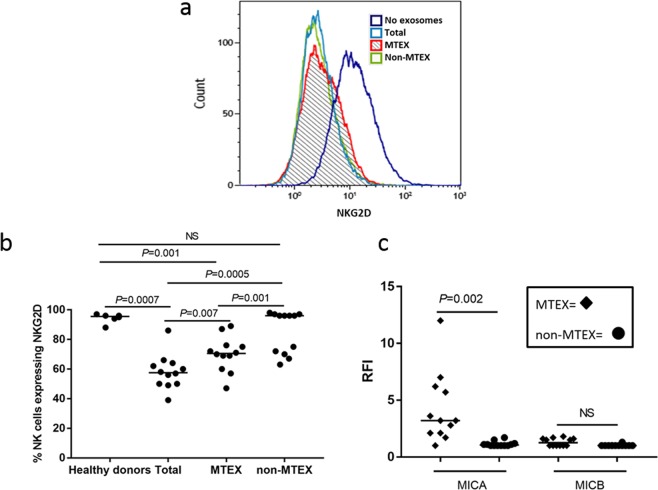
Melanoma patients' plasma contains exosomes produced by malignant and normal cells. Plasma exosomes were isolated and separated by immunocapture into two fractions: melanoma cell-derived exosomes (MTEX) and normal cell-derived exosomes (non-MTEX). Immunosuppressive effects of MTEX on primary human immune cells were evaluated. Exosomes were isolated from plasma of 12 melanoma patients and six healthy donors (HDs). Expression levels of 19 immunoregulatory proteins in MTEX, non-MTEX and HDs exosomes were evaluated by on-bead flow cytometry. Functional/phenotypic changes induced in CD8+ T or natural killer (NK) cells by MTEX or non-MTEX were compared. Plasma protein levels were higher in patients than HDs (P < 0.0009). In patients, MTEX accounted for 23-66% of total exosomes. MTEX were enriched in immunosuppressive proteins (P = 0.03). MTEX, but not HDs exosomes, inhibited CD69 expression (P ≤ 0.0008), induced apoptosis (P ≤ 0.0009) and suppressed proliferation (P ≤ 0.002) in CD8+ T cells and downregulated NKG2D expression in NK cells (P = 0.001). Non-MTEX were enriched in immunostimulatory proteins (P = 0.002) and were only weakly immunosuppressive. Elevated MTEX/total exosome ratios and, surprisingly, non-MTEX ability to induce apoptosis of CD8+ T cells emerged as positive correlates of disease stage. MTEX emerge as the major mechanism of tumor-induced immune suppression and as an underestimated barrier to successful melanoma immunotherapy.


Conflict of interest statement

The authors declare no competing interests.

Figures

Figure 1
Figure 1
The RFI scores for: (a) MAAs, (b) immunostimulatory proteins and (c) immunosuppressive proteins carried by total exosomes from plasma of HDs, and by MTEX and non-MTEX from plasma of melanoma patients. In (d) the stimulatory/suppressive (stim/supp) ratio for HDs exosomes and for MTEX and non-MTEX are shown. The MAA RFI score includes CSPG4, TYRP2, MelanA, Gp100, and VLA4; the immunostimulatory RFI score includes CD40, CD40L, CD80, OX40, and OX40L; the immunosuppressive RFI score includes PDL-1, CD39, CD73, FasL, LAP-TGFβ, TRAIL, and CTLA-4. Wilcoxon signed-rank tests were used to evaluate differences between MTEX and non-MTEX; Wilcoxon-Mann-Whitney tests were used to evaluate differences between patients and healthy controls. Horizontal bars indicate median values. NS: no significant difference.
Figure 2
Figure 2
The RFI values for: (a) individual MAAs, (b) individual immunostimulatory proteins and (c) individual immunosuppressive proteins as determined by on-bead flow cytometry for all 12 melanoma patients. Horizontal bars indicate median values. NS: no significant difference. Wilcoxon signed-rank tests were used to evaluate differences between MTEX and non-MTEX.
Figure 3
Figure 3
Exosome-mediated effects on CD69 expression levels in CD8+ T cells. MTEX were immunocaptured from patients’ plasma as described in Methods and were co-incubated with primary human activated CD8+ T cells for 6 h at 37 °C. CD69 expression levels on T cells were measured by flow cytometry. (a) Representative exosome-mediated downregulation of CD69 expression levels on activated CD8+ T cells after co-incubation with exosomes as indicated. (b) % of CD69 + CD8+ T cells after co-incubation with HDs’ exosomes or total exosomes, MTEX or non-MTEX obtained from melanoma patients’ plasma. Horizontal bars indicate median values. All data are normalized to no exosome (PBS) control for each patient/HD individually. Wilcoxon signed-rank tests were used to evaluate differences between paired samples (e.g., between MTEX and non-MTEX); Wilcoxon-Mann-Whitney tests were used to evaluate differences between patients and HDs. NS: no significant difference.
Figure 4
Figure 4
Exosome-induced suppression of CD8+ T cell proliferation. (a) Representative CSFE-assay for exosomes from one patient showing proliferation inhibition induced in CD8+ T cells by total exosomes, MTEX and non-MTEX. No suppression was seen with non-MTEX. (b) CD8+ T cell proliferation after co-incubation with HDs exosomes or total exosomes, MTEX or non-MTEX obtained from melanoma patients’ plasma. Note that non-MTEX in 4/12 patients mediated suppression. Data were normalized to no exosome (PBS) control. Horizontal bars indicate median values. NS: no significant difference. Wilcoxon signed-rank tests were used to evaluate differences between paired samples; Wilcoxon-Mann-Whitney tests were used to evaluate differences between patients and HDs. Results for blocking of MTEX-induced suppression of CD8+ T cell proliferation are presented in SFig. 8.
Figure 5
Figure 5
Exosome-mediated apoptosis in primary activated CD8+ T cells. (a) Representative flow cytometry data for PI/Annexin V binding in CD8+ T cells co-incubated for 6 h with total exosomes, MTEX or non-MTEX isolated from plasma of one melanoma patient. Control cells were co-incubated with PBS. In (b), % apoptosis of CD8+ T cells after co-incubation with exosomes from HDs plasma or total exosomes, MTEX or non-MTEX from melanoma patients’ plasma. In (c), % apoptosis of CD8+ T cell after co-incubation with total exosomes, MTEX or non-MTEX from melanoma patients in the presence of anti-Fas mAb. Isotype control Ab was used in place of anti-Fas Ab (see SFig. 9b), and all data are normalized to no exosome (PBS) control. Horizontal bars indicate median values. NS: no significant difference. Wilcoxon signed-rank tests were used to evaluate differences between paired samples; Wilcoxon-Mann-Whitney tests were used to evaluate differences between patients and healthy donors.
Figure 6
Figure 6
Exosome-mediated effects on NKG2D receptor expression levels on human primary NK cells. In (a), total exosomes, MTEX or non-MTEX were co-incubated with human activated NK cells for 24 h. Representative data of exosome-mediated suppression of NKG2D levels by exosomes from one melanoma patient are shown. In (b), % of NK cells expressing NKG2D after co-incubation with exosomes of HDs or total exosomes, MTEX or non-MTEX from melanoma patients’ plasma. In (c), the presence of MICA or MICB (determined as RFI) on MTEX and non-MTEX. All data were normalized to no exosome (PBS) controls. Blocking of MTEX-mediated downregulation of NKG2D on NK cells by neutralizing anti-TGF-β Ab is shown in SFig. 8. Horizontal bars indicate median values. NS: no significant difference. Wilcoxon signed-rank tests were used to evaluate differences between paired samples; Wilcoxon-Mann-Whitney tests were used to evaluate differences between patients and healthy donors.

References

    1. Guy GP, Jr., et al. Vital signs: melanoma incidence and mortality trends and projections - United States, 1982–2030. MMWR Morb. Mortal. Wkly. Rep. 2015;64:591–596. - PMC - PubMed
    1. Gajewski TF, et al. Immune resistance orchestrated by the tumor microenvironment. Immunol. Rev. 2006;213:131–145. doi: 10.1111/j.1600-065X.2006.00442.x. - DOI - PubMed
    1. Passarelli A, Mannavola F, Stucci LS, Tucci M, Silvestris F. Immune system and melanoma biology: a balance between immunosurveillance and immune escape. Oncotarget. 2017;8:106132–106142. doi: 10.18632/oncotarget.22190. - DOI - PMC - PubMed
    1. Chen G, et al. Exosomal PD-L1 contributes to immunosuppression and is associated with anti-PD-1 response. Nature. 2018;560:382–386. doi: 10.1038/s41586-018-0392-8. - DOI - PMC - PubMed
    1. Ottaviano M, De Placido S, Ascierto PA. Recent success and limitations of immune checkpoint inhibitors for cancer: a lesson from melanoma. Virchows Arch. 2019;474:421–432. doi: 10.1007/s00428-019-02538-4. - DOI - PubMed

Publication types