. 2020 Jan 9;11(1):153.
doi: 10.1038/s41467-019-14008-1.

Cardiac muscle thin filament structures reveal calcium regulatory mechanism

Affiliations

Cardiac muscle thin filament structures reveal calcium regulatory mechanism

Yurika Yamada et al. Nat Commun. .

Abstract

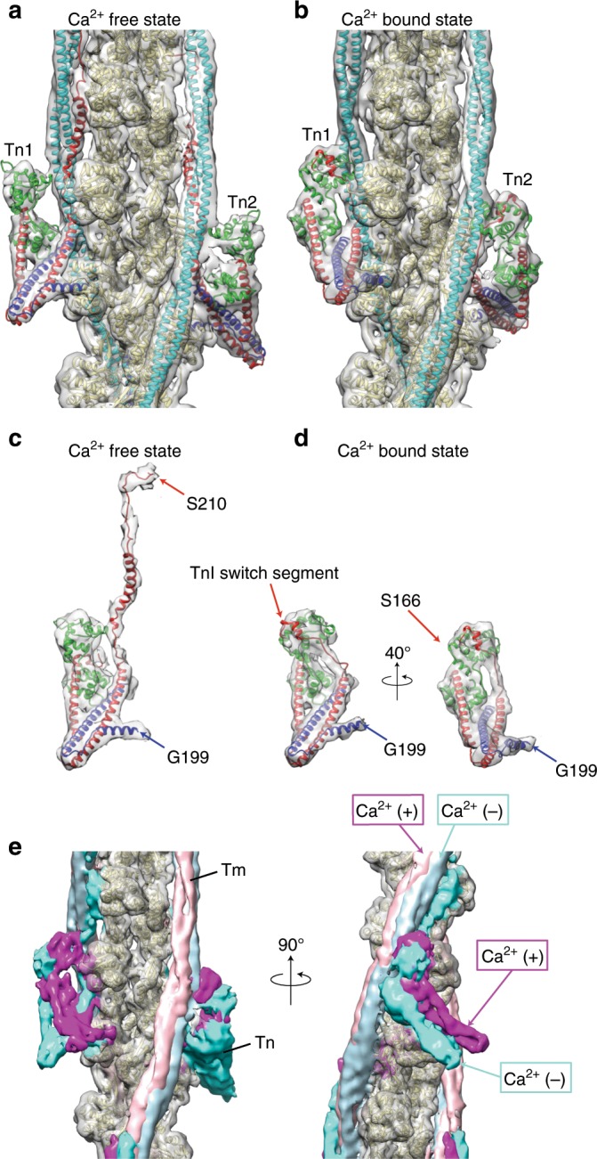
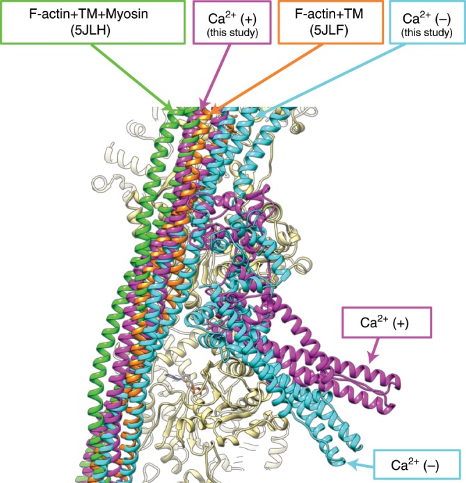
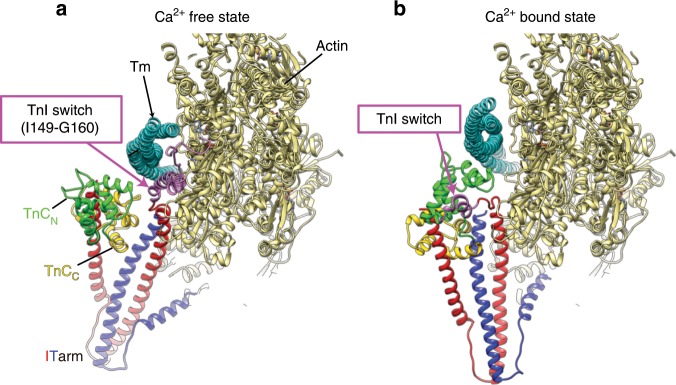
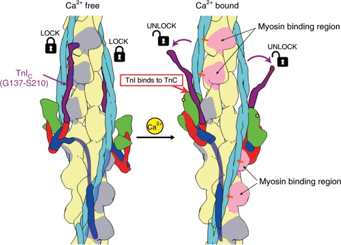
Contraction of striated muscles is driven by cyclic interactions of myosin head projecting from the thick filament with actin filament and is regulated by Ca2+ released from sarcoplasmic reticulum. Muscle thin filament consists of actin, tropomyosin and troponin, and Ca2+ binding to troponin triggers conformational changes of troponin and tropomyosin to allow actin-myosin interactions. However, the structural changes involved in this regulatory mechanism remain unknown. Here we report the structures of human cardiac muscle thin filament in the absence and presence of Ca2+ by electron cryomicroscopy. Molecular models in the two states built based on available crystal structures reveal the structures of a C-terminal region of troponin I and an N-terminal region of troponin T in complex with the head-to-tail junction of tropomyosin together with the troponin core on actin filament. Structural changes of the thin filament upon Ca2+ binding now reveal the mechanism of Ca2+ regulation of muscle contraction.


Conflict of interest statement

The authors declare no competing interests.

Figures

Fig. 1
Fig. 1. Structure of human cardiac thin filament in the Ca2+ free state.
a a typical cryoEM image of the thin filament with periodic Tn binding along actin filament. b The model of the thin filament fitted into the cryoEM density map in two different views. Approximately 12 actin subunits are shown with a pair of Tm coiled coils and a pair of the Tn ternary complexes consisting of the Tn core, TnIC extended upward from the core and TnTN attached closely to the head–tail junction of Tm near the bottom. c Magnified images of TnTN complexed with the head–tail junction of Tm. The models are colored as: actin, beige; Tn, purple; and Tm, light blue and orange. Actin filament is oriented with its pointed end top including all the other figures.
Fig. 2
Fig. 2. The entire structures of the Tn complex with two different conformations.
a The entire structure of the Tn ternary complex is shown for each of the Tn pair, labeled Tn1 and Tn2, in front and back of the thin filament. The difference maps for TnIC and TnTN are colored red and dark blue above and below the Tn core, respectively. The difference in the relative positions of the Tn core and TnTN between the pair is clear while the conformations of the TnIC chains are the same. b The difference map for the entire Tn ternary complex is contoured at a lower level to show the continuous volume of Tn in pink, for each of the Tn pair, again in front and back of the thin filament.
Fig. 3
Fig. 3. Structural changes of the thin filament and Tn complex upon Ca2+ binding to TnC.
a, c Ca2+ free state. b, d Ca2+ bound state. c, d The Tn ternary complex extracted from the thin filament to show the good fit of the model into the difference map. Models are colored as: actin, beige; Tm, light blue; TnI, magenta; TnT, dark blue; and TnC, green. e 3D volumes of the thin filament in the two states are superimposed with Tn and Tm in blue and light blue for the Ca2+ free state and in magenta and light pink for the Ca2+ bound state and actin in beige for both. Two different views are presented.
Fig. 4
Fig. 4. Variation in the shift of Tm coiled coil on the surface of actin filament.
Tm coiled coils are colored blue and magenta for the Ca2+ free and bound states, respectively. Positions of corresponding Cα atoms are indicated by open circles.
Fig. 5
Fig. 5. Positions of Tm coiled coil in the four different states.
Models are colored as: actin, beige; Tm and Tn in the Ca2+ free state, blue; Tm and Tn in the Ca2+ bound state, magenta; Tm in the actin filament–Tm structure, orange; and Tm in the actomyosin filament–Tm structure, green. The model 5JLH is a human actomyosin–tropomyosin complex composed of the motor domain of human non-muscular myosin-2C, cytoplasmic γ-actin, and cytoplasmic tropomyosin 3.1, and the model 5JLF is composed of rabbit skeletal muscle actin and recombinant α-tropomyosin from Mus musculus with an alanine–serine extension in its N-terminus.
Fig. 6
Fig. 6. Structural changes of the thin filament and Tn complex upon Ca2+ binding to TnC.
a Ca2+ free state, b Ca2+ bound state. Model colors are the same as in Fig. 3 except for TnCC colored brown. The models are viewed obliquely from the pointed end of actin filament.
Fig. 7
Fig. 7. Schematic diagram of the Ca2+ regulatory mechanism of muscle contraction.
The structural changes between Ca2+ free and bound states are shown. In the Ca2+ free state, TnIC (dark purple) binds to actin (beige) and Tm (light blue) above the Tn core (green, red and blue), covering the myosin head binding sites. But entire TnIC dissociates from them upon Ca2+ binding to TnCN by the binding of a short N-terminal portion of TnIC to TnCN. This causes Tm to move around on the actin filament surface (short orange arrows) together with TnTN near the head-to-tail junction of Tm, thereby exposing some of the myosin head binding sites shaded in light gray to allow actin–myosin interactions (light pink).

References

    1. Huxley. AF, Niedergerke R. Structural changes in muscle during contraction: interference microscopy of living muscle fibres. Nature. 1954;173:971–973. doi: 10.1038/173971a0. - DOI - PubMed
    1. Huxley H, Hanson J. Changes in the cross-striations of muscle during contraction and stretch and their structural interpretation. Nature. 1954;173:973–976. doi: 10.1038/173973a0. - DOI - PubMed
    1. Lymn RW, Taylor EW. Mechanism of adenosine triphosphate hydrolysis by actomyosin. Biochemistry. 1971;10:461–4624. doi: 10.1021/bi00801a004. - DOI - PubMed
    1. Ebashi S, Endo M, Otsuki I. Control of muscle contraction. Q. Rev. Biophys. 1969;2:351–384. doi: 10.1017/S0033583500001190. - DOI - PubMed
    1. Lehman W, Craig R, Vibert P. Ca(2+)-induced tropomyosin movement in Limulus thin filaments revealed by three-dimensional reconstruction. Nature. 1994;368:65–67. doi: 10.1038/368065a0. - DOI - PubMed

Publication types