. 2020 Jan 9;11(1):160.
doi: 10.1038/s41467-019-13979-5.

The nuclear pore complex prevents sister chromatid recombination during replicative senescence

Affiliations

The nuclear pore complex prevents sister chromatid recombination during replicative senescence

Paula Aguilera et al. Nat Commun. .

Abstract

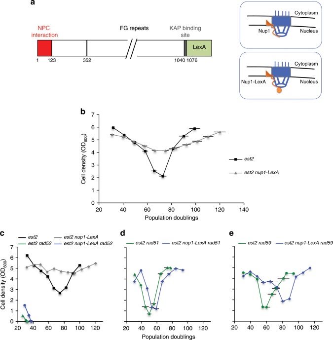
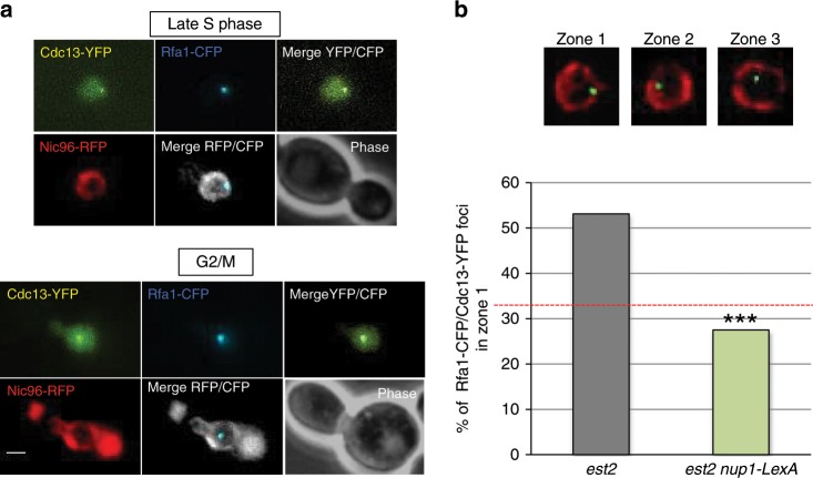
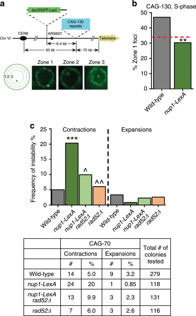
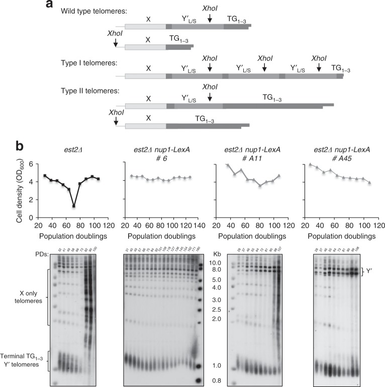
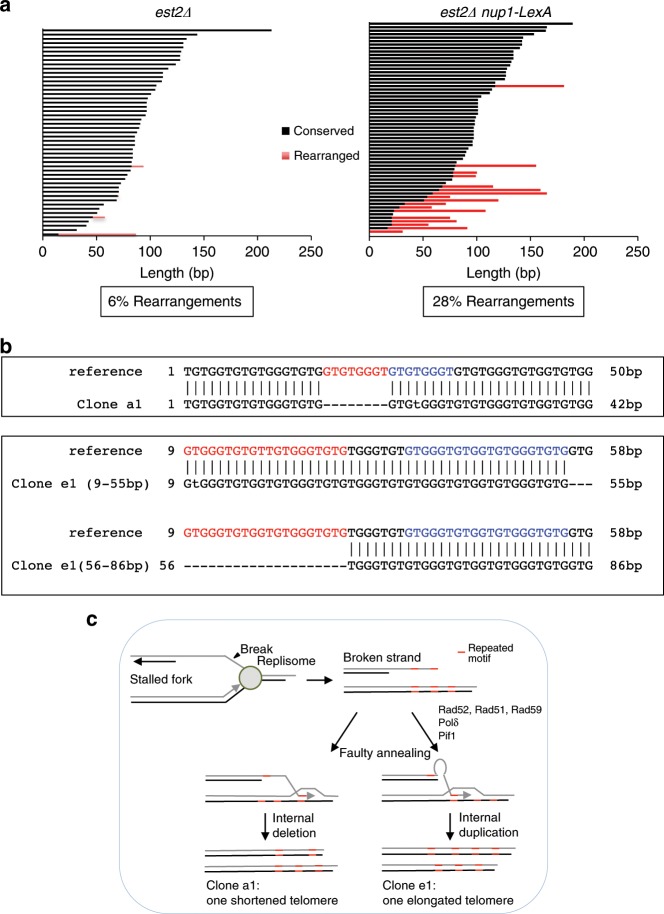
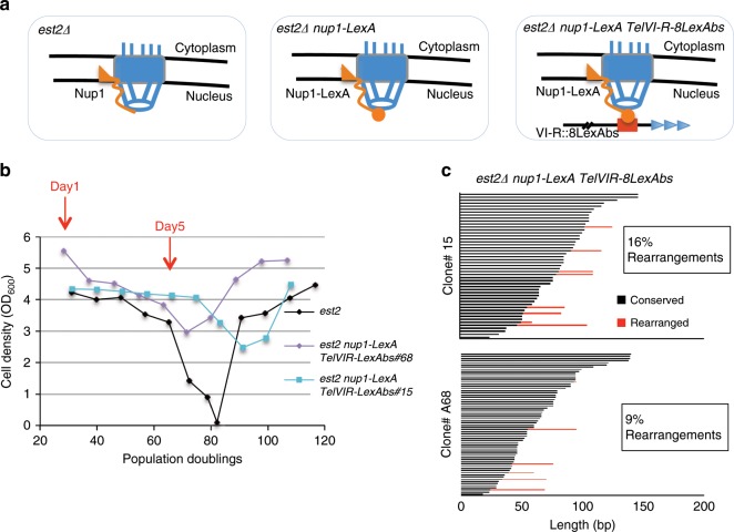
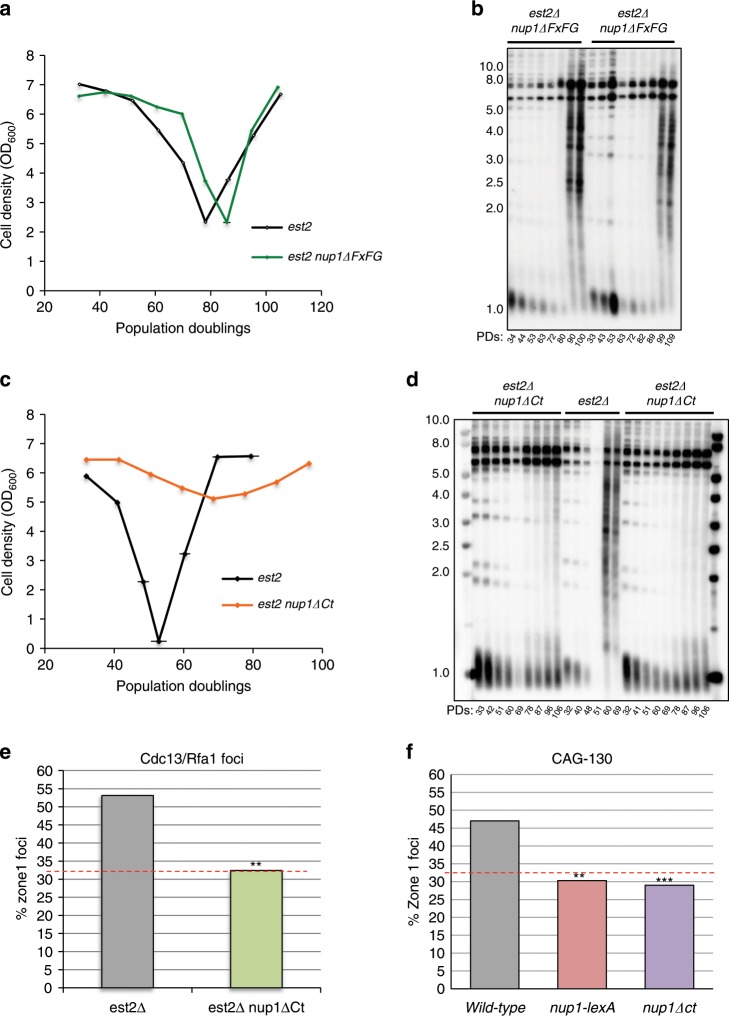
The Nuclear Pore Complex (NPC) has emerged as an important hub for processing various types of DNA damage. Here, we uncover that fusing a DNA binding domain to the NPC basket protein Nup1 reduces telomere relocalization to nuclear pores early after telomerase inactivation. This Nup1 modification also impairs the relocalization to the NPC of expanded CAG/CTG triplet repeats. Strikingly, telomerase negative cells bypass senescence when expressing this Nup1 modification by maintaining a minimal telomere length compatible with proliferation through rampant unequal exchanges between sister chromatids. We further report that a Nup1 mutant lacking 36 C-terminal residues recapitulates the phenotypes of the Nup1-LexA fusion indicating a direct role of Nup1 in the relocation of stalled forks to NPCs and restriction of error-prone recombination between repeated sequences. Our results reveal a new mode of telomere maintenance that could shed light on how 20% of cancer cells are maintained without telomerase or ALT.


Conflict of interest statement

The authors declare no competing interests.

Figures

Fig. 1
Fig. 1. Attenuation of senescence in est2∆ nup1-LexA cells.
a Schematic structure of the nuclear basket protein Nup1 fused to LexA. b Mean senescence profiles of the est2∆ (n = 22) and est2∆ nup1-LexA (n = 28) clones analyzed in the course of this study. Each clone was isolated by sporulation of a heterozygous diploid strain. Each spore colony was then propagated in liquid culture through daily serial dilutions. OD600 was measured every day to estimate the cell density reached in 24 h. PD numbers were estimated from the initial spores. c Mean senescence profiles of est2∆ and est2∆ nup1-LexA clones in the absence of RAD52 (n = 4 and 8, respectively). Control est2∆ (n = 7) and est2∆ nup1-LexA (n = 5) curves are from the clones used in this specific experiment. d Mean senescence profiles of est2∆ and est2∆ nup1-LexA clones in the absence of RAD51 (n = 4 and 7, respectively). e Mean senescence profiles of est2∆ and est2∆ nup1-LexA clones in the absence of RAD59 (n = 4 and 6, respectively). Error bars indicate SD.
Fig. 2
Fig. 2. Nup1-LexA alters the localization of damaged telomeres during senescence.
a Damaged telomeres were detected as foci containing both Cdc13-YFP and Rfa1-CFP and localized relative to Nic96-RFP that marks the nuclear periphery. Representative images of late S (upper panel) and G2/M (lower panel) cells are shown. Scale bar: 2 μm. b The localization of the Cdc13-YFP/Rfa1-CFP foci in the three zones of equal area of the nucleus marked by Nic96-RFP (upper panel) was scored in late S and G2/M cells. Cells were imaged after one restreak from spore colonies (about 35 PDs from the initial spores). The percentages of zone 1 foci are from analysis of two independently isolated est2∆ (n = 96 cells) and est2∆ nup1-LexA (n = 120 cells) clones. The analysis of the repartition of Rfa1/Cdc13 foci between the three equal zones of the nucleus is shown for individual clones in Supplementary Fig. 2a. Statistical differences were determined by a Fisher’s exact test (***p = 0.0001).
Fig. 3
Fig. 3. Nup1-LexA affects localization and stability of CAG trinucleotide repeats.
a Yeast chromosome VI containing 130 integrated CAG repeats and a lacO array that binds the LacI-GFP protein. S-phase cells were imaged, and the location of the foci was scored into one of three zones of equal area, using the GFP-Nup49 signal to mark the nuclear periphery. b Percent of zone 1 foci for CAG-130 S-phase cells (283 cells for wild-type, 142 cells for nup1-LexA). Statistical differences were determined by a Fisher’s exact test (**p = 0.0012). c Frequency of contractions and expansions for CAG-70 repeats in wild-type and nup1-LexA cells. The frequency was determined by analysis of PCR amplicon length on a high-resolution fragment analyzer gel system, using PCR primers that flank the CAG-70 tract located on a YAC (see Fig. S6A); (***) p = 0.0001 compared with wild-type by Fisher’s exact test; (^) p = 0.0313; (^^) p = 0.0017 compared with nup1-LexA by Fisher’s exact test.
Fig. 4
Fig. 4. Telomere length is maintained at a minimal length in nup1-LexA cells.
a Schematic representation of wild-type, type I and type II telomeres. All telomeres contain one X element in the subtelomeric region. In addition, about two-third of the telomeres contain from one to four long (L) or short (S) subtelomeric sequences called Y′ separated by short interstitial TG1–3 repeats, both being amplified in type I survivors. Positions of the XhoI sites are shown. b Senescence profiles of one est2∆ clone (black) and three independent representative est2∆ nup1-LexA clones (gray). Telomere length and survivor formation in the same replicative senescence experiment were monitored by TG1–3 probed Southern blots of XhoI-digested DNA.
Fig. 5
Fig. 5. Analysis of telomeres by DNA sequencing.
a Telomeres VI-R were amplified and sequenced from clonal populations of est2∆ and est2∆ nup1-LexA cells using a specific primer after about 65 population doublings from the initial spores (Day 5) and align with the reference sequence obtained at Day 1 (see Supplementary Fig.  4a for the senescence profiles). Each bar represents an individual telomere. Bars are sorted by the length of the centromere-proximal sequence that is identical to the reference sequence (black). The red bars show the length of the distal rearranged sequence that cannot be continuously aligned with the reference. b Examples of rearrangements. Upper panel, deletion: TelVI-R from the clone a1 aligns with the reference provided that a 8-bp-long gap is introduced after the 17th nucleotide. Note the repeated motif (in red and blue) at and after the gap. Lower panel, insertion: The 55 first nucleotides of the clone e1 align perfectly with the reference. The last 60 nucleotides (56–86 are shown) cannot be aligned with the reference except if starting at position 29 of the reference instead of 56. This suggests that part of the sequence has been duplicated. Note the presence of the motif (in blue) at the end of the conserved sequence that is repeated just upstream the point of misalignment (in red). c Model of non-conservative HR-dependent repair between sister chromatids. The sequence of the telomeres contains motifs that are repeated several times. Thus a 3′ terminal end with such a motif can anneal at different positions in the sister chromatid to initiate repair leading to internal deletion (left part) or duplication (right part).
Fig. 6
Fig. 6. Tethering of TelVI-R to Nup1-LexA prevents SCR.
a Schematic of the three est2∆ strains expressing (right panels) or not (left panel) nup1-LexA and bearing either wild-type TelVI-R or modified TELVI-R with eight LexA DNA-binding sites inserted 1.2 kb away from the TG1–3 repeats. b Senescence profiles of the two est2∆ nup1-LexA TelVI-R-8lexAbs clones (#15 and #A68) used for sequencing. The est2∆ profile is from Supplementary Fig.  4 and is shown for comparison. D1 and D5 are highlighted in red. c TelVI-R sequence analysis of the clones est2∆ nup1-LexA TelVI-R-8LexAbs #15 and #A68 at Day 5 (see Supplementary Fig. 5 for the statistics of comparison with WT TelVI-R).
Fig. 7
Fig. 7. nup1Ct phenocopies the phenotypes of nup1-LexA.
a Mean senescence profiles of est2∆ (n = 5) and est2∆ nup1∆FxFG (n = 5) clones. Error bars are SD. b Telomere length analysis of two representative est2∆ nup1∆FxFG clones. Southern blot of XhoI-digested DNA prepared from samples of senescing cells was revealed with a TG1–3 probe. c Mean senescence profiles of est2∆ (n = 4) and est2∆ nup1∆Ct (n = 8) clones. Error bars are SD. d Telomere length analysis of representative clones of the indicated genotypes. e The localization of the Cdc13-YFP/Rfa1-CFP foci in the three zones of equal area of the nucleus marked by Nic96-RFP was scored in late S and G2/M est2∆ and est2∆ nup1∆Ct cells as described in Fig. 2. The percentages of zone 1 foci are from three independently isolated est2∆ nup1∆Ct (n = 139 cells) clones. WT cells are from Fig. 3. Statistical differences were determined by a Fisher’s exact test (**p = 0.0019). f Percent of zone 1 foci for CAG-130 S-phase in nup1∆Ct cells (n = 143). Statistical differences were determined by a Fisher’s exact test (***p = 0,0006). WT and nup1-LexA cells are from Fig. 3. The repartition of the Rfa1/Cdc13 and CAG-130 foci between the three zones of the nucleus is shown in the Supplementary Fig. 8.
Fig. 8
Fig. 8. Model for the role of the NPC in the repair of fork stalled at telomeres.
Replication forks stalled at telomeres relocalize to the NPC and are mainly repaired by a conservative pathway that allows replication to resume (left). When this pathway fails, the stalled forks engage in error-prone Rad51-dependent SCR that maintain a telomere length compatible with continuous proliferation.

References

    1. Larrivee M, LeBel C, Wellinger RJ. The generation of proper constitutive G-tails on yeast telomeres is dependent on the MRX complex. Genes Dev. 2004;18:1391–1396. doi: 10.1101/gad.1199404. - DOI - PMC - PubMed
    1. Stewart JA, Chaiken MF, Wang F, Price CM. Maintaining the end: roles of telomere proteins in end-protection, telomere replication and length regulation. Mutat. Res. 2012;730:12–19. doi: 10.1016/j.mrfmmm.2011.08.011. - DOI - PMC - PubMed
    1. Pardo B, Marcand S. Rap1 prevents telomere fusions by nonhomologous end joining. EMBO J. 2005;24:3117–3127. doi: 10.1038/sj.emboj.7600778. - DOI - PMC - PubMed
    1. Martina M, et al. A balance between Tel1 and Rif2 activities regulates nucleolytic processing and elongation at telomeres. Mol. Cell. Biol. 2012;32:1604–1617. doi: 10.1128/MCB.06547-11. - DOI - PMC - PubMed
    1. Bonetti D, et al. Shelterin-like proteins and Yku inhibit nucleolytic processing of Saccharomyces cerevisiae telomeres. PLoS Genet. 2010;6:e1000966. doi: 10.1371/journal.pgen.1000966. - DOI - PMC - PubMed

Publication types

MeSH terms