Review
. 2020 Sep;34(9):1515-1534.
doi: 10.1038/s41433-019-0741-3. Epub 2020 Jan 9.

Management of nystagmus in children: a review of the literature and current practice in UK specialist services

Collaborators, Affiliations
Review

Management of nystagmus in children: a review of the literature and current practice in UK specialist services

J E Self et al. Eye (Lond). 2020 Sep.

Erratum in

Abstract

Nystagmus is an eye movement disorder characterised by abnormal, involuntary rhythmic oscillations of one or both eyes, initiated by a slow phase. It is not uncommon in the UK and regularly seen in paediatric ophthalmology and adult general/strabismus clinics. In some cases, it occurs in isolation, and in others, it occurs as part of a multisystem disorder, severe visual impairment or neurological disorder. Similarly, in some cases, visual acuity can be normal and in others can be severely degraded. Furthermore, the impact on vision goes well beyond static acuity alone, is rarely measured and may vary on a minute-to-minute, day-to-day or month-to-month basis. For these reasons, management of children with nystagmus in the UK is varied, and patients report hugely different experiences and investigations. In this review, we hope to shine a light on the current management of children with nystagmus across five specialist centres in the UK in order to present, for the first time, a consensus on investigation and clinical management.

摘要: 眼球震颤是一种眼球运动障碍以单眼或双眼异常的、不自主的节律性摆动为特征, 其发病缓慢。在英国的发病率不低, 通常就诊于小儿眼科和成人普通/斜视门诊。眼球震颤在某些情况下单发, 也可伴发多系统疾病、严重视力损害或神经系统疾病。同样, 在一些病例中, 患者视力正常, 另一些病例中, 患者视力严重下降。而且其对视力的影响远超过对静态视敏度的影响, 几乎检测不到, 而且可能在每分钟、每天或每月的基础上不断变化。基于以上原因, 在英国, 针对儿童眼球震颤的管理是多样的, 而且患者的症状与调查报告也有很大的差别。我们希望本文能从英国五个专科中心的眼球震颤儿童的现行的管理有所启发, 以便首次达成调查与临床管理共识。.


Conflict of interest statement

The authors declare that they have no conflict of interest.

Figures

Fig. 1
Fig. 1. How to draw a pedigree diagram whilst taking a family history.
This pedigree is consistent with X-linked inheritance with variable penetrance in females (typical in FRMD7 gene related INS).
Fig. 2
Fig. 2. Diagnostic use of OCT in INS.
An algorithm adapted from ‘potential of hand-held optical coherence tomography to determine cause of INS in children by using foveal morphology’ [31].
Fig. 3
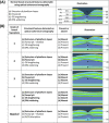
Fig. 3. Foveal tomograms obtained from children, which demonstrate some of the diagnostic features seen in INS on OCT imaging.
(Adapted from [16]). a Typical foveal hypoplasia, where there is continuation of the normally absent IRLs (outlined in white) in a case of albinism. b Atypical foveal hypoplasia in achromatopsia where, in addition to foveal hypoplasia (IRLs outlined in white), there is Inner segment ellipsoid band (ISE) disruption and a hypo-reflective zone (white circle). c Normal foveal morphology. d Retinal dystrophy consisting of: absent rod photoreceptors and ISE (small white arrows), ONL thinning and abnormal lamination of the inner retinal layers seen in a case of microcephaly lymphoedema and chorioretinal dysplasia.
Fig. 4
Fig. 4. Grading foveal hypoplasia.
An algorithm for grading foveal hypoplasia on the basis of OCT findings. Adapted from [33].
Fig. 5
Fig. 5. Examples of the use of eye tracking in clinical cases.
a Schematic of idealised horizontal jerk nystagmus waveforms showing (top row) accelerating slow phases (ASP’s) that are almost pathognomonic for infantile nystagmus (INS), and bottom row decelerating slow phases (DSP’s), which are typically seen in FMNS and acquired gaze-evoked nystagmus. b Example from a 6-year-old boy referred with apparent recent onset of gaze-evoked nystagmus. Urgent brain MRI was normal and there were no other neurological signs. EOM recording revealed a conjugate horizontal jerk nystagmus in lateral gaze with clear ASPs (top panel). In primary position, nystagmus was not evident clinically, but recordings showed a very fine nystagmus with frequent ASPs (bottom panel). Conclusion was INS since infancy that had been undetected due to broad null around primary position, and MRI was had not been necessary. c A 15-year-old female presented with spasms of oscillopsia and blurred vision that were correlated with clinically visible flutter-like episodes. EOM recording showed sporadic bursts of back-to-back saccadic oscillations that were predominantly horizontal. Episodes were not post-saccadic oscillations, as typically seen in ocular flutter, but were associated with spontaneous convergence and conjugate depression. Upon questioning, patient demonstrated ability to generate voluntary nystagmus with convergence at will. Precautionary brain MRI and chest X-ray were normal. Conclusion was ‘involuntary’ voluntary nystagmus or ‘eye movement tics’ [4].
Fig. 6
Fig. 6. The use of visual electrodiagnostics in diagnosing nystagmus.
A schema summarising the role of paediatric visual electrodiagnostic for aiding diagnosis in infants and children presenting with nystagmus. It is imporant to note that visual electrodiagnostic testing has other roles besides diagnosis such as evaluating potential for vision which is discussed elsewhere.
Fig. 7
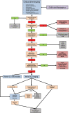
Fig. 7. A diagnostic workflow that forms the basis of our clinical practice across a number of specialist paediatric nystagmus services in the UK.
It is important to note that most cases will require additional evaluation for visual prognosis and/or monitoring (e.g. electrodiagnostics in the case with optic nerve hypoplasia) and this pathway is meant as a guide to seeking an initial diagnosis only.

References

    1. Sarvananthan N, Surendran M, Roberts EO, Jain S, Thomas S, Shah N, et al. The prevalence of nystagmus: the Leicestershire nystagmus survey. Investig Ophthalmol Vis Sci. 2009;50:5201–6. - PubMed
    1. Gottlob I, Zubcov A, Catalano RA, Reinecke RD, Koller HP, Calhoun JH, et al. Signs distinguishing spasmus nutans (with and without central nervous system lesions) from infantile nystagmus. Ophthalmology. 1990;97:1166–75. - PubMed
    1. Leigh RJ, Zee DS. The neurology of eye movements. New York: Oxford University Press; 2006.
    1. Casteels I, Harris CM, Shawkat F, Taylor D. Nystagmus in infancy. Br J Ophthalmol. 1992;76:434–7. - PMC - PubMed
    1. Osborne D, Theodorou M, Lee H, Ranger M, Hedley-Lewis M, Shawkat F, et al. Supranuclear eye movements and nystagmus in children: a review of the literature and guide to clinical examination, interpretation of findings and age-appropriate norms. Eye. 2019;33:261–73. - PMC - PubMed