Review
. 2020 Mar;18(3):177-189.
doi: 10.1038/s41579-019-0306-2. Epub 2020 Jan 9.

Plasmodium asexual growth and sexual development in the haematopoietic niche of the host

Affiliations
Review

Plasmodium asexual growth and sexual development in the haematopoietic niche of the host

Kannan Venugopal et al. Nat Rev Microbiol. 2020 Mar.

Abstract

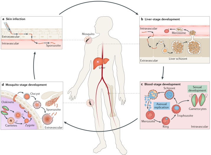
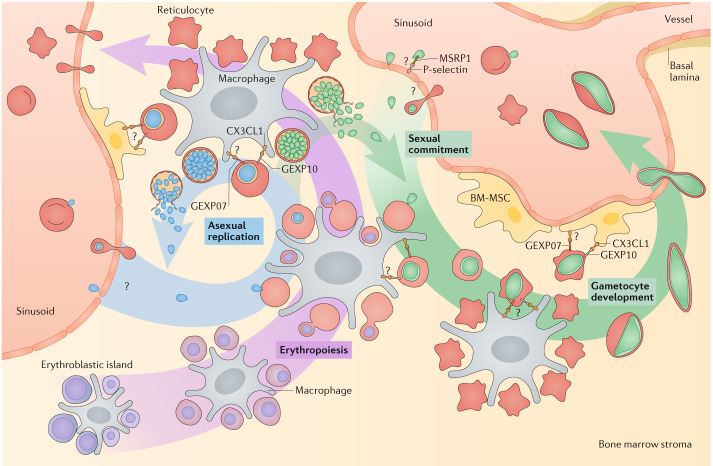
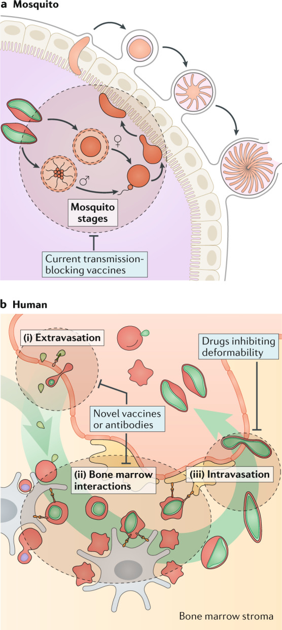
Plasmodium spp. parasites are the causative agents of malaria in humans and animals, and they are exceptionally diverse in their morphology and life cycles. They grow and develop in a wide range of host environments, both within blood-feeding mosquitoes, their definitive hosts, and in vertebrates, which are intermediate hosts. This diversity is testament to their exceptional adaptability and poses a major challenge for developing effective strategies to reduce the disease burden and transmission. Following one asexual amplification cycle in the liver, parasites reach high burdens by rounds of asexual replication within red blood cells. A few of these blood-stage parasites make a developmental switch into the sexual stage (or gametocyte), which is essential for transmission. The bone marrow, in particular the haematopoietic niche (in rodents, also the spleen), is a major site of parasite growth and sexual development. This Review focuses on our current understanding of blood-stage parasite development and vascular and tissue sequestration, which is responsible for disease symptoms and complications, and when involving the bone marrow, provides a niche for asexual replication and gametocyte development. Understanding these processes provides an opportunity for novel therapies and interventions.


Conflict of interest statement

The authors declare no competing interests.

Figures

Fig. 1
Fig. 1. Life cycle of Plasmodium falciparum in humans and mosquitoes.
a | P. falciparum sporozoites (orange) are injected into the skin during the blood meal of an infected mosquito. They will migrate to and enter a blood capillary. b | Through the bloodstream, the sporozoites reach the liver sinusoids and there they leave the blood circulation to invade a hepatocyte, after multiple transmigration events. In the hepatocyte, they undergo one asexual replication cycle that results in a liver schizont containing thousands of merozoites (yellow). The merozoites enter the bloodstream in membrane-bound structures termed merosomes. Once released, merozoites infect red blood cells (red) to initiate the intra-erythrocytic parasite cycle. c | In the blood, P. falciparum parasites undergo cycles of asexual replication (blue). After invasion of a red blood cell, they develop from ring stages to trophozoites and then to schizonts. Mature schizonts burst to release merozoites that initiate another replication cycle. A subpopulation of parasites commits to produce male and female sexual progeny or gametocytes (green). d | A female Anopheles mosquito picks up gametocytes while feeding on an infected human. Male and female gametocytes undergo gametogenesis within the midgut of the mosquito. The gametes then fertilize to form a zygote (orange), which further develops into motile ookinetes. Ookinetes cross the midgut epithelium to form an oocyst beneath the basal lamina. In the oocyst, thousands of sporozoites form, which upon bursting of the oocyst wall enter the haemolymph to invade the salivary gland. From there, sporozoites are transmitted to the next human during the subsequent mosquito bite, closing the complex life cycle of the parasite.
Fig. 2
Fig. 2. Sexual development of Plasmodium falciparum.
A subset of schizonts commit to the sexual cycle, producing sexual merozoites. Merozoites and young gametocytes (green) home to the bone marrow, leave the sinusoids and enter the parenchyma. Alternatively, the gametocytes form in the parenchyma from committed schizonts. In the bone marrow parenchyma, gametocytes develop from stage I to stage IV. Remodelling of the membrane of the host red blood cell (red) results in transient deposition of surface antigens (orange) and a reversible increase in cellular rigidity (purple). Restored deformability during maturation to stage V gametocytes triggers their release back into the bloodstream, where they can be taken up during another mosquito bite. Asexual replication in the bone marrow parenchyma most likely contributes to the accumulation of asexual parasites and sexual commitment in this compartment.
Fig. 3
Fig. 3. Intravascular sequestration of Plasmodium falciparum.
Trophozoite and schizont stages of asexual P. falciparum parasites (blue) sequester in the capillaries of several organs, including the brain, lung, spleen and bone marrow. Cytoadherence of infected red blood cells (red) to endothelial cells and to uninfected red blood cells (rosetting) facilitates sequestration. The main parasite ligand is P. falciparum erythrocyte membrane protein 1 (PfEMP1), which is exposed on the surface of infected red blood cells by knob-like structures. The different variants of PfEMP1 interact with diverse endothelial cell receptors, such as endothelial protein C receptor (EPCR), intercellular adhesion molecule 1 (ICAM1), platelet and endothelial cell adhesion molecule 1 (PECAM1) and CD36. In pregnant women, P. falciparum also sequesters in the placenta through the interaction of the PfEMP1 variant var2CSA and the placental receptor chondroitin sulfate A (CSA). Ligand receptor interactions involved in rosetting are not clearly defined, but likely involve repetitive interspersed families of polypeptides (RIFIN) and subtelomeric variant open reading frame (STEVOR) as well as PfEMP1.
Fig. 4
Fig. 4. Plasmodium falciparum development in the haematopoietic niche of the bone marrow.
In the haematopoietic niche of the bone marrow, erythropoiesis occurs in erythroblastic islands, consisting of a central macrophage surrounded by erythroid cells (purple). Coinciding with maturation of the erythroid cells from polychromatic to orthochromatic nucleated red blood cell precursors and then to reticulocytes, erythroblastic islands move closer to the sinusoids. When the reticulocytes have lost their cell nucleus, they are eventually released and enter (either through or between endothelial cells) into the sinusoidal lumen. The asexual parasite cycle in the bone marrow parenchyma is likely established both by influx of asexual merozoites (blue) or ring stages from the sinusoids and by a genuine asexual cycle in the bone marrow. In the parenchyma, asexual parasites may invade and develop in association with erythroblastic islands, or with other cell types. The exported gametocyte-exported parasite proteins (GEXP07 and GEXP10) on the surface of infected red blood cells interact with the host chemokine fractalkine (CX3CL1), which is expressed on different host cells including bone marrow mesenchymal stem cells (BM-MSCs), providing a potential mechanism by which parasites are retained in the bone marrow parenchyma. Gametocytes (green) mature in the bone marrow and are derived either from extravasated sexual merozoites or rings, or from extravascular schizonts that commit to produce sexual progeny in the bone marrow environment. Most gametocytes associate with erythroblastic islands. Stage I and II gametocytes express GEXP07 and GEXP10, which might contribute to bone marrow retention by interacting with other cell types, such as the nursing macrophages and BM-MSCs. Stages III and IV are retained in the bone marrow by their high rigidity, preventing passage through the endothelium. Ultimately, mature stage V gametocytes enter the sinusoids. MSRP1, merozoite surface protein 7-related protein 1.
Fig. 5
Fig. 5. Revisiting interventions to block Plasmodium falciparum transmission.
a | Current transmission-blocking vaccines target parasite processes in the mosquito, posing formidable technical challenges. Antibodies are taken up with the few microlitres of a mosquito blood meal and require high titres in the human blood. Many of the target proteins are not expressed during human infection, and hence there is no natural boosting of the immune response. Finally, efficacy testing requires mosquito feeding assays, which are cumbersome. b | Alternatively, transmission can be blocked by targeting gametocytes (green) with stage V density in the circulating blood as a readout. Many antimalarials that are active against asexual blood-stage parasites (blue) are also active against immature gametocytes, including artemisinin and its derivatives. A few antimalarials are mostly active against stage V gametocytes, in particular primaquine. Identification of the bone marrow as a reservoir for asexual parasites and gametocytes opens new opportunities for interventions targeting both asexual parasite burden and transmission: (i) vaccines or human monoclonal antibodies could target receptor–ligand interactions required for parasite homing and extravasation, thereby blocking establishment of bone marrow infection and gametocyte development; (ii) vaccines or antibodies could also inhibit interactions at the erythroblast island or with mesenchymal stem cells, possibly triggering premature parasite release into the circulation and subsequent clearance in the spleen; and (iii) drugs that inhibit the deformability switch may lead to the accumulation of mature gametocytes in the bone marrow parenchyma and hence prevent their release into the circulation and their transmission.

References

    1. WHO. World Malaria Report 2018. https://www.who.int/malaria/publications/world-malaria-report-2018/repor... (2018).
    1. Tavares J, et al. Role of host cell traversal by the malaria sporozoite during liver infection. J. Exp. Med. 2013;210:905–915. doi: 10.1084/jem.20121130. - DOI - PMC - PubMed
    1. Amino R, et al. Quantitative imaging of Plasmodium transmission from mosquito to mammal. Nat. Med. 2006;12:220–224. doi: 10.1038/nm1350. - DOI - PubMed
    1. Mota MM, et al. Migration of Plasmodium sporozoites through cells before infection. Science. 2001;291:141–144. doi: 10.1126/science.291.5501.141. - DOI - PubMed
    1. Sturm A, et al. Manipulation of host hepatocytes by the malaria parasite for delivery into liver sinusoids. Science. 2006;313:1287–1290. doi: 10.1126/science.1129720. - DOI - PubMed

Publication types